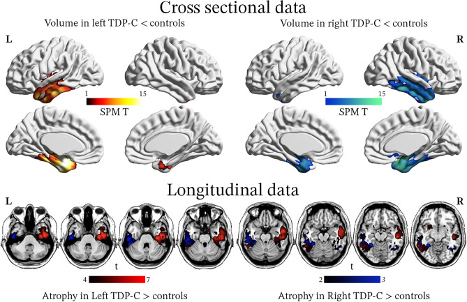
Fig. 4.
Gray matter atrophy associated with FTLD-TDP type C: left and right variants. All patients had pathology-proven TDP-C but were classified as having either a left- (n = 18) or right- (n = 12) predominant pattern of atrophy on ante mortem MRIs. At baseline, both groups show asymmetric but bilateral volume reduction in the temporal lobes, with a strong predominance in anterior and medial areas. In patients with multiple MRIs (n = 13 left- and 4 right-predominant cases), longitudinal analyses revealed that in both subgroups, atrophy progressed to the contralateral hemisphere and to more posterior temporal areas. Adapted with permission from Borghesani 2020 [179]

