Abstract
Introduction
The purpose of this study was twofold. First, the efficacy of arthroscopic repair in patients with full thickness, isolated subscapularis tendon tears (I-STTs) or combined subscapularis tendon tears (C-STTs) involving the rotator cuff tendons was evaluated. Second, the outcomes between these two groups were compared. The influence of age and gender on the cohort clinical outcomes was also analysed. Our hypothesis was that satisfactory functional results could be obtained arthroscopically in both groups without any influence of age or gender.
Material and methods
Seventy-nine patients were enrolled: 15 with I-STTs and 64 with C-STTs. The clinical outcomes were assessed using Constant and Disabilities of the Arm, Shoulder and Hand (DASH) scores, Numeric Rating Scale (NRS) for pain and Visual Analogue Scale (VAS) for satisfaction. The subscapularis strength was assessed using a comparative dynamometric bear-hug test. Group outcomes were compared, including statistical analysis.
Results
For each group, there were no differences regarding the subscapularis strength of the operated and non-operated shoulders. A comparison of the post- with the pre-operative outcomes showed an increase in the Constant score and a decrease in the NRS. Comparing the two groups, we found no difference in strength of the operated and non-operated shoulders, but a significant difference in relation to pre-operative Constant score and pre-operative NRS. Age was negatively correlated with both pre-operative and post-operative Constant scores. No association was found between gender and the outcomes, although the DASH score was higher in women.
Conclusions
Arthroscopic repair of STTs provided functional restoration, pain relief and patient satisfaction in both groups. Age and gender did not affect the clinical outcomes achieved by arthroscopic STT repair.
Keywords: arthroscopy, Constant score, rotator cuff, subscapularis tendon tears, subscapularis repair
Introduction
Symptomatic rotator cuff tears (RCTs) are one of the major origins of shoulder pain and functional impairment. The subscapularis is as strong as the other three muscles of the rotator cuff together, and its integrity is therefore crucial in maintaining net joint reaction force and glenohumeral stability [1].
Subscapularis tendon tears (STTs) are not as uncommon as they were thought to be in the past [2], with an estimated prevalence from 27.4% [3] to 43% [4] among all RCTs, and incidence reported to be up to 37% [5]. In the literature, the relative prevalence of isolated STTs (I-STTs) is reported from 17% [6] to 25% [7], while the combined STTs (C-STTs) are the most common, occurring in conjunction with supraspinatus tears, such as anterosuperior RCTs or as massive RCTs [6, 7]. There are many options for the treatment of SSTs. Initially, most tears should be treated conservatively, especially if the tear is partial or has a chronic onset. Conservative solutions are activity modifications, physiotherapy, nonsteroidal anti-inflammatory drugs (NSAIDs) and steroid injections. Surgery should be reserved for cases of persistent pain and/or loss of function despite 3–6-month accurate conservative management. Surgical treatment can be performed either by an open approach or by arthroscopic techniques [8]. Arthroscopic surgery for subscapularis reparation was first described in 2002 by Burkhart and Therany and showed brilliant short-term results [9]. Some years later, Lafosse et al. introduced a 5-type classification of STTs and described a surgical procedure for the arthroscopic repair of these lesions depending on their classification [10]. Additional studies have confirmed satisfactory mid- and long-term functional outcomes [11–14].
The influence of age on the outcomes of RCTs has been analysed by several authors, concluding that good results could be achieved even in the elderly [15, 16]. Adams et al. reported that none of their post-operative outcome evaluations was significantly associated with age [11]. Regarding gender as a prognostic factor for RCTs, the published studies have used distinct scores reporting different conclusions [17, 18]. However, no studies have investigated the influence of gender on STTs.
The primary aim of the present, prospective study was first to evaluate the efficacy of arthroscopic repair in two groups of patients with full thickness I-STTs or C-STTs involving the rotator cuff tendons and secondly, to compare post-operative outcomes between these two patient groups. Further, the influence of age and gender on the cohort functional outcomes was assessed. Our hypothesis was that satisfactory functional results could be obtained arthroscopically in both groups without any influence of age or gender.
Material and methods
Patients
Between January 2010 and December 2014, a consecutive series of Caucasian patients with diagnosis of unilateral, full thickness STT, resistant to conservative treatment, were prospectively enrolled in this study in our hospital and divided into two groups according to injury type: I-STTs and C-STTs. All subjects participating in this study received a thorough explanation of the risks and benefits of inclusion and gave their oral and written informed consent to publish the data. This study was performed in accordance with the ethical standards of the 1964 Declaration of Helsinki as revised in 2000 and those of Good Clinical Practice after approval by the local ethical committee.
Inclusion and exclusion criteria
The inclusion criteria were as follows: the pre-operative diagnosis of at least a full thickness tendon tear of the superior third of the subscapularis (Lafosse II to V [10]) on the basis of physical examination and confirmed by magnetic resonance imaging (MRI); failure of conservative treatment, involving a period of relative rest, nonsteroidal anti-inflammatory medication, physiotherapy (at least 3 months), corticosteroid injections (more than 2); functional impairment or unacceptable pain for a minimum of 3 months; arthroscopic treatment at our institution by the same shoulder surgeon; a minimum follow-up period of 24 months; and ability to provide informed consent for participation. Specific patients’ exclusion criteria were partial superior third lesion of the subscapularis tendon (Lafosse I), severe glenohumeral osteoarthritis, neurological diseases, presence of tumours, infections and previous surgery or revision repairs on the ipsilateral shoulder.
Pre-operative evaluation
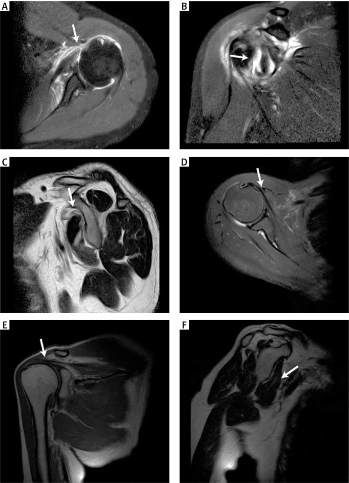
For each patient, a detailed clinical pre-operative examination was performed, including inspection, range of movement (ROM), internal rotation (IR) strength and a special test of both shoulders. Passive and active ROM of the shoulder (IR, external rotation and forward flection) were assessed, noting any deficiencies relative to a normal contralateral shoulder. The clinical suspicion of a tear was confirmed in all patients by pre-operative radiological evaluation, including X-rays and MRI of the affected shoulder (Figure 1). The Constant score was completed, and a final score between 0 (no functionality) and 100 (full functionality) was obtained [19]. It consisted of individual scores for pain (0–15 points), activity of daily living (0–20 points), movement (0–40 points) and strength (0–25) [19]. Pain was rated using a numeric rating scale (NRS, range 15–0 points), according to the scheme of the Constant score. All patients received a standard pre-operative assessment by standard radiographs, including anteroposterior views (with the arm in internal rotation, external rotation, and neutral rotation with the patient standing) as well as an axillary view. Cuff tears were evaluated with MRI scans, including oblique coronal, oblique sagittal and axial T2-weighted spin-echo sequences. Final confirmation of the lesion was made intraoperatively.
Figure 1.
Magnetic resonance images of subscapular tears highlighted by the white arrow. A–C – Acute traumatic isolated subscapularis tear (Lafosse IV) and perilesional oedema. D–F – Subscapularis (Lafosse II) and supraspinatus tears in degenerated tendons with marked muscular hypotrophy
Surgical technique
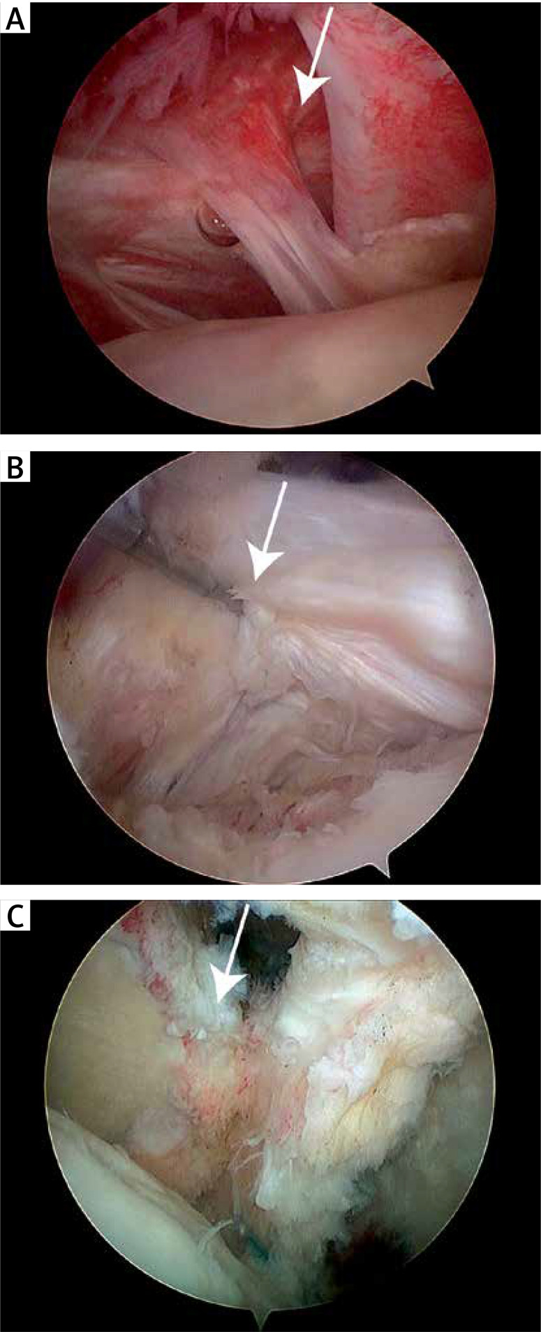
All of the arthroscopic operations were carried out by the same experienced shoulder surgeon, the senior author, in day surgery using the Burkhart technique [9]. General anaesthesia associated with an interscalene block was performed. All procedures were carried out with patients in the beach-chair position with 1–2 kg of traction applied to the arm to be operated on. Joint irrigation was provided by 4 l saline bags using an arthroscopic pump. The first surgical step consisted in a rigorous anatomic and intra-articular assessment performed in each case through a standard posterior viewing portal. Manipulating the arm in internal-external rotation and in abduction, the joint was examined to identify the status of the long head of the biceps tendon and STT, allowing arthroscopic diagnosis of its lesion according to the Lafosse classification (Figure 2) [10]. The STT was repaired as the first stage in the repair sequence before repair of any other tendons. Four portals were used for the procedure: an anterior one was used for placement of the anchor into the lesser tuberosity and suture passage; this step was carried out in association with an accessory portal between the two portals; the anterolateral portal was used as a working portal for subscapularis mobilisation, preparation of the bone bed, traction sutures, coracoplasty instrumentation, release, repair and as a viewing portal for complete tendon lesions; while the posterior portal was used for the arthroscope. The reinsertion of the subscapularis tendon was performed by either 1 or 2 screw-type anchors (TWINFIX Ultra HA Suture Anchor 5.5 mm × 20 mm, Smith & Nephew) depending on the inferior extension of the tear, fitted with 2 double-mounted sutures. In order to enhance tendon-to-bone healing, a previous adequate spongialization of the footprint was carried out [20]. After the reinsertion, coracoplasty was performed in a minority of the cases with a coracohumeral distance of less than 7 mm to prevent coracoid impingement syndrome [21–24]. Long head biceps tenotomy was routinely performed in each operation, not only in cases of complete intra-articular luxation of the tendon [25, 26]. Finally, in C-STTs, reinsertion of the posterosuperior cuff was performed using the single row technique with one to three anchors, depending on the size of the tear.
Figure 2.
Arthroscopic images of subscapularis tendon tears (highlighted by the arrows) classified according to Lafosse. A – Lafosse II, B – Lafosse III, C – Lafosse IV
Post-operative protocol and rehabilitation
All patients followed the same post-operative protocol and were checked in the same standardised manner by one of the senior authors not involved in the surgical procedure. An abduction shoulder brace was applied immediately after the operation and maintained 24 h/day for the first 3 weeks post-operatively, with the exception of the physiotherapeutic sessions. The rehabilitation of wrist and elbow was started immediately. From the second post-operative day, passive-assisted physiotherapeutic mobilisation without load was started at home with flexion and extension exercises lasting for 30 min, gradually increasing and continuing after discharge for a month. External rotation was avoided during the first 3 weeks, and then the ROM in external rotation was gradually increased. After 6 weeks, active mobilisation was allowed, and patients were encouraged to continue self-rehabilitation with the only limitation being to avoid holding heavy objects for an additional 6 weeks. Strengthening exercises were introduced 12 weeks after the operation. After that, no specific additional physiotherapy was suggested before resuming daily activities. During this post-operative period, analgesic therapy of etoricoxib (90–60 mg, 1 cp/day) was prescribed for 2 weeks, also for heterotopic ossification prophylaxis. In case of persistent pain, paracetamol/codeine phosphate (1 g, max × 2/day) was suggested. All patients were checked during the post-operative period according to our follow-up aftercare algorithm. They were seen in our out-patient clinic once a week for the first 2 weeks after discharge. The first visit was 8 days after surgery for suture medication, while the stitches were removed after 2 weeks. The shoulder brace was removed definitively 3 weeks after surgery.
Post-operative evaluation
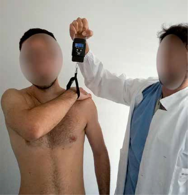
Data collection was performed at our institution by two external and independent investigators, the junior authors, who were not involved in the patients’ treatment. Patients’ anthropometric data were collected from the electronic database of the hospital, including gender, body mass index (BMI), affected side, dominant arm, trauma, comorbidities and the American Society of Anaesthesiologists (ASA) classification to globally estimate surgical risk, trauma or degenerative disease. The strength of the subscapularis muscle was assessed during the bear-hug test (BHT) using a dynamometer as follows: the dynamometer was connected to an armband, and the patient was then asked to resist as the author pulled up the dynamometer with a perpendicular force to the wrist (Figure 3). The minimal force required to lift the palm from the opposite shoulder was read on the dynamometer and recorded. For each subject, the test was repeated 5 times for each side; the mean dynamometer reading of the strength of the subscapularis for each shoulder was then calculated. The subscapularis strength of the operated shoulder was compared to the subscapularis strength of the non-operated one by the comparative dynamometric bear-hug test [27].
Figure 3.
Bear-hug test setup: test was performed with the palm of the involved side placed on the opposite shoulder. A dynamometer was connected with an armband to the patient’s wrist. The patient was then asked to hold that position (resisting internal rotation) as the physician tried to pull the patient’s hand from the shoulder with an external rotation force applied perpendicularly to the wrist. The minimal force required to lift the palm from the shoulder was read on the dynamometer
Clinical function was assessed based on the Constant score, including the Numeric Rating Scale (NRS) for pain as it is depicted in the Constant score [19]. Furthermore, patient satisfaction with regard to the surgery was evaluated using the Visual Analogue Scale (VAS) with a numeric scale ranging from 0 (unsatisfied) to 10 (very satisfied) [28]. Specifically, 0–3 was considered unsatisfied, 4–6 barely satisfied, 7–8 satisfied and 9–10 very satisfied. Finally, patients were asked to complete the Disabilities of the Arm, Shoulder and Hand (DASH) self-assessing questionnaire [29]. Rating 30 different symptoms and disabilities in daily living, a final score between 0 (no disability) and 100 (full disability) was obtained. To assess the influence of age on the post-operative outcome, the population was divided into three categories depending on the age at the time of surgery: < 50, ≥ 50 ≤ 65, > 65.
Statistical analysis
An a priori power analysis to calculate the sample size was performed using the G power program, Version 3.1.9.4 (Heinrich-Heine-Universität Düsseldorf, Düsseldorf, Germany) [30] with an allocation rate of 1 : 4 for the two groups, based on the literature [9, 31, 32]. We performed a two-tailed Mann-Whitney test, with a power of 80%, an α of 0.05 and an effect size of 0.8, resulting in 17 patients for the I-SST group and 67 patients for the C-STT group, for a total of 85 patients.
Statistical analyses were performed by an independent statistician from the Department of Statistics at our university, using SAS 9.2 (SAS Institute Inc., Cary, NC, USA) for Windows. In all statistical analyses, scores and VAS numbers were treated as quantitative variables. The Shapiro-Wilk test was used to determine whether quantitative data were distributed normally. As normality was rejected for almost all clinical and functional quantitative features and taking into account the limited sample size in post hoc analyses, the statistical comparisons were based on non-parametric techniques. Consistently, mean ± standard deviation (SD) with median, first and third quartiles was used to summarise the variables in the tables. Age at time of surgery was analysed as a continuous variable (in years) or divided into three categories (< 50, 50–65, > 65 years). Depending on the study design, the comparison of mean values of the quantitative outcomes after and before surgery, or between the operated and non-operated shoulder, was performed by applying the Wilcoxon signed-rank test. Meanwhile, the comparison of the mean values between two independent groups (women vs. men or isolated vs. multiple lesions) was performed using the Mann-Whitney test. When evaluating the association of the age groups or Lafosse classification and the clinical outcomes, the Kruskal-Wallis test was used to compare mean values of continuous variables. Spearman’s correlations were performed to analyse the linear association between quantitative variables. Multivariable linear regression models using generalised linear models were built to identify the potential predictors of the post-surgery Constant score. The models provided the estimate of parameters and adjusted p-values. For qualitative/grouped variables, frequency distributions were compared by means of the χ2 test or Fisher’s exact test, as appropriate. The significance level (p) was set at 0.05 (p value < 0.05).
Results
Patient data
From 2010 to 2014, 121 patients (125 shoulders) with a full thickness STT underwent arthroscopic surgery repair. We could not evaluate 42 patients: 5 of the I-STT group (it was not possible to contact 2 patients, 1 patient moved to another country, 1 died and 1 refused to participate in the post-operative follow-up), and 37 of the C-STT group (it was not possible to contact 12 patients, 2 died, 14 patients refused to participate in the follow-up visit because they were fully recovered, 2 moved to another city, 2 were involved in car accidents with clavicle or acromion fractures of the operated shoulder, 1 underwent arm amputation for bone cancer, and 2 were diagnosed with neurological disorders). Hence, 79 patients completed the post-operative clinical evaluation at a minimum follow-up of 2 years (mean follow-up time: 53 months; range 24–74 months). At the time of surgery, the mean age of the patients was 60.3 years (range: 38–76 years) (Table I). The mean body mass index (BMI) was 26.36 (range: 21.32–30.87). There were 33 women included in the study (42%) and 46 men (52%). In our cohort, the main cause of tendon tear was age-related degeneration (42 patients, 53% of all patients), while a traumatic injury was found in the medical history of 47% of cases. The STTs were isolated in 15 (19%) cases, while in 64 (81%) cases, other tendons were also involved (19 involved the subscapularis and supraspinatus (24%), and 45 were massive, including infraspinatus (57%)). According to the Lafosse classification, in our sample 10 type II, 24 type III, 31 type IV and 14 type V lesions were found. The subscapularis tendon was repaired in all of the cases (100%). Long head biceps tenotomy was performed in 79 shoulders (100%), while coracoplasty was performed on 18 (23%) shoulders.
Table I.
Demographic and clinical data of patient series (n = 79)
| Variable | Value |
|---|---|
| Age, mean ± SD [years] | 60.3 ±10.0 |
| Sex, n (%): | |
| Male | 46 (58) |
| Female | 33 (42) |
| BMI, mean ± SD [kg/m2] | 26.36 ±3.87 |
| Affected side, n (%): | 60 (76) |
| Right | 19 (24) |
| Left | |
| Dominant extremity, n (%): | |
| Right | 73 (92.4) |
| Left | 6 (7.6) |
| Aetiology, n (%): | |
| Injury | 37 (47) |
| No injury | 42 (53) |
| Type of rotator cuff lesion, n (%): | |
| Isolated STTs | 15 (19) |
| Multiple rotator cuff tears | 64 (81) |
| Type of STT lesion (according to Lafosse), n (%): | |
| II | 10 (13) |
| III | 24 (30) |
| IV | 31 (39) |
| V | 14 (18) |
| Coracoplasty, n (%) | 18 (23) |
| Co-morbidities, n (%): | |
| Diabetes mellitus | 10 (13) |
SD – standard deviation, BMI – body mass index, STT – subscapularis tendon tear.
Functional outcomes of the cohort
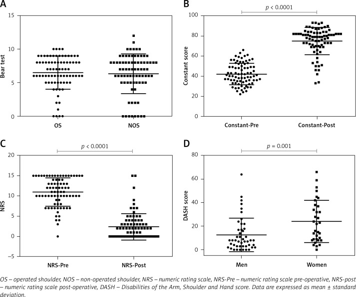
The mean post-operative strength of the operated shoulder measured by BHT was 6.49 ±2.46 kg, whereas the mean strength of the non-operated shoulder was 6.33 ±2.92 kg. The difference was not significant (p = 0.69) (Table II, Figure 4). Post-operatively, at follow-up, the mean Constant score (Constant-Post) was 74.8 ±13.5 points (range: 39–93). The difference between the mean pre-operative Constant score (Constant-Pre) and the mean Constant-Post (Constant-Dif) was on average 32.7 ±10.6 points (p < 0.0001). The mean DASH score was 17.4 ±16.8. The DASH score of women was higher (24.1 ±18.0) compared to that of the men (12.6 ±14.2) (p = 0.001). The NRS score decreased significantly by 8.5 ±4.2 points (NRS-Dif), from 10.9 ±3.5 points pre-operatively (NRS-Pre) to 2.4 ±3.2 points post-operatively (NRS-Post) (p < 0.0001). Concerning the satisfaction of the patients, 55 out of 79 patients were very satisfied (69.6%), 21 were satisfied (26.6%), 1 was barely satisfied and 2 were unsatisfied. These results are summarised in Table II. No statistically significant differences were found comparing the pre- and post-operative scores, dividing the patients according to the Lafosse classification.
Table II.
Clinical and functional outcome results
| Parameter | Overall mean values ± SD | Median (Q1–Q3) | P-value |
|---|---|---|---|
| Dynamometric bear-hug test [kg]: | |||
| Operated shoulder | 6.49 ±2.46 | 6.5 (5–8) | 0.69* |
| Non-operated shoulder | 6.33 ±2.92 | 7.0 (5–8) | |
| Difference | 0.16 ±2.59 | 0.0 (–1 to 1) | |
| Constant score: | |||
| Pre-operative | 42.1 ±10.6 | 41 (35–50) | < 0.0001* |
| Post-operative | 74.8 ±13.5 | 79 (70–83) | |
| Difference (post-pre) | 32.7 ±10.6 | 35 (25–40) | |
| NRS: | |||
| Pre-operative | 10.9 ±3.5 | 11 (9–14) | < 0.0001* |
| Post-operative | 2.4 ±3.2 | 2 (0–4) | |
| Difference (post-pre) | –8.5 ±4.2 | –9 (–12 to –6) | |
| VAS Satisfaction | 8.7 ±1.4 | 9 (8–10) | |
| DASH score: | |||
| Men and women | 17.4 ±16.8 | 11 (4–27) | 0.001 (men vs. women)‡ |
| Women (n = 33) | 24.1 ±18.0 | 22 (9–37) | |
| Men (n = 46) | 12.6 ±14.2 | 8 (3–17) | |
NRS – Numeric Rating Scale, DASH – Disabilities of the Arm, Shoulder and Hand score.
P-value provided by Wilcoxon’s signed-rank test.
P-value provided by Mann-Whitney test.
Figure 4.
Clinical outcomes. A – Bear-hug test, B – Constant score, C – NRS and D – DASH score of enrolled patients
Functional outcomes of the I-STT group
In analysing I-STT patients, no significant differences in the strength between the operated and the non-operated shoulder were found (p = 0.719). The Constant-Post was significantly increased compared to the Constant-Pre (p = 0.001). The NRS-Post was significantly decreased compared to the NRS-Pre (p = 0.001).
Functional outcomes of the C-STT group
The strength between the operated and the non-operated shoulder of the C-STT patients was comparable (p = 0.929), while there was an increase of the Constant-Post compared to the Constant-Pre (p < 0.0001). The NRS-Post decreased significantly compared to NRS-Pre (p < 0.0001).
Comparison between I-STT and C-STT groups
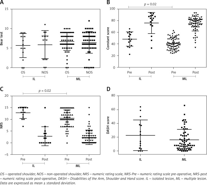
Comparing patients with I-STTs and patients with C-STTs, the Constant-Pre and NRS-Pre were significantly higher in the first group (p = 0.02 and p = 0.02, respectively). The mean Constant-Dif of the isolated lesions was 27.7 ±8.9 points, whereas the mean Constant-Dif of the combined lesions was 33.8 ±10.7 points. This difference, depending on the lesion pattern, was significant (p = 0.04). In contrast, no significant difference was found comparing the Constant-Post (p = 0.17), the NRS-Post (p = 0.92), the NRS-Dif (p = 0.10), the BHT of the operated shoulder (p = 0.27), the BHT of the non-operated shoulder (p = 0.96), the BHT-Dif (p = 0.69), the satisfaction (p = 0.15) or the DASH score (p = 0.57) between the two groups of patients. These results are summarised in Table III and Figure 5.
Table III.
Clinical and functional outcomes in isolated (I-STTs) and multiple lesions (C-STTs)
| Parameter | I-STTs (n = 15) | C-STTs (n = 64) | P-value | ||||
|---|---|---|---|---|---|---|---|
| Mean ± SD | Median (Q1–Q3) | Mean ± SD | Median (Q1–Q3) | ||||
| Dynamometric bear-hug test [kg]: | |||||||
| Operated shoulder | 5.7 ±3.1 | 6 (4–8) | 6.7 ±2.3 | 7 (6–8) | 0.27‡ | ||
| Non-operated shoulder | 5.9 ±3.6 | 7.5 (2–8) | 6.4 ±2.8 | 6 (5–8) | 0.96‡ | ||
| Difference | –0.2 ±2.1 | 0 (0–3) | 0.2 ±2.7 | 0 (–1 to 1) | 0.69* | ||
| Constant score: | |||||||
| Pre-operative | 48.3 ±12.3 | 48 (37–59) | 40.7 ±9.8 | 39.5 (33–49) | 0.02 ‡ | ||
| Post-operative | 75.9 ±18.1 | 84 (71–88) | 74.5 ±12.3 | 79 (69–82) | 0.17‡ | ||
| Difference (post-pre) | 27.7 ±8.9 | 28 (11–36) | 33.8 ±10.7 | 36 (25–41) | 0.04* | ||
| NRS: | |||||||
| Pre-operative | 12.9 ±2.4 | 13 (11–15) | 10.5 ±3.6 | 10 (8–13) | 0.02 ‡ | ||
| Post-operative | 2.8 ±4.1 | 2 (0–4) | 2.3 ±3.1 | 2 (0–3) | 0.92‡ | ||
| Difference (post-pre) | –10.1 ±4.1 | –10 (–14 to –8) | –8.2 ±4.2 | –8 (–11 to –4) | 0.10* | ||
| VAS satisfaction | 8.7 ±1.7 | 9 (9–9) | 8.7 ±1.4 | 9 (8–10) | 0.82‡ | ||
| DASH score: | |||||||
| Men and women | 22.7 ±22.3 | 14 (3–28) | 16.2 ±15.2 | 9.5 (4.5–23.5) | 0.57‡ | ||
NRS – Numeric Rating Scale, DASH – Disabilities of the Arm, Shoulder and Hand score.
P-value provided by Wilcoxon’s signed-rank test.
P-value provided by Mann Whitney test.
Figure 5.
Clinical outcomes. A – Bear-hug test, B – constant score, C – NRS and D – DASH score of enrolled patients divided according to isolated or multiple lesions
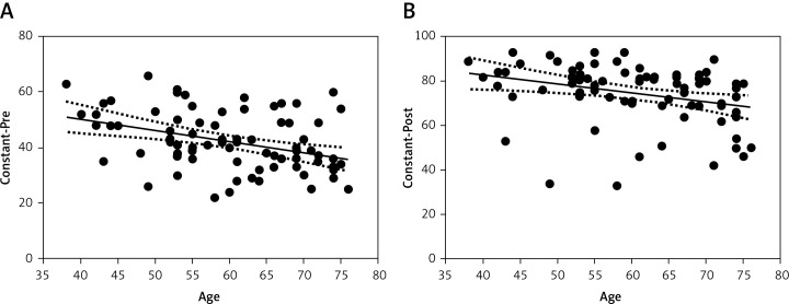
Age was negatively correlated with the Constant-Pre (Spearman’s r = –0.37, p = 0.007) and the Constant-Post (Spearman’s r = –0.39, p = 0.0003) (Figure 6). Furthermore, the patient’s age at the time of surgery was significantly associated with the number of torn tendons and lesion pattern (p < 0.001). The relationship between the three variables that are significantly correlated with each other (the type of lesion and age were the independent variables and the Constant-Post the dependent variable) was studied by a multivariable analysis showing that the type of lesion had a low influence on the Constant-Post score, which is instead significantly influenced by the patient’s age (p = 0.007). For every increase of 1 year, the Constant-Post decreases on average by 0.41 points and an average of 2 points every 5 years.
Figure 6.
Correlations between age and Constant score. A – Pre-operative constant score was negatively correlated with age (r = –0.37, p = 0.007). B – Post-operative constant score was negatively correlated with age (r = –0.39, p = 0.0003)
Even dividing the study population into 3 categories depending on the age at the time of surgery (< 50 years, 50–65 years, > 65 years), the age categories were associated with the lesion pattern (p = 0.003), BHT of the non-operated shoulder (p = 0.03), the Constant-Pre (p = 0.03) and the Constant-Post (p = 0.03) (Table IV). In contrast, no significant associations were found between age categories and BHT of the operated shoulder (p = 0.053), BHT-Dif (p = 0.69), Constant-Dif (p = 0.11), NRS-Post (p = 0.50), NRS-Dif (p = 0.28), satisfaction (p = 0.74) or DASH score (p = 0.84).
Table IV.
Clinical and functional outcomes by age categories
| Parameter | < 50 (n = 12) | ≥ 50 and ≤ 65 (n = 38) | > 65 (n = 29) | P-value | |||
|---|---|---|---|---|---|---|---|
| Lesion pattern: | |||||||
| Isolated lesions | 6 (50%) | 8 (21%) | 1 (3%) | 0.002 † | |||
| Multiple lesions | 6 (50%) | 30 (79%) | 28 (97%) | ||||
| Parameter | Mean ± SD | Median (Q1–Q3) | Mean ± SD | Median (Q1–Q3) | Mean ± SD | Median (Q1–Q3) | |
| Dynamometric BHT [kg]: | |||||||
| Operated shoulder | 7.4 ±2.8 | 9 (5–9) | 6.6 ±2.5 | 7.5 (5–8) | 6.0 ±2.0 | 6 (5–7) | 0.053¶ |
| Non-operated shoulder | 7.8 ±2.9 | 8 (7–9) | 6.2 ±3.1 | 6.5 (5–8) | 5.9 ±2.6 | 6 (5–7) | 0.03 ¶ |
| Difference | –0.4 ±1.9 | 0 (–2 to 0) | 0.4 ±2.8 | 0 (–1 to 1) | 0.1 ±2.6 | 0 (–1 to 0) | 0.69¶ |
| Constant score: | |||||||
| Pre-operative | 49.1 ±11.5 | 50 (43–56) | 42.0 ±10.2 | 42 (36–49) | 39.5 ±9.9 | 36 (33–49) | 0.03 ¶ |
| Post-operative | 77.2 ± 17.4 | 83 (74–88) | 77.0 ± 12.5 | 81 (73–84) | 71.0 ± 12.6 | 75 (64–80) | 0.03 ¶ |
| Difference | 28.1 ±9.0 | 27 (25–36) | 35.0 ±9.7 | 36 (28–41) | 31.5 ±11.8 | 32 (21–41) | 0.11¶ |
| NRS: | |||||||
| Pre-operative | 11.2 ± 3.6 | 12 (7–15) | 11.5 ± 3.0 | 12 (10–15) | 10.2 ± 4.0 | 11 (7–13) | 0.31¶ |
| Post-operative | 3.4 ± 4.4 | 2 (0.5–5) | 2.2 ± 2.7 | 2 (0–3) | 2.3 ± 3.4 | 0 (0–4) | 0.50¶ |
| Difference | –7.8 ± 4.4 | –7.5 (–11 to –5.5) | –9.3 ± 4.1 | –10 (–12 to –7) | –7.8 ± 4.2 | –8 (–11 to –5) | 0.28¶ |
| VAS satisfaction | 8.3 ± 2.0 | 9 (7.25– 10.00) | 8.8 ± 1.2 | 9 (8–10) | 8.8 ± 1.5 | 9 (8–10) | 0.74¶ |
| DASH score: | |||||||
| Men + women | 19.4 ± 20.3 | 9.5 (5.5–31.5) | 16.3 ± 17.5 | 9 (3–28) | 17.9 ± 14.5 | 14 (7–24) | 0.84¶ |
NRS – Numeric Rating Scale, DASH – Disabilities of the Arm, Shoulder and Hand score.
P-values from one-way Kruskal-Wallis test.
P-value from Fisher’s exact test only for comparison of lesion pattern.
No association was found between gender and other outcome measures: Constant-Pre (p = 0.33), Constant-Post (p = 0.09), Constant-Dif (p = 0.16), NRS-Post (p = 0.07), NRS-Dif (p = 0.54), satisfaction (p = 0.22) (Table IV).
Complications
One patient required surgery for revision rotator cuff repair due to trauma during the post-operative period. There was one superficial wound infection resolved with antibiotic treatment. There were no instances of deep wound infection or implant failure in the study group.
Discussion
Since the subscapularis (SSC) is the largest and strongest rotator cuff muscle, playing an essential role in shoulder function and stability [2–5], the repair of a tear of this tendon represents an essential factor for surgical outcomes [3, 33]. The arthroscopic technique has rapidly developed since Gerber et al. first described open repair of isolated STTs in 1996 [34], showing good clinical results [11–14, 35, 36]. The main aim of this study was to evaluate medium- to long-term clinical and functional outcomes of a consecutive series of patients with diagnosis of isolated or combined STTs treated arthroscopically. The efficacy of arthroscopic subscapularis repair was then evaluated by comparing the post-operative results among I-STT and C-STT patient groups. Finally, the influence of age and gender on cohort clinical outcomes was analysed.
This study showed how the majority of STTs, isolated or combined, can be successfully repaired arthroscopically with a low rate of complications. In our cohort, with a predominance of type III and type IV lesions, good and excellent functional and clinical outcomes were reported at a mean follow-up of 53 months. Previously, encouraging arthroscopic results have also been reported by several other authors [6, 10, 37–39] with comparable results [6, 9–14, 37–39], even when the uncommon I-STTs were analysed [40]. However, for some clinical aspects, our functional results were even better than those reported in the literature. The mean improvement of the Constant score was higher (32.7 points) than that reported by Lafosse et al. (26.6), Toussaint et al. (25.8–27.2), and Seppel et al. (26.4) in their studies [10, 14, 41]. In particular, Ide et al. described a series of 20 patients with combined traumatic RCTs, including the subscapularis [38]. At a mean 3-year follow-up, functional outcome improved in a similar way to that of the previously mentioned reports. At a mean follow-up of nearly 9 years, functional outcome was maintained compared with previous reports [9, 11], and rates of patient satisfaction and return to activity were both over 90%. In our series, even the reduction of pain was on average greater than in many other studies [10, 12]. The majority of patients were very pleased with the operation. This overall satisfaction rate is higher than those reported by Adams et al. (88%), Denard et al. (92.4%) and Seppel et al. (88.2%) [11, 12, 41].
Technically, we found that the number of anchors used for STT repair did not influence the tendon stability and force, or the outcomes of our series. Also dividing our cohort into two groups according to the extension of the tears and the number of anchors used (Lafosse II and Lafosse III repaired by 1 anchor; Lafosse IV and V repaired by 2 anchors), there was no statistical association between the number of these sutures and post-operative outcomes. Despite the significant increase in publications concerning this topic, there is still controversy regarding the influence of the different number of anchors and repair configurations proposed (single row, double row, suture bridge) on the biomechanical and clinical results [42–45].
The routinely performed adjuvant surgical procedures during arthroscopy, such as biceps tenotomy and acromioplasty (in some cases), may have positively influenced the pain perception and clinical outcomes. Several studies have shown that patients with irreparable rotator cuffs benefit from these adjuvant procedures, in particular when the long head biceps tenotomy is systematically performed [46], rather than from the repair of the rotator cuff itself [47].
A significant association was found between the pattern of the tear (isolated or combined subscapularis), and the mean Constant-Dif, unlike Gerhardt et al. and Miškulin et al. [35, 36]. The clinical improvement was greater in the C-STT group than in the I-STT group. This is probably due to the worse pre-operative anatomical damage and consequently worse clinical condition of the multiple lesions compared to the isolated ones. The mean Constant-Pre of the I-STT group was higher (48.3) than the mean Constant-Pre of the C-STT group (40.7). This is in accordance with the study of Kukkonen et al., in which intraoperatively detected tear size correlated significantly with pre-operative Constant score (r = –0.20, p < 0.0001), but there was an even stronger and more significant correlation between tear size and final post-operative Constant score (r = –0.36, p < 0.0001) [48].
As described by Lafosse et al. [13], a dynamometric BHT was used to measure the subscapularis strength, finding that the operated side showed significantly lower strength than the non-operated side (p = 0.031). In contrast, no substantial difference comparing the strength of the operated shoulder with the strength of the non-operated one was found in our cohort (p = 0.69). This difference can be explained by the different number of enrolled patients (23 vs. 79) and different follow-up time-points (32 vs. 53). Another reason could be found in the aetiology of the rotator cuff tendon pathology, as this condition is mainly caused by the intrinsic degeneration of the tendons or by repetitive traumas. Hence, we hypothesized that, after some years, patients develop a pathological subscapularis on the non-operated shoulder due to the same underlying degenerative pathology that is affecting both shoulders. This “metachronous” rotator cuff degeneration of the contralateral shoulder was perhaps silent in many cases in the study population of Lafosse et al. because of the relative short follow-up time. This opinion is supported by several recent studies [8, 49–51], such as that of Moosmayer et al., which shows that a substantial percentage of asymptomatic RCTs became symptomatic and underwent anatomic deterioration during a relatively short-term follow-up [52]. Hence, we can affirm that in terms of strength, the operated subscapularis tendon recovers after the operation to the same degree as the contralateral subscapularis, proving the efficacy of an arthroscopic restoration of the subscapularis anatomy.
A negative correlation between age and Constant score values, both pre- and post-operatively, was found. Further, our analysis showed a significant association between the three age categories and both Constant values, pre- and post-operatively. Since shoulder function worsens with age, the Constant score is affected by age. In contrast, the Constant-Dif was not influenced by age at the time of surgery, suggesting that a good relative clinical improvement can also be achieved in elderly people. Previous studies concerning RCTs have claimed that clinical improvement does not depend on different age categories [15, 16, 53–56] despite the higher rate of rotator cuff re-tears and the lower rate of tendon healing at 6-month MRI follow-up found in the elderly [16, 57]. To the best of our knowledge, only two studies have been published investigating the influence of age on the clinical outcomes of patients with STTs divided by injury type [11, 58]. Adams et al. did not find any correlation between age and clinical outcomes (UCLA, VAS and ASES scores) [11], while Rhee et al. found a correlation with Constant, ASES, and Simple Shoulder Test scores [58].
The hypothetical worse post-operative recovery of RCTs in women is still controversial in the literature [17, 37, 59, 60]. Our work consolidates the hypothesis that men and women have similar clinical improvement. In fact, the Constant score was not affected by gender. However, we report that women showed on average a DASH score two times lower than men (24.1 vs. 12.6).
Although the main strengths of our study are the large population (79 cases) and the long mean follow-up time (53 months), it presents some limitations. First, as most of the STTs were associated with other RCTs (C-STTs), this bias could have affected the results but not its clinical relevance as most STTs were not isolated. Second, we did not have comparable strength measurements of the affected shoulder pre- and post-operatively, because we performed the dynamometric BHT only post-operatively, as well as the DASH score. Third, another weakness was the lack of a control group, which would have been useful to compare the results of this arthroscopic technique. Finally, we did not routinely perform MRI or ultrasound of the affected shoulder in the follow-up. Thus, we lacked information about the tendon healing rate and the recovery of muscle atrophy and fatty infiltration.
In conclusion, our results showed that arthroscopic repair technique of STTs is an effective procedure, providing pain relief, function restoration and patient’s satisfaction for both categories of lesions. Comparing the clinical aspects of the shoulder before and after surgery, a statistically significant improvement among patients of the C-STT group with respect to patients of the I-STT group was found. Furthermore, the comparative dynamometric BHT of subscapularis strength did not show significant differences between the operated shoulder and the non-operated one. The patient’s age was associated with the presence of C-STTs, and inversely correlated with the Constant score. Finally, gender did not affect the objective clinical improvement achieved arthroscopically, even if the DASH score was lower among women.
Conflict of interest
The authors declare no conflict of interest.
References
- 1.Keating JF, Waterworth P, Shaw-Dunn J, Crossan J. The relative strengths of the rotator cuff muscles. A cadaver study. J Bone Joint Surg Br. 1993;75:137–40. doi: 10.1302/0301-620X.75B1.8421011. [DOI] [PubMed] [Google Scholar]
- 2.Deutsch A, Altchek DW, Veltri DM, Potter HG, Warren RF. Traumatic tears of the subscapularis tendon. Clinical diagnosis, magnetic resonance imaging findings, and operative treatment. Am J Sports Med. 1997;25:13–22. doi: 10.1177/036354659702500104. [DOI] [PubMed] [Google Scholar]
- 3.Arai R, Sugaya H, Mochizuki T, Nimura A, Moriishi J, Akita K. Subscapularis tendon tear: an anatomic and clinical investigation. Arthroscopy. 2008;24:997–1004. doi: 10.1016/j.arthro.2008.04.076. [DOI] [PubMed] [Google Scholar]
- 4.Adams CR, Schoolfield JD, Burkhart SS. Accuracy of pre-operative magnetic resonance imaging in predicting a subscapularis tendon tear based on arthroscopy. Arthroscopy. 2010;26:1427–33. doi: 10.1016/j.arthro.2010.02.028. [DOI] [PubMed] [Google Scholar]
- 5.Garavaglia G, Ufenast H, Taverna E. The frequency of subscapularis tears in arthroscopic rotator cuff repairs: a retrospective study comparing magnetic resonance imaging and arthroscopic findings. Int J Shoulder Surg. 2011;5:90–4. doi: 10.4103/0973-6042.91000. [DOI] [PMC free article] [PubMed] [Google Scholar]
- 6.Bennett WF. Arthroscopic repair of isolated subscapularis tears: a prospective cohort with 2- to 4-year follow-up. Arthroscopy. 2003;19:131–43. doi: 10.1053/jars.2003.50053. [DOI] [PubMed] [Google Scholar]
- 7.Barth J, Audebert S, Toussaint B, et al. Diagnosis of subscapularis tendon tears: are available diagnostic tests pertinent for a positive diagnosis? Orthop Traumatol Surg Res. 2012;98(8 Suppl):S178–85. doi: 10.1016/j.otsr.2012.09.008. [DOI] [PubMed] [Google Scholar]
- 8.Mall NA, Chahal J, Heard WM, et al. Outcomes of arthroscopic and open surgical repair of isolated subscapularis tendon tears. Arthroscopy. 2012;28:1306–14. doi: 10.1016/j.arthro.2012.02.018. [DOI] [PubMed] [Google Scholar]
- 9.Burkhart SS, Tehrany AM. Arthroscopic subscapularis tendon repair: technique and preliminary results. Arthroscopy. 2002;18:454–63. doi: 10.1053/jars.2002.30648. [DOI] [PubMed] [Google Scholar]
- 10.Lafosse L, Jost B, Reiland Y, Audebert S, Toussaint B, Gobezie R. Structural integrity and clinical outcomes after arthroscopic repair of isolated subscapularis tears. J Bone Joint Surg Am. 2007;89:1184–93. doi: 10.2106/JBJS.F.00007. [DOI] [PubMed] [Google Scholar]
- 11.Adams CR, Schoolfield JD, Burkhart SS. The results of arthroscopic subscapularis tendon repairs. Arthroscopy. 2008;24:1381–9. doi: 10.1016/j.arthro.2008.08.004. [DOI] [PubMed] [Google Scholar]
- 12.Denard PJ, Jiwani AZ, Ladermann A, Burkhart SS. Long-term outcome of a consecutive series of subscapularis tendon tears repaired arthroscopically. Arthroscopy. 2012;28:1587–91. doi: 10.1016/j.arthro.2012.02.031. [DOI] [PubMed] [Google Scholar]
- 13.Lafosse L, Lanz U, Saintmard B, Campens C. Arthroscopic repair of subscapularis tear: surgical technique and results. Orthop Traumatol Surg Res. 2010;96(8 Suppl):S99–108. doi: 10.1016/j.otsr.2010.09.009. [DOI] [PubMed] [Google Scholar]
- 14.Toussaint B, Audebert S, Barth J, et al. Arthroscopic repair of subscapularis tears: preliminary data from a prospective multicentre study. Orthop Traumatol Surg Res. 2012;98(8 Suppl):S193–200. doi: 10.1016/j.otsr.2012.10.004. [DOI] [PubMed] [Google Scholar]
- 15.Osti L, Papalia R, Del Buono A, Denaro V, Maffulli N. Comparison of arthroscopic rotator cuff repair in healthy patients over and under 65 years of age. Knee Surg Sports Traumatol Arthrosc. 2010;18:1700–6. doi: 10.1007/s00167-010-1081-9. [DOI] [PubMed] [Google Scholar]
- 16.Robinson PM, Wilson J, Dalal S, Parker RA, Norburn P, Roy BR. Rotator cuff repair in patients over 70 years of age: early outcomes and risk factors associated with re-tear. Bone Joint J. 2013;95-B:199–205. doi: 10.1302/0301-620X.95B2.30246. [DOI] [PubMed] [Google Scholar]
- 17.Cho CH, Ye HU, Jung JW, Lee YK. Gender affects early postoperative outcomes of rotator cuff repair. Clin Orthop Surg. 2015;7:234–40. doi: 10.4055/cios.2015.7.2.234. [DOI] [PMC free article] [PubMed] [Google Scholar]
- 18.Razmjou H, Davis AM, Jaglal SB, Holtby R, Richards RR. Disability and satisfaction after rotator cuff decompression or repair: a sex and gender analysis. BMC Musculoskelet Dis. 2011;12:66. doi: 10.1186/1471-2474-12-66. [DOI] [PMC free article] [PubMed] [Google Scholar]
- 19.Constant CR, Murley AH. A clinical method of functional assessment of the shoulder. Clin Orthop Relat Res. 1987;214:160–4. [PubMed] [Google Scholar]
- 20.Ficklscherer A, Serr M, Loitsch T, et al. The influence of different footprint preparation techniques on tissue regeneration in rotator cuff repair in an animal model. Arch Med Sci. 2017;13:481–8. doi: 10.5114/aoms.2016.60581. [DOI] [PMC free article] [PubMed] [Google Scholar]
- 21.Lo IK, Burkhart SS. The etiology and assessment of subscapularis tendon tears: a case for subcoracoid impingement, the roller-wringer effect, and TUFF lesions of the subscapularis. Arthroscopy. 2003;19:1142–50. doi: 10.1016/j.arthro.2003.10.024. [DOI] [PubMed] [Google Scholar]
- 22.Lo IK, Parten PM, Burkhart SS. Combined subcoracoid and subacromial impingement in association with anterosuperior rotator cuff tears: an arthroscopic approach. Arthroscopy. 2003;19:1068–78. doi: 10.1016/j.arthro.2003.10.016. [DOI] [PubMed] [Google Scholar]
- 23.Park JY, Lhee SH, Oh KS, Kim NR, Hwang JT. Is arthroscopic coracoplasty necessary in subcoracoid impingement syndrome? Arthroscopy. 2012;28:1766–75. doi: 10.1016/j.arthro.2012.06.013. [DOI] [PubMed] [Google Scholar]
- 24.Richards DP, Burkhart SS, Campbell SE. Relation between narrowed coracohumeral distance and subscapularis tears. Arthroscopy. 2005;21:1223–8. doi: 10.1016/j.arthro.2005.06.015. [DOI] [PubMed] [Google Scholar]
- 25.Friedman JL, FitzPatrick JL, Rylander LS, Bennett C, Vidal AF, McCarty EC. Biceps tenotomy versus tenodesis in active patients younger than 55 years: is there a difference in strength and outcomes? Orthop J Sports Med. 2015;3:2325967115570848. doi: 10.1177/2325967115570848. [DOI] [PMC free article] [PubMed] [Google Scholar]
- 26.Gurnani N, van Deurzen DF, Janmaat VT, van den Bekerom MP. Tenotomy or tenodesis for pathology of the long head of the biceps brachii: a systematic review and meta-analysis. Knee Surg Sports Traumatol Arthrosc. 2016;24:3765–71. doi: 10.1007/s00167-015-3640-6. [DOI] [PubMed] [Google Scholar]
- 27.Barth JRH, Burkhart SS, De Beer JF. The bear-hug test: a new and sensitive test for diagnosing a subscapularis tear. Arthroscopy. 2006;22:1076–84. doi: 10.1016/j.arthro.2006.05.005. [DOI] [PubMed] [Google Scholar]
- 28.Winemaker MJ, Amendola A. Comparison of bioabsorbable pins and Kirschner wires in the fixation of chevron osteotomies for hallux valgus. Foot Ankle Int. 1996;17:623–8. doi: 10.1177/107110079601701007. [DOI] [PubMed] [Google Scholar]
- 29.Germann G, Wind G, Harth A. The DASH (Disability of Arm-Shoulder-Hand) Questionnaire – a new instrument for evaluating upper extremity treatment outcome. Handchir Mikrochir Plast Chir. 1999;31:149–52. doi: 10.1055/s-1999-13902. [DOI] [PubMed] [Google Scholar]
- 30.Faul F, Erdfelder E, Lang AG, Buchner A. G*Power 3: a flexible statistical power analysis program for the social, behavioral, and biomedical sciences. Behav Res Met. 2007;39:175–91. doi: 10.3758/bf03193146. [DOI] [PubMed] [Google Scholar]
- 31.Barth JR, Burkhart SS, De Beer JF. The bear-hug test: a new and sensitive test for diagnosing a subscapularis tear. Arthroscopy. 2006;22:1076–84. doi: 10.1016/j.arthro.2006.05.005. [DOI] [PubMed] [Google Scholar]
- 32.Kim TK, Rauh PB, McFarland EG. Partial tears of the subscapularis tendon found during arthroscopic procedures on the shoulder: a statistical analysis of sixty cases. Am J Sports Med. 2003;31:744–50. doi: 10.1177/03635465030310051801. [DOI] [PubMed] [Google Scholar]
- 33.Desai VS, Camp CL, Boddapati V, Dines JS, Brockmeier SF, Werner BC. Increasing numbers of shoulder corticosteroid injections within a year preoperatively may be associated with a higher rate of subsequent revision rotator cuff surgery. Arthroscopy. 2019;35:45–50. doi: 10.1016/j.arthro.2018.07.043. [DOI] [PubMed] [Google Scholar]
- 34.Gerber C, Hersche O, Farron A. Isolated rupture of the subscapularis tendon. J Bone Joint Surg. 1996;78:1015–23. doi: 10.2106/00004623-199607000-00005. [DOI] [PubMed] [Google Scholar]
- 35.Gerhardt C, Bartl C, Voigt C, et al. Recovery of subscapularis and shoulder function following arthroscopic treatment of isolated anterior and combined anterosuperior rotator cuff lesions. Arch Orthop Trauma Surg. 2016;136:75–81. doi: 10.1007/s00402-015-2334-3. [DOI] [PubMed] [Google Scholar]
- 36.Miskulin M, Vrgoc G, Sporis G, Dulic O, Gavrilovic G, Milanovic Z. Single-row arthroscopic cuff repair with double-loaded anchors provides good shoulder function in long-term follow-up. Int Orthop. 2015;39:233–40. doi: 10.1007/s00264-014-2557-x. [DOI] [PubMed] [Google Scholar]
- 37.Bennett WF. Arthroscopic repair of anterosuperior (supraspinatus/subscapularis) rotator cuff tears: a prospective cohort with 2- to 4-year follow-up. Classification of biceps subluxation/instability. Arthroscopy. 2003;19:21–33. doi: 10.1053/jars.2003.50023. [DOI] [PubMed] [Google Scholar]
- 38.Ide J, Tokiyoshi A, Hirose J, Mizuta H. Arthroscopic repair of traumatic combined rotator cuff tears involving the subscapularis tendon. J Bone Joint Surg Am. 2007;89:2378–88. doi: 10.2106/JBJS.G.00082. [DOI] [PubMed] [Google Scholar]
- 39.Kim SH, Oh I, Park JS, Shin SK, Jeong WK. Intra-articular repair of an isolated partial articular-surface tear of the subscapularis tendon. Am J Sports Med. 2005;33:1825–30. doi: 10.1177/0363546505278259. [DOI] [PubMed] [Google Scholar]
- 40.Saltzman BM, Collins MJ, Leroux T, et al. Arthroscopic repair of isolated subscapularis tears: a systematic review of technique-specific outcomes. Arthroscopy. 2017;33:849–60. doi: 10.1016/j.arthro.2016.10.020. [DOI] [PubMed] [Google Scholar]
- 41.Seppel G, Plath JE, Volk C, et al. Long-term results after arthroscopic repair of isolated subscapularis tears. Am J Sports Med. 2017;45:759–66. doi: 10.1177/0363546516676261. [DOI] [PubMed] [Google Scholar]
- 42.Denard PJ, Ladermann A, Burkhart SS. Double-row fixation of upper subscapularis tears with a single suture anchor. Arthroscopy. 2011;27:1142–9. doi: 10.1016/j.arthro.2011.02.033. [DOI] [PubMed] [Google Scholar]
- 43.Cole BJ, ElAttrache NS, Anbari A. Arthroscopic rotator cuff repairs: an anatomic and biomechanical rationale for different suture-anchor repair configurations. Arthroscopy. 2007;23:662–9. doi: 10.1016/j.arthro.2007.02.018. [DOI] [PubMed] [Google Scholar]
- 44.Pauly S, Gerhardt C, Chen J, Scheibel M. Single versus double-row repair of the rotator cuff: does double-row repair with improved anatomical and biomechanical characteristics lead to better clinical outcome? Knee Surg Sports Traumatol Arthrosc. 2010;18:1718–29. doi: 10.1007/s00167-010-1245-7. [DOI] [PubMed] [Google Scholar]
- 45.Saridakis P, Jones G. Outcomes of single-row and double- row arthroscopic rotator cuff repair: a systematic review. J Bone J Surg Am. 2010;92:732–42. doi: 10.2106/JBJS.I.01295. [DOI] [PubMed] [Google Scholar]
- 46.Biz C, Vinanti GB, Rossato A, Arnaldi E, Aldegheri R. Prospective study of three surgical procedures for long head biceps tendinopathy associated with rotator cuff tears. Muscles Ligaments Tendons J. 2012;2:133–6. [PMC free article] [PubMed] [Google Scholar]
- 47.Godeneche A, Freychet B, Lanzetti RM, Clechet J, Carrillon Y, Saffarini M. Should massive rotator cuff tears be reconstructed even when only partially repairable? Knee Surg Sports Traumatol Arthrosc. 2017;25:2164–73. doi: 10.1007/s00167-016-4105-2. [DOI] [PubMed] [Google Scholar]
- 48.Kukkonen J, Kauko T, Virolainen P, Aarimaa V. The effect of tear size on the treatment outcome of operatively treated rotator cuff tears. Knee Surg Sports Traumatol Arthrosc. 2015;23:567–72. doi: 10.1007/s00167-013-2647-0. [DOI] [PubMed] [Google Scholar]
- 49.Keener JD, Galatz LM, Teefey SA, et al. A prospective evaluation of survivorship of asymptomatic degenerative rotator cuff tears. J Bone Joint Surg Am. 2015;97:89–98. doi: 10.2106/JBJS.N.00099. [DOI] [PMC free article] [PubMed] [Google Scholar]
- 50.Tashjian RZ. Epidemiology, natural history, and indications for treatment of rotator cuff tears. Clin Sports Med. 2012;31:589–604. doi: 10.1016/j.csm.2012.07.001. [DOI] [PubMed] [Google Scholar]
- 51.Yamaguchi K, Tetro AM, Blam O, Evanoff BA, Teefey SA, Middleton WD. Natural history of asymptomatic rotator cuff tears: a longitudinal analysis of asymptomatic tears detected sonographically. J Shoulder Elbow Surg. 2001;10:199–203. doi: 10.1067/mse.2001.113086. [DOI] [PubMed] [Google Scholar]
- 52.Moosmayer S, Tariq R, Stiris M, Smith HJ. The natural history of asymptomatic rotator cuff tears: a three-year follow-up of fifty cases. J Bone Joint Surg Am. 2013;95:1249–55. doi: 10.2106/JBJS.L.00185. [DOI] [PubMed] [Google Scholar]
- 53.Gouda M, Halawh A, Singer M, Shawky M. Outcome of arthroscopic rotator cuff repair in different age groups. Egyptian Orthop J. 2013;48:51–5. [Google Scholar]
- 54.Rebuzzi E, Coletti N, Schiavetti S, Giusto F. Arthroscopic rotator cuff repair in patients older than 60 years. Arthroscopy. 2005;21:48–54. doi: 10.1016/j.arthro.2004.09.019. [DOI] [PubMed] [Google Scholar]
- 55.Sperling JW, Cofield RH, Schleck C. Rotator cuff repair in patients fifty years of age and younger. J Bone Joint Surg Am. 2004;86-a:2212–5. doi: 10.2106/00004623-200410000-00012. [DOI] [PubMed] [Google Scholar]
- 56.Verma NN, Bhatia S, Baker CL, 3rd, et al. Outcomes of arthroscopic rotator cuff repair in patients aged 70 years or older. Arthroscopy. 2010;26:1273–80. doi: 10.1016/j.arthro.2010.01.031. [DOI] [PubMed] [Google Scholar]
- 57.Cho NS, Rhee YG. The factors affecting the clinical outcome and integrity of arthroscopically repaired rotator cuff tears of the shoulder. Clin Orthop Surg. 2009;1:96–104. doi: 10.4055/cios.2009.1.2.96. [DOI] [PMC free article] [PubMed] [Google Scholar]
- 58.Rhee YG, Lee YS, Park YB, Kim JY, Han KJ, Yoo JC. The outcomes and affecting factors after arthroscopic isolated subscapularis tendon repair. J Shoulder Elbow Surg. 2017;26:2143–51. doi: 10.1016/j.jse.2017.05.017. [DOI] [PubMed] [Google Scholar]
- 59.Oh JH, Kim SH, Ji HM, Jo KH, Bin SW, Gong HS. Prognostic factors affecting anatomic outcome of rotator cuff repair and correlation with functional outcome. Arthroscopy. 2009;25:30–9. doi: 10.1016/j.arthro.2008.08.010. [DOI] [PubMed] [Google Scholar]
- 60.O’Holleran JD, Kocher MS, Horan MP, Briggs KK, Hawkins RJ. Determinants of patient satisfaction with outcome after rotator cuff surgery. J Bone Joint Surg Am. 2005;87:121–6. doi: 10.2106/JBJS.C.01316. [DOI] [PubMed] [Google Scholar]


