Abstract
Background
Minimally invasive Ivor Lewis esophagectomy (MIILE) provides better outcomes than open techniques, particularly in terms of post-operative recovery and pulmonary complications. However, in addition to requiring advanced technical skills, thoracoscopic access makes it hard to perform esophagogastric anastomosis safely, and the reported rates of anastomotic leak vary from 5 to 16%. Several minimally invasive esophago-gastric anastomotic techniques have been described, but to date strong evidence to support one technique over the others is still lacking. We herein report the technical details and preliminary results of a new robot-assisted hand-sewn esophago-gastric anastomosis technique.
Methods
From January 2018 to December 2020, 12 cases of laparoscopic/thoracoscopic Ivor Lewis esophagectomy with robot-assisted hand-sewn esophago-gastric anastomosis were performed. The gastric conduit was prepared and tailored taking care of vascularization with a complete resection of the gastric fundus. The anastomosis consisted of a robot-assisted, hand-sewn four layers of absorbable monofilament running barbed suture (V-lock). The posterior outer layer incorporated the gastric and esophageal staple lines.
Results
The post-operative course was uneventful in nine cases. Two patients developed chyloperitoneum, one patient a Sars-Cov-2 infection, and one patient a late anastomotic stricture. In all cases, there were no anastomotic leaks or delayed gastric conduit emptying. The median post-operative stay was 13 days (min 7, max 37 days); the longest in-hospital stay was recorded in patients who developed chyloperitoneum.
Conclusion
Despite the small series, we believe that our technique looks to be promising, safe, and reproducible. Some key points may be useful to guarantee a low complications rate after MIILE, particularly regarding anastomotic leaks and delayed emptying: the resection of the gastric fundus, the use of robot assistance, the incorporation of the staple lines in the posterior aspect of the anastomosis, and the use of barbed suture. Further cases are needed to validate the preliminary, but very encouraging, results.
Supplementary Information
The online version contains supplementary material available at 10.1007/s00464-021-08715-4.
Keywords: Ivor Lewis esophagectomy, Esophageal cancer, Robotic anastomosis, Esophagogastric anastomosis, Minimally invasive surgery
Minimally invasive Ivor Lewis esophagectomy (MIILE) for esophageal cancer, which consists of laparoscopic and thoracoscopic phases, has been introduced to overcome the frequent complications that generally occur after thoracotomy, above all, pulmonary infections. Moreover, MIILE provides some other advantages over the open approach, such as fast recovery, less postoperative pain, and shorter in-hospital stays without compromising oncological results [1, 2]. However, in addition to requiring advanced technical skills, the thoracoscopic approach makes it hard to perform esophagogastric anastomosis safely, as the reported rates of anastomotic leak vary from 5 to 16% in the best of cases, depending on the surgeon’s experience and the technique adopted. This complication is associated with an unfavorable postoperative course due to longer in-hospital stays, arduous fistula healing, possible onset of potentially lethal mediastinitis, and implied high management costs. Anastomotic leakage after Ivor Lewis esophagectomy leads to three-times higher mortality and also to a lower survival rate at 5 years [3].
Several minimally invasive esophago-gastric anastomotic techniques have been described, such as end-to-side circular stapled, end-to-side double stapling, side-to-side linear stapled, or hand-sewn anastomosis technique [4–7]. To date, however, strong evidence to support one technique over the others is still lacking; thus, the anastomotic technique usually depends on the surgeon’s choice [8, 9].
In this regard, as Robot-Assisted Surgery (RAS) has been developed to overcome some of the main drawbacks of pure laparoscopy, especially the bi-dimensional vision and the kinematics limitations; by allowing a finer dissection into mediastinum and more precise suturing, it is expected to bring advantages also for fashioning esophago-gastric anastomosis [10–15]. However so far, no data are available to confirm if this expectation for RAS does translate into tangible results for esophageal surgery.
We herein describe, and video-report, the technical details of a new robot-assisted hand-sewn esophagogastric anastomotic technique (Pavia Technique, PT) during MIILE.
Furthermore, as the details to be taken into account start early from the abdominal phase, we describe the steps of the entire operation, subsequently focusing and commenting only on the relevant key points of PT.
Our preliminary experience, reporting the intraoperative and postoperative results of patients who had undergone MIILE with this esophagogastric anastomotic PT, is also reported.
Materials and methods
Twelve consecutive laparoscopic/thoracoscopic Ivor Lewis esophagectomies with the PT, performed from January 2018 to December 2020, were retrospectively included in the study.
In the study period, all patients with mid-distal esophagus cancer referred to our center were considered for a minimally invasive Ivor Lewis [16] with the PT. The only exclusion criteria were major vascular involvement, previous major open surgery on the supra-mesocolic area, previous right thoracotomy, and general contraindications to pneumoperitoneum or pneumothorax. All patients had been previously discussed within a multidisciplinary setting and had undergone neo-adjuvant treatment, if indicated.
All the procedures were performed by a single surgeon experienced in laparoscopic and robotic surgery (> 100 operation both).
Abdominal laparoscopic phase
The patient is placed in supine position with the legs apart; one 11-mm assistant port is placed immediately above the umbilicus, two 12-mm trocars are placed symmetrically about 2–4 cm above the transverse umbilical line, along the mid-clavicular line, and two 5-mm trocars are placed symmetrically on the anterior axillary line in the upper abdomen (Fig. 1).
Fig. 1.
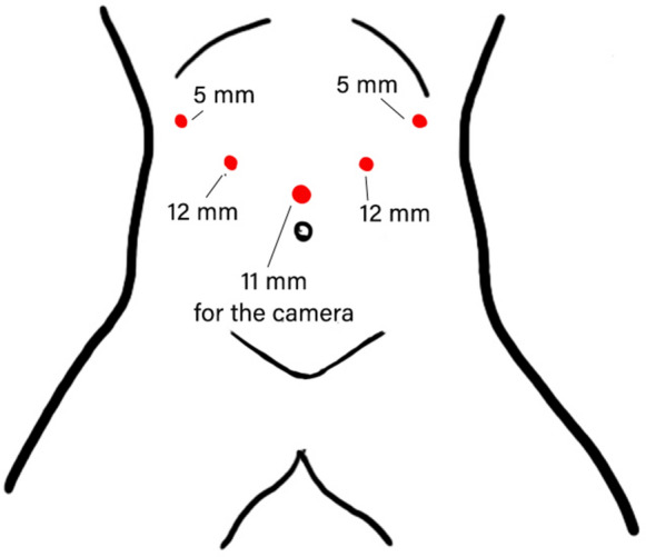
Patient’s and ports position during the abdominal phase of MIILE
The stomach is mobilized, taking care to preserve the right gastroepiploic vessels and the arcade along the greater curvature. The gastric tube is then created by stapling it from the incisura angularis toward the gastric fundus, creating a 4–5 cm wide gastric conduit. The first staple firing is made perpendicular to the long axis of the stomach in order to maintain the necessary conduit diameter. Multiple firings of 60 mm endostapler (Johnson and Johnson, ECHELON FLEX ENDOPATH STAPLER) using green and gold cartridges with absorbable buttress material (GORE® SEAMGUARD® Bioabsorbable Staple Line Reinforcement) are used in order to reduce the risk of bleeding from the staple line. The gastric section is not completed at the level of the fundus, leaving nearly 3 cm of tissue that will be divided during the thoracic phase. Pyloroplasty is not routinely performed. The D2 lymphadenectomy is usually performed. The inferior mediastinum is dissected as much as possible, up to performing the partial section of the right pillar and the opening of the right pleura. At the end of the procedure, a drain is placed from the abdomen into the chest through the right pleura opening, avoiding the intercostal space along with the related post-operative discomfort.
Thoracic phase (thoracoscopic/robot assisted)
Once the abdominal laparoscopic phase is completed, the patient is placed in a semi-prone position. The semi-prone position is obtained by placing the patient on their left flank and tilting the operatory bed to the extreme left. Three ports (12 mm) are placed aligned on the posterior axillary line; a 12-mm trocar for the assistant is placed in the right anterior chest, where the mini-thoracotomy is performed for specimen removal (Fig. 2).
Fig. 2.
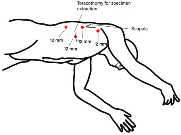
Patient’s and ports position during thoracic phase of MIILE
The esophagus is mobilized by monopolar hook or energy devices, including all surrounding nodes; then, the azygos vein is sectioned with a vascular endostapler and the mobilization of the esophagus is conducted 3–5 cm above the carina. Subcarinal nodes are resected as well.
The esophagus is sectioned with a linear 60 mm stapler at the level of the azygos vein, or above depending on the location of the tumor; the staple line is usually 4–5 cm wide (Fig. 3).
Fig. 3.

Intrathoracic proximal section of the esophagus
The gastric conduit is then pulled up into the chest through the hiatus, taking care to have the gastric staple line toward the surgeon, to prevent rotation of the conduit (Fig. 4).
Fig. 4.
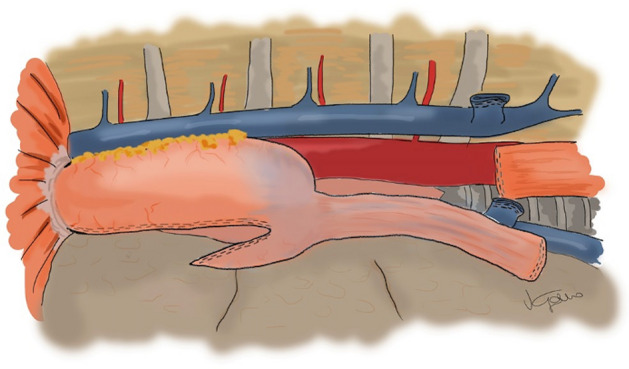
Gastric conduit’s pull-through
Thus, the distance between the esophageal stump and the gastric conduit is evaluated; the gastric fundus is completely transected where blood perfusion is optimal (Figs. 5, 6).
Fig. 5.
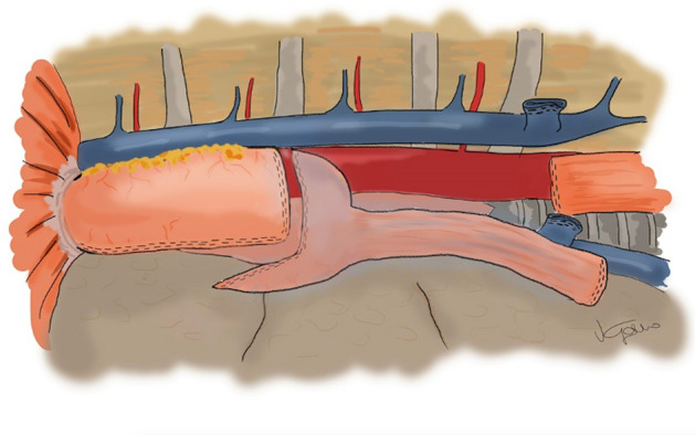
Tailored transection of gastric conduit
Fig. 6.
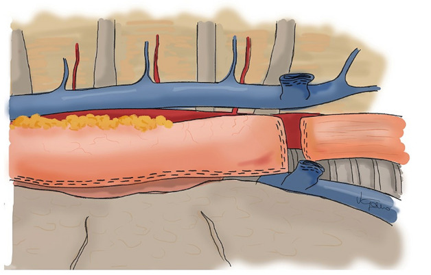
The two stapled lines obtained are approximated without tension
The specimen is finally extracted through a mini-thoracotomy, whose length is related to specimen volume, usually obtained by slightly enlarging the access of the assistant trocar. The oncological margins are checked before continuing on with the reconstructive phase.
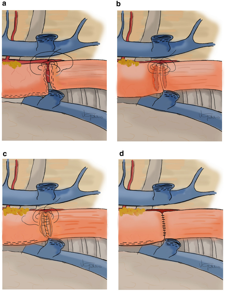
The gastro-esophageal anastomosis is then performed after setting up the da Vinci Si robotic system with trocar-in-trocar technique. Following this, a four layered robot-assisted hand-sewn anastomosis, with 3/0 monofilament absorbable running barbed sutures (Medtronic V-Loc™ Wound Closure Device), is performed.
The posterior side is accomplished by suturing together the gastric and esophageal stumps, incorporating staple lines (Fig. 7a).
Fig. 7.
Esophagogastric anastomotic phases. A 1st posterior layer, incorporating staple lines. B 2nd inner mucosal posterior layer. C 3rd inner mucosal anterior layer. D 4th outer sero-muscular layer
After completing the posterior external layer, monopolar scissors are used to create an incision of about 2.5 cm on both the esophageal and gastric sides. Then, the posterior mucosal layer of running barbed suture is performed (Fig. 7b). The anastomosis is closed with a third anterior layer of sero-muscular 3/0 barbed running suture (Fig. 7c). Before closing this anterior running suture, a gastric tube is passed into the conduit, through the anastomosis, under direct vision. Finally, the anastomosis is completed with the fourth, anterior sero-muscular outer layer performed with transverse stitches which gently approximate tissues and avoid esophageal fiber rupture (Fig. 7d).
To finish, we leave only the transabdominal drain along the conduit near the anastomosis (video).
A hydrosoluble medium contrast swallow X-ray is usually performed on the 6th or 7th post-operative day, in order to rule out any anastomotic leaks; an oral diet is then resumed and drains removed.
A feeding jejunostomy is not routinely performed, but this solution is tailored according to the patients’ comorbidities and Nutritional Index Score.
Informed consent and ethical approval
All patients provided informed consent for surgery and for anonymous use of videos and photographs of the procedures, for scientific or training purposes.
All procedures performed in studies involving human participants were in accordance with the ethical standards of the institutional and/or national research committee and with the 1964 Helsinki declaration and its later amendments or comparable ethical standards. For this type of study, formal consent is not required.
Results
Ten patients were male (83%), while 2 patients were female (16%), with a mean age of 69.3 years (min 55, max 77 years). Mean BMI was 27.5 (min 23.1, max 34.4). ASA score was 2 in 9 patients and 3 in 3 patients. Six patients underwent neoadjuvant therapy. All lesions were in the mid or distal esophagus: 4/12 were located at the esophagogastric junction, 4/12 between 34 and 37 cm from the incisors, and the remaining 4 between 30 and 33 cm from the incisors. The mean operative time was 467 ± 71 min (min 280, max 560 min). All the PT anastomosis were successfully performed. The only intraoperative complication was recorded in a patient affected by chronic bullous emphysema: at the end of the surgical procedure, an air leak was identified due to the rupture of a bulla on the upper pole of the right lung, which required an atypical lung resection. There was no anastomotic leakage or delayed conduit emptying. The post-operative course was uneventful in nine cases. In two patients, an asymptomatic chyloperitoneum was detected and successfully managed with a Lipiodol injection in inguinal nodes; the remaining patient, who had undergone surgery in November 2020, developed Sars-Cov-2 infection during the hospitalization. One patient developed an anastomotic stricture 5 months after surgery, which was treated by pneumatic dilation of the anastomosis.
The median post-operative hospital stay was 13 days (min 7, max 37 days); the longest in-hospital stay was recorded in patients who developed chyloperitoneum. There were no differences between patients underwent or not neoadjuvant therapy in terms of in-hospital stay, operative time, or complications. Free-resection margin was achieved in all patients; 7 out of 12 lesions were histologically defined as adenocarcinomas, 2/12 as adeno-squamous carcinomas of esophago-gastric junction, 1/12 as squamous carcinoma, 1/12 as neuroendocrine carcinoma, and the remaining one as neuroendocrine/adenocarcinoma. The “T” stage according to the TNM staging system was classified as T3 in 9/12 lesions and as T2 in 3/12 lesions. A mean of 32.5 ± 18.3 nodes was harvested (min 7, max 66).
Discussion
Minimally invasive and open esophagectomy show comparable oncological outcomes and survival [1, 2], but with the thoracoscopic approach, significant advantages in terms of post-operative pain, short in-hospital stay, recovery time, and morbidity, particularly regarding pulmonary infections, are reported [1, 2, 10]. However, in spite of these advantages, its widespread use is still limited mainly because of the technical challenges of the reconstructive phase, which can lead to high risk of fistula and therefore failure of the entire operation. In this setting, it is well recognized that the exploitation of robotic technology could play a role, by overcoming some kinematic limitation of pure laparoscopic surgery, and therefore, its application in the field of esophageal surgery has caused interest to rise [17].
RAS can facilitate both traditional stapled esophago-gastric anastomotic reconstructive techniques performed during a mini-invasive approach, as well as completely hand-sewn ones [12, 18, 19]. Indeed, in the case of linear stapling (LSEA), the robot can be used to ease the suture of the esophago-gastrotomy, where the stapler is inserted, while with circular stapling techniques (CEEA), the robotic platform allows easy insertion of the anvil into the esophagus stump and to fix it in place with hand-sewn purse-string suture [19].
In contrast, the minimally invasive hand-sewn anastomosis takes the form of an adaptation of thoracotomic hand-sewn anastomosis, which is preferred by many surgeons as it seems to be associated with the lowest rate of anastomotic leaks [20]. For this reason, the hand-sewn anastomosis was translated in minimally invasive field. However, the complex technical suturing skills required in performing the suture by non-articulated instruments inside a rigid anatomical compartment, the chest, have limited the worldwide diffusion of this technique, so far.
In this scenario, RAS may really play a pivotal role, allowing surgeons to perform hand-sewn anastomosis easier, by utilizing the wide degrees of movements that robotic wristed instruments possess.
A recent review by Plat et al. [18] reported only five articles describing robot-assisted hand-sewn anastomosis technique. The authors generally agreed with double-layered technique for both anterior and posterior side of the anastomosis, but all the described techniques differed in many details, depending on surgeons’ habits.
For these reasons, we aimed to report our personal successful experience so far, and also video report the details of our PT.
In our series, the first operator was already experienced in performing advanced laparoscopic and robotic surgery, as well as upper GI surgery and bariatrics, so that the PT could be considered a technique derived from this background. Furthermore, since all cases were operated with the da Vinci Si platform, we choose to utilize the robot only for the execution of the reconstructive phase, as in our view, the other phases of the operation can be well performed with pure laparo/thoracoscopy, and this hybrid approach avoids downtime related to multiple dockings.
We acknowledge that RAS is able to accomplish both the abdominal and thoracic phases of the esophagectomy and therefore it can be utilized also to perform the entire operation, although with a less than expected cost–benefit ratio. Furthermore, we also acknowledge that most hospitals are now equipped with the Xi instead of a Si model and the innovations introduced by the da Vinci Xi could get over these limitations. Indeed, aside from having a shorter docking time, the Xi is more able to cope with multi-quadrant surgery due to its intrinsic flexibility and provides effective energy devices and staplers [21]. These characteristics could allow the replacement of the laparoscopic phase with the robot, thereby performing a fully robot-assisted procedure, with implications on the workflow, operating time, and even costs [22].
However, as the present paper is particularly focused on the Achilles heel of the entire operation, represented by the reconstructive phase, we think that in this respect, our experience should be meaningful both for Si and Xi robot, and our considerations should not significantly be affected by this bias.
As it is widely known, the failure of the esophago-gastric anastomosis during an Ivor Lewis esophagectomy brings disastrous consequences, hence why we think that every technique detailing successful tips and tricks deserves to be shared.
Reporting our experience, we believe that in addition to RAS, the success of esophago-gastric anastomosis depends also on the attention paid to the other described details.
Indeed, other key points of our presented hand-sewn PT are as follows:
The resection of gastric fundus,
The incorporation of esophageal and gastric staple lines in the posterior outer layer of the anastomosis,
The use of 3/0 barbed suture for all the four layers of the anastomosis.
The tubularization of the stomach often results in a reduced blood supply of gastric fundus. In fact, even if vascularization seems to be sufficient during the ICG test, it is often impaired and largely dependent on intramural micro-vessels, the patient’s specific course, and the distribution of the gastroepiploic arcade. This is why, when the length of the gastric conduit is satisfactory, we prefer to resect the gastric fundus, in order to guarantee a perfect vascularization. In addition, the resection of the gastric fundus allows the tailoring of the length of gastric conduit in order to prevent its kinking on the diaphragm, which can represent one of the causes of delayed emptying after Ivor Lewis esophagectomy.
Incorporating staple lines in the posterior outer layer provides a reinforcement of the posterior wall of the anastomosis as well as a tension reduction on the whole anastomosis. Moreover, the risk of bleeding from staple lines and the risk of posterior leakages can be reduced as well.
The use of barbed sutures has proven to be safe in general and upper GI surgery [14, 23–25]. In our experience, these sutures show the required tensile strength and allow the consistency of the necessary tension after each passage, overcoming the main drawback of RAS, which is absence of tactile feedback.
Some authors believe that a robotic hand-sewn approach increases operative time and makes this technique difficult to standardize, as it requires a long learning curve and advanced technical skills. Nevertheless, we are convinced that on the contrary, robotic assistance could help surgeons create an easier and reliable anastomosis, additionally increasing the confidence in preforming a completely hand-sewn anastomosis and possibly the widespread adoption of the minimally invasive approach in esophageal surgery.
The main limitations of our study are the retrospective nature, the small sample size, including the learning curve (as the reported cases are the first performed at our institution), the absence of long-term follow-up, and finally the lack of a control group.
Overall though, the good results obtained in spite of these limitations, and of the inclusion also of locally advanced tumors, as demonstrated by the half series who had undergone neo-adjuvant chemo are, in our opinion, further points in favor of the technique.
We also acknowledge that the short-term follow-up limits our conclusions with regard to some complications such as strictures, dysphagia, or gastroesophageal reflux disease. However, as the most dangerous complication of this anastomosis is the fistula, which generally occur in the post-operative course, we think that our results already represent an encouraging outcome to be reported in view of the zero rate of leakage recorded in these patients.
Conclusion
Robotic hand-sewn esophago-gastric anastomosis can be fashioned in different ways and many variants have been described concerning the type of suture (running or interrupted sutures), number of layers (single or double), and suture material. As we believe in the relevance of every single technical detail to realize a safe and replicable anastomosis, we have illustrated our PT.
Despite our small series, we believe that our PT may be a promising, safe, and a replicable technique. Some key points may be useful to guarantee a low complication rate after MIILE, such as anastomotic leaks and delayed emptying. Further cases are needed to validate these preliminary, but very encouraging, results.
Supplementary Information
Below is the link to the electronic supplementary material.
Declarations
Disclosures
Dr. A. Peri, Dr. N. Furbetta, Dr. J. Viganò, Dr. L. Pugliese, Dr. G. Di Franco, Prof. F.S. Latteri, Dr. N. Mineo, Dr. F.C. Bruno, Dr. V. Gallo, Prof. L. Morelli, and Prof. A. Pietrabissa have no conflicts of interest or financial ties to disclose.
Footnotes
Publisher's Note
Springer Nature remains neutral with regard to jurisdictional claims in published maps and institutional affiliations.
References
- 1.Meredith K, Blinn P, Maramara T, Takahashi C, Huston J, Shridhar R. Comparative outcomes of minimally invasive and robotic-assisted esophagectomy. Surg Endosc. 2020;34:814–820. doi: 10.1007/s00464-019-06834-7. [DOI] [PubMed] [Google Scholar]
- 2.Li B, Yang Y, Toker A, Yu B, Kang CH, Abbas G, Soukiasian HJ, Li H, Daiko H, Jiang H, Fu J, Yi J, Kernstine K, Migliore M, Bouvet M, Ricciardi S, Chao Y-K, Kim Y-H, Wang Y, Yu Z, Abbas AE, Sarkaria IS, Li Z. International consensus statement on robot-assisted minimally invasive esophagectomy (RAMIE) J Thorac Dis. 2020;12:7387–7401. doi: 10.21037/jtd-20-1945. [DOI] [PMC free article] [PubMed] [Google Scholar]
- 3.Gujjuri RR, Kamarajah SK, Markar SR. Effect of anastomotic leaks on long-term survival after oesophagectomy for oesophageal cancer: systematic review and meta-analysis. Dis Esophagus. 2020 doi: 10.1093/dote/doaa085. [DOI] [PubMed] [Google Scholar]
- 4.Kukar M, Ben-David K, Peng JS, Attwood K, Thomas RM, Hennon M, Nwogu C, Hochwald SN. Minimally invasive Ivor Lewis esophagectomy with linear stapled anastomosis associated with low leak and stricture rates. J Gastrointest Surg. 2020;24:1729–1735. doi: 10.1007/s11605-019-04320-y. [DOI] [PMC free article] [PubMed] [Google Scholar]
- 5.Laxa BU, Harold KL, Jaroszewski DE. Minimally invasive esophagectomy: esophagogastric anastomosis using the transoral orvil for the end-to-side Ivor-Lewis technique. Innov Technol Tech Cardiothorac Vasc Surg. 2009;4:319–325. doi: 10.1097/IMI.0b013e3181c4fc8b. [DOI] [PubMed] [Google Scholar]
- 6.Charalabopoulos A, Davakis S, Syllaios A, Lorenzi B. Intrathoracic hand-sewn esophagogastric anastomosis in prone position during totally minimally invasive two-stage esophagectomy for esophageal cancer. Dis Esophagus. 2020 doi: 10.1093/dote/doaa106. [DOI] [PubMed] [Google Scholar]
- 7.Bartella I, Fransen LFC, Gutschow CA, Bruns CJ, van Berge Henegouwen ML, Chaudry MA, Cheong E, Cuesta MA, Van Daele E, Gisbertz SS, van Hillegersberg R, Hölscher A, Mercer S, Moorthy K, Nafteux P, Nilsson M, Pattyn P, Piessen G, Räsanen J, Rosman C, Ruurda JP, Schneider PM, Sgromo B, Nieuwenhuijzen GA, Luyer MDP, Schröder W. Technique of open and minimally invasive intrathoracic reconstruction following esophagectomy—an expert consensus based on a modified Delphi process. Dis Esophagus. 2021 doi: 10.1093/dote/doaa127. [DOI] [PubMed] [Google Scholar]
- 8.Gao HJ, Mu JW, Pan WM, Brock M, Wang ML, Han B, Ma K. Totally mechanical linear stapled anastomosis for minimally invasive Ivor Lewis esophagectomy: operative technique and short-term outcomes. Thorac Cancer. 2020;11:769–776. doi: 10.1111/1759-7714.13339. [DOI] [PMC free article] [PubMed] [Google Scholar]
- 9.Honda M, Kuriyama A, Noma H, Nunobe S, Furukawa TA. Hand-sewn versus mechanical esophagogastric anastomosis after esophagectomy: a systematic review and meta-analysis. Ann Surg. 2013;257:238–248. doi: 10.1097/SLA.0b013e31826d4723. [DOI] [PubMed] [Google Scholar]
- 10.Diez Del Val I, Loureiro Gonzalez C, LarburuEtxaniz S, BarrenetxeaAsua J, Leturio Fernandez S, Ruiz Carballo S, EtxebarriaBeitia E, Perez de Villarreal P, Hierro-Olabarria L, Bilbao Axpe JE, Mendez Martin JJ. Contribution of robotics to minimally invasive esophagectomy. J Robot Surg. 2013;7:325–332. doi: 10.1007/s11701-012-0391-y. [DOI] [PubMed] [Google Scholar]
- 11.Wee JO, Bravo-Iñiguez CE, Jaklitsch MT. Early experience of robot-assisted esophagectomy with circular end-to-end stapled anastomosis. Ann Thorac Surg. 2016;102:253–259. doi: 10.1016/j.athoracsur.2016.02.050. [DOI] [PubMed] [Google Scholar]
- 12.Wang Z, Zhang H, Wang F, Wang Y. Robot-assisted esophagogastric reconstruction in minimally invasive Ivor Lewis esophagectomy. J Thorac Dis. 2019;11:1860–1866. doi: 10.21037/jtd.2019.05.29. [DOI] [PMC free article] [PubMed] [Google Scholar]
- 13.van der Sluis PC, Tagkalos E, Hadzijusufovic E, Babic B, Uzun E, van Hillegersberg R, Lang H, Grimminger PP. Robot-assisted minimally invasive esophagectomy with intrathoracic anastomosis (Ivor Lewis): promising results in 100 consecutive patients (the european experience) J Gastrointest Surg. 2020 doi: 10.1007/s11605-019-04510-8. [DOI] [PMC free article] [PubMed] [Google Scholar]
- 14.Wang F, Zhang H, Zheng Y, Wang Z, Geng Y, Wang Y. Intrathoracic side-to-side esophagogastrostomy with a linear stapler and barbed suture in robot-assisted Ivor Lewis esophagectomy. J Surg Oncol. 2019;120:1142–1147. doi: 10.1002/jso.25698. [DOI] [PMC free article] [PubMed] [Google Scholar]
- 15.de Groot EM, Möller T, Kingma BF, Grimminger PP, Becker T, van Hillegersberg R, Egberts JH, Ruurda JP. Technical details of the hand-sewn and circular-stapled anastomosis in robot-assisted minimally invasive esophagectomy. Dis Esophagus. 2020 doi: 10.1093/dote/doaa055. [DOI] [PubMed] [Google Scholar]
- 16.Griffin SM, Jones R, Kamarajah SK, Navidi M, Wahed S, Immanuel A, Hayes N, Phillips AW. Evolution of esophagectomy for cancer over 30 years: changes in presentation, management and outcomes. Ann Surg Oncol. 2020 doi: 10.1245/s10434-020-09200-3. [DOI] [PMC free article] [PubMed] [Google Scholar]
- 17.Clark J, Sodergren MH, Purkayastha S, Mayer EK, James D, Athanasiou T, Yang GZ, Darzi A. The role of robotic assisted laparoscopy for oesophagogastric oncological resection; an appraisal of the literature. Dis Esophagus. 2011;24:240–250. doi: 10.1111/j.1442-2050.2010.01129.x. [DOI] [PubMed] [Google Scholar]
- 18.Plat VD, Stam WT, Schoonmade LJ, Heineman DJ, van der Peet DL, Daams F. Implementation of robot-assisted Ivor Lewis procedure: robotic hand-sewn, linear or circular technique? Am J Surg. 2020;220:62–68. doi: 10.1016/j.amjsurg.2019.11.031. [DOI] [PubMed] [Google Scholar]
- 19.Zhang H, Wang Z, Zheng Y, Geng Y, Wang F, Chen LQ, Wang Y. Robotic side-to-side and end-to-side stapled esophagogastric anastomosis of Ivor Lewis esophagectomy for cancer. World J Surg. 2019;43:3074–3082. doi: 10.1007/s00268-019-05133-5. [DOI] [PubMed] [Google Scholar]
- 20.Castro PMV, Ribeiro GFP, de Freitas Rocha A, Mazzurana M, Alvarez GA Hand-sewn versus stapler esophagogastric anastomosis after esophageal ressection: systematic review and meta-analysis. Arq Bras Cir Dig. 2014;27:216–221. doi: 10.1590/s0102-67202014000300014. [DOI] [PMC free article] [PubMed] [Google Scholar]
- 21.Alhossaini RM, Altamran AA, Choi S, Roh CK, Seo WJ, Cho M, Son T, Il KH, Hyung WJ. Similar operative outcomes between the da Vinci Xi ® and da Vinci Si ® systems in robotic gastrectomy for gastric cancer. J Gastric Cancer. 2019;19:165–172. doi: 10.5230/jgc.2019.19.e13. [DOI] [PMC free article] [PubMed] [Google Scholar]
- 22.Morelli L, Di Franco G, Lorenzoni V, Guadagni S, Palmeri M, Furbetta N, Gianardi D, Bianchini M, Caprili G, Mosca F, Turchetti G, Cuschieri A. Structured cost analysis of robotic TME resection for rectal cancer: a comparison between the da Vinci Si and Xi in a single surgeon’s experience. Surg Endosc. 2019;33:1858–1869. doi: 10.1007/s00464-018-6465-9. [DOI] [PubMed] [Google Scholar]
- 23.Wiggins T, Majid MS, Markar SR, Loy J, Agrawal S, Koak Y. Benefits of barbed suture utilisation in gastrointestinal anastomosis: a systematic review and meta-analysis. Ann R Coll Surg Engl. 2020;102:153–159. doi: 10.1308/rcsann.2019.0106. [DOI] [PMC free article] [PubMed] [Google Scholar]
- 24.Manigrasso M, Velotti N, Calculli F, Aprea G, Di Lauro K, Araimo E, Elmore U, Vertaldi S, Anoldo P, Musella M, Milone M, Maria Sosa Fernandez L, Milone F, Domenico De Palma G. Barbed suture and gastrointestinal surgery. A retrospective analysis. Open Med. 2019;14:503–508. doi: 10.1515/med-2019-0055. [DOI] [PMC free article] [PubMed] [Google Scholar]
- 25.Morelli L, Furbetta N, Gianardi D, Guadagni S, Di Franco G, Bianchini M, Palmeri M, Masoni C, Di Candio G, Cuschieri A. Use of barbed suture without fashioning the “classical” Wirsung-jejunostomy in a modified end-to-side robotic pancreatojejunostomy. Surg Endosc. 2020 doi: 10.1007/s00464-020-07991-w. [DOI] [PMC free article] [PubMed] [Google Scholar]
Associated Data
This section collects any data citations, data availability statements, or supplementary materials included in this article.


