Abstract
Motor neuron disease (MND) comprises a group of fatal neurodegenerative diseases with no effective cure. As progressive motor neuron cell death is one of pathological characteristics of MND, molecules which protect these cells are attractive therapeutic targets. Accumulating evidence indicates that EphA4 activation is involved in MND pathogenesis, and inhibition of EphA4 improves functional outcomes. However, the underlying mechanism of EphA4’s function in MND is unclear. In this review, we first present results to demonstrate that EphA4 signalling acts directly on motor neurons to cause cell death. We then review the three most likely mechanisms underlying this effect.
Keywords: receptors, Eph, EphA4, motor neurons, motor neuron death, neurodegenerative disease, motor neuron disease, pathogenesis
1. Introduction
Motor neuron disease (MND) refers to a group of neurodegenerative diseases, which have the shared characteristic of the progressive loss of upper and/or lower motor neurons [1,2]. Disease onset is insidious, with patients gradually losing control of their voluntary muscles, resulting in relatively late diagnosis [3]. Due to the nature of the disease and the lack of an effective treatment, MND patients usually die within 2 to 3 years following diagnosis, largely because of the loss of respiratory function [1,2,3]. Riluzole, the only drug approved in Australia, only prolongs the median life expectancy by 2 to 3 months [4,5,6,7]. New and effective therapeutic treatments are therefore urgently needed.
It is widely accepted that MND is a complex disease, with both genetic and environmental factors co-contributing to its pathogenesis [2,3,8]. Although the details of its pathogenesis are unclear, motor neuron cell death is regarded as the hallmark. Preventing this death is the primary therapeutic aim [9,10,11]. Here we discuss the importance of EphA4 signalling in motor neuron cell death, the mechanisms of signalling and the potential for ameliorating MND by blocking its signalling.
EphA4 belongs to the A subgroup of Eph receptors and is well known as a pan-receptor that can widely bind, albeit with varying affinities, to all ephrin ligands, including five glycosyl phosphatidylinositol (GPI)-linked cell membrane-bound type A ephrins and three transmembrane type-B ephrins [12,13,14]. This striking feature indicates that EphA4 interaction with both A- and B-type ephrins regulates both normal and pathophysiological functions [15]. This suggests that blocking EphA4 signalling could be more efficacious than blocking other Ephs or ephrins. Well established as a guidance molecule involved in the development of the corticospinal tract [16,17], EphA4 has also been shown to inhibit axon regeneration following spinal cord injury (SCI) [18,19,20]. The fact that EphA4 represses the axonal regrowth of motor neurons after SCI suggested that it may also contribute to the differential vulnerability of motor neurons in MND through a similar mechanism [21,22]. In support of this view, emerging research has identified that EphA4 is indeed associated with MND pathogenesis [21,22,23,24,25]. In two zebrafish models of MND, overexpressing human mutant SOD1 and TDP-43, knockdown of the zebrafish paralogue of EphA4, Rtk2, rescued both mutant SOD1-induced axonopathy and axonal outgrowth defects caused by the mutant TDP-43. The same study also examined the effect of EphA4 in rodent SOD1G93A models of MND. EphA4+/− mice were crossed with SOD1G93A mice to obtain EphA4+/− SOD1G93A mice. Compared with EphA4+/+ SOD1G93A controls, the heterozygous deletion of EphA4 significantly increased motor performance and survival. The administration of EphA4-blocking peptide to SOD1G93A rats also delayed disease onset and enhanced survival. Finally, in MND patients, lower levels of expression of EphA4 mRNA in whole blood samples correlated with prolonged disease progression [21]. This study showed for the first time that EphA4 is involved in the disease progression of MND, and inhibiting EphA4 expression or activation can affect disease progression, making it an attractive target for MND therapies. Subsequently, a novel isoform of full-length EphA4 (EphA4-FL) was identified in mice and humans, EphA4-N, which contained the extracellular domains and transmembrane domain of EphA4-FL. EphA4-N was alternatively transcribed from the EphA4-FL gene, successfully translated into a functional protein, and was able to function as an endogenous dominant-negative inhibitor in terms of its repressive effect on EphA4-FL signalling in vitro. It has been shown that there was a lower level of expression of the inhibitory EphA4-N in human MND patients, compared to healthy controls, allowing more aggressive signalling by EphA4-FL. In SOD1G93A mice, there was an increase in EphA4-FL expression in the pre-symptomatic phase, indicating that EphA4–FL signalling was dominant in the early pathogenesis of MND [23]. In previous studies, we generated a wildtype EphA4-Fc (a recombinant fusion protein derived from the extracellular domain of wildtype EphA4 and the Fc domain of human IgG) which effectively blocks EphA4-ephrin interaction in vitro and demonstrated that it improves functional performance in mice and rats after SCI by increasing the number of axons reaching and crossing the lesion site compared with saline-treated controls [18,19,23]. More recently, to reduce glycosylation, we mutated both the human and mouse EphA4-Fc (mEphA4-Fc) at three glycosylation sites, N235, N340 and N408. This resulted in significantly prolonging the half-life of human mEphA4-Fc from less than 24 h to 31.1 h in healthy Wistar rats following a single intravenous dose, while maintaining comparable binding and blocking ability to the ephrin ligands [26]. Results of the toxicokinetic analysis of human mEphA4-Fc cells in healthy Sprague-Dawley rats following 5× weekly repeat intravenous dosing showed that the terminal elimination half-life ranged from 52.8 h to 77.5 h. More importantly, using this glycosylation mutant of mouse mEphA4-Fc in the SOD1G93A model significantly improved motor performance, including rotarod and hind-limb grip strength tests [22]. Briefly, SOD1G93A mice were treated with either the mouse mEphA4-Fc or a saline control. Functional behavioural tests were monitored on a weekly basis from week 8 to the end of disease. The balance and motor coordination of mice were assessed by means of the rotarod test, whereas hind-limb grip strength was also monitored. The SOD1G93A mice receiving the mEphA4-Fc treatment exhibited improved performance in the rotarod test compared to control SOD1G93A mice from week 17 to week 23, with the differences at weeks 19–21 being statistically significant. Consistent with this result, mEphA4-Fc-treated SOD1G93A mice also showed better hind-limb grip strength from week 8 to week 22 compared with the vehicle control group, with the differences at weeks 9 and 18–21 reaching statistical significance. Given the substantial loss of induced motor function in this model, these results suggest that mEphA4-Fc is a promising therapeutic treatment for MND and EphA4 activation is involved in the disease pathogenesis [22].
2. EphA4 Signalling Acts Directly on Motor Neurons to Cause Cell Death
To further investigate whether EphA4 signalling functions at the level of motor neurons to cause death in MND, SOD1G93A mice with specific EphA4 gene deletion in choline acetyltransferase (ChAT)-expressing cells were obtained by crossing them with EphA4flox/flox and ChAT-CreKI/KI mice on a SOD1G93A genetic background [22]. ChAT is preferentially expressed in cholinergic neurons from postnatal day 5 (P5) throughout adulthood, and given that motor neurons are cholinergic neurons, the Cre-mediated deletion of the EphA4 gene was restricted to ChAT-expressing motor neurons in this mouse model [27,28]. However, as ChAT expression does not occur until P5, EphA4 expression remains normal during the critical embryonic period, allowing the normal patterning and development of the central nervous system (CNS). The results showed that heterozygous deletion of EphA4 in motor neurons significantly increased the number of surviving motor neurons in the spinal cord of the SOD1G93A mice, compared to the homozygous deletion group or normal SOD1G93A mice at 17 weeks of age. This was also the timepoint at which the improvement in two behavioural tasks, rotarod performance and hind-limb grip strength, reached statistical significance in the mEphA4-Fc-treated SOD1G93A mice, compared with the control group. In addition, at the same timepoint, the morphology of the post-synaptic endplates of neuromuscular junctions (NMJs) in the tibialis anterior (TA) muscle were also better maintained in the heterozygous deletion group at the same timepoint, and more fragmented post-synaptic endplates of NMJs and debris of damaged endplates were observed in another two control groups [22]. This is the first evidence to reveal that EphA4 exerts direct negative effects on motor neuron survival in MND. Notably, only SOD1G93A mice with specific heterozygous deletion of EphA4 in motor neurons exhibited a significant protective effect on cell survival, with the motor neuron death in the homozygous deletion group being similar to that in normal SOD1G93A controls [22]. However, SOD1G93A mice with homozygous germline deletion of EphA4 showed reduced viability due to a low birth rate and body weight [21]. Taken together, these data suggest that although a certain expression level of EphA4 is essential for motor neuron development and survival, a lower level of expression of EphA4 signalling could protect motor neurons from death in MND.
To further investigate the direct effect of EphA4 on motor neurons, we have first estimated the expression level of EphA4 in motor neurons using a human-induced pluripotent stem cell (iPSC)-derived motor neuron system [18,29]. Fibroblasts were obtained via skin biopsy from a symptomatic familial MND patient harbouring a SOD1E101G mutation and a healthy control [30]. The methods were carried out in accordance with the guidelines set out in the National Statement on Ethical Conduct in Research Involving Humans and informed consent was obtained from all donors [31]. All experimental protocols were approved by the University of Wollongong Human Research Ethics Committee (approval HREC 13/272, most recently reviewed and approved on 29 June 2021). Fibroblasts were reprogrammed into iPSCs and confirmed as pluripotent, as previously described [29]. The iPSCs were cultured on Matrigel-coated 6-cm tissue culture plates in TeSR-E8 (Stem Cell Technologies) at 37 °C, 5% CO2, 3% O2 in a humidified incubator (hypoxic conditions). The iPSCs were differentiated into motor neurons as previously described [29], with some modifications. Neuronal precursor cells were treated with increasing concentrations of retinoic acid over three days (0.1 µM to 0.3 µM), and on the third day 2 µM of purmorphamine was also added to the medium [32]. BrainPhys (Stemcell Technologies) was used as the basal medium to generate motor neuron precursor cells and mature motor neurons [33].
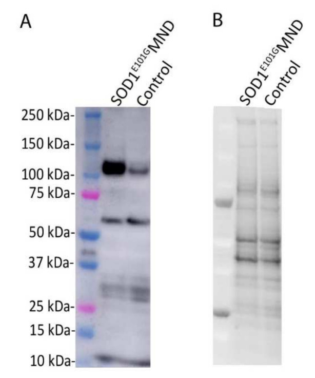
After one week of proliferation and three weeks of maturation under hypoxic conditions, the relative levels of EphA4 protein were compared between MND and healthy control iPSC-derived motor neurons using Western blotting. The BCA and Western blot assays were conducted as described previously [23], except that 20 µg of protein was used for the Western blotting. Each sample measurement was normalised to its corresponding total protein value and the relative level of EphA4 protein was five times higher in iPSC-derived motor neurons from the SOD1E101G MND patient compared to motor neurons derived from the healthy control (Figure 1).
Figure 1.
(A) Relative levels of EphA4 (110 kDa) are 5× higher in iPSC-derived motor neurons from a SOD1E101G MND patient compared to iPSC-derived motor neurons from a healthy control. (B) Corresponding total protein blot from the SOD1E101G MND patient and healthy control. Four-week-old iPSC-derived motor neurons were harvested by rinsing in 1× PBS followed by manual scraping in RIPA buffer (50mM Tris-HCl pH 7.4, 1% NP40, 0.25% Na-deoxycholate, 150 Mm NaCl, 1mM EDTA, phosphate (PhosSTOP, Roche) and protease (complete protease inhibitor cocktail, Roche) inhibitors). Cellular debris was pelleted by means of centrifugation at 10,000× g for 10 min at 4 °C. The cleared protein lysate was quantified via BCA and 20 µg of protein was used for Western blotting. Proteins were denatured by boiling at 95 °C for 5 min in Laemmli buffer with 5% β-mercaptoethanol and loaded on a Criterion 4–20% stain-free gel (Biorad). Samples were electrophoresed in SDS-PAGE buffer and transferred onto a PVDF membrane. The membrane was imaged using the Criterion stain-free gel imaging system (Biorad) to obtain total protein values for quantification. The membrane was blocked in 5% skim milk in TBS for 1 h and incubated in EphA4 antibody (1:1000 in 5% skim milk in Tris-buffered saline solution, mouse anti-EphA4, ECM Bioscience) overnight at 4 °C. The membrane was then incubated in an HRP-conjugated secondary antibody and EphA4 (approximate molecular weight 110 kDa) was detected using Pierce ECL Plus Western Blotting substrate (Thermo Fischer) and the chemiluminescence function on the Amersham GE 600 Imager (GE Life Sciences). Densitometry analysis was conducted using Image Studio Lite version 5.2 and each sample measurement was normalised to its corresponding total protein value.
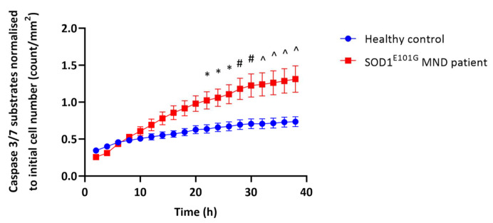
To gain insight into the role of highly expressed EphA4 in iPSC-derived motor neuron death in MND, we next determined whether the iPSC-derived motor neurons from the MND patient carrying a SOD1E101G mutation were more vulnerable to oxidative stress, the most common stress observed under MND conditions, than iPSC-derived motor neurons from a healthy control [34,35]. As previously described, iPSC-derived neurons transitioned from hypoxic to normoxic culture conditions can be used to assess the impact of oxidative stress on caspase 3/7 activity, as a marker of apoptosis [30]. After a maturation phase under hypoxic conditions, iPSC-derived motor neurons were transferred to a humidified incubator at 37 °C, 5% CO2 and 21% O2 (normoxic conditions). At the time of transfer, NucView 488 caspase 3 enzyme substrate (which fluoresces in response to caspase 3/7 activity in cells, Gene Target Solutions) and Reddot 1 (live cell marker, Gene Target Solutions) were added to the motor neuron culture. Caspase 3/7 activity was assessed every two hours for 38 h using an Incucyte (Essen Bioscience) instrument. From 22 h, the levels of caspase 3/7 activity were significantly higher in the iPSC-derived motor neurons from the MND patient compared to the iPSC-derived motor neurons from the healthy control (Figure 2). This suggests that the iPSC-derived motor neurons from the MND patient harbouring a SOD1E101G mutation were more vulnerable to apoptotic cell death under these oxidative stress conditions compared to control. Together, these findings led us to ask if the increased EphA4 expression could contribute to the vulnerability of MND motor neurons under stress conditions.
Figure 2.
IPSC-derived motor neurons from a SOD1E101G MND patient are more vulnerable to oxidative stress then iPSC-derived motor neurons from a healthy control. IPSC-derived motor neurons from a SOD1E101G MND patient and a healthy control were matured for three weeks under hypoxic conditions (3% oxygen) and then transferred to a normoxic incubator (21% oxygen) for 38 h in order to induce oxidative stress. Apoptosis was assessed by quantifying the number of caspase 3/7 substrates every 2 h using an Incucyte. Two-way ANOVA revealed that there were significant main effects of time (F(18,144) = 36.53, p < 0.0001), cell line (F(1.8) = 8.677, p = 0.0185), and time × cell line interaction (F(18.112) = 8.830, p < 0.0001). Sidak’s test for multiple comparisons showed that iPSC-derived motor neurons from the SOD1E101G MND patient showed a significant increase in the number of caspase 3/7 substrates per mm2 from 22 h onwards compared to the healthy control. Data presented as mean ± SEM of n = 5 technical replicates; * p < 0.05; # p < 0.01; ^ p < 0.001.
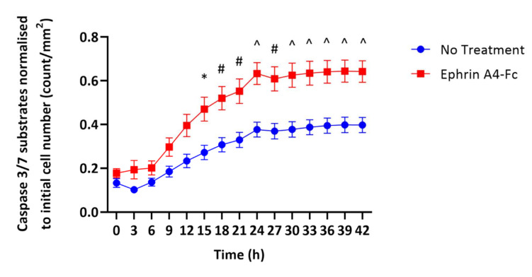
To address this possibility, we next investigated whether the activation of the EphA4 induced by ephrin ligands led to apoptosis in MND motor neurons. Four-week-old iPSC-derived motor neurons from the SOD1E101G MND patient were transferred from hypoxic (3% O2) to normoxic (21% O2) conditions. At the time of transfer, NucView 488 caspase 3 substrate and Reddot 1 were added to the cells as described above, either with or without ephrin A4-Fc ligand (10 µg/mL). Caspase 3/7 activity was assessed every 3 h for 42 h using the Incucyte. From 15 h, the levels of caspase 3/7 activation were significantly higher in the iPSC-derived motor neurons from the SOD1E101G MND patient treated with ephrin A4-Fc compared to motor neurons without treatment (Figure 3). This suggests that EphA4 activation may directly contribute to motor neuron death in MND. In addition, lactate dehydrogenase (LDH) levels in the cell culture medium were measured using a Pierce LDH Cytotoxicity Assay Kit (Thermo Scientific, catalog #88953). LDH cytotoxicity (A490 nm–A680 nm and normalised to the total protein values from each line) was two times higher in the ephrin A4-Fc-treated motor neurons compared to those with no treatment (0.08 vs. 0.04). Altogether, these data suggest that the activation of overexpressed EphA4 in iPSC-derived motor neurons from an MND patient directly results in increased motor neuron death.
Figure 3.
Treatment with the EphA4 ligand, ephrin A4-Fc results in increased cell death in iPSC-derived motor neurons from a SOD1E101G MND patient. IPSC-derived motor neurons from a SOD1E101G MND patient were matured for four weeks under hypoxic conditions (3% oxygen) and then transferred to a normoxic incubator (21% oxygen) for 42 h, with or without the EphA4 ligand ephrin A4-Fc (10 µg/mL). Apoptosis was assessed by quantifying the number of caspase 3/7 substrates every 3 h using an Incucyte. Two-way ANOVA revealed that there were significant main effects of time (F (14.112) = 219.2, p < 0.0001), treatment (F (1.8) = 11.86, p = 0.0088), and time × treatment interaction (F (14.112) = 14.46, p < 0.0001). Sidak’s test for multiple comparisons showed that iPSC-derived motor neurons from the SOD1E101G MND patient treated with ephrin A4-Fc showed a significant increase in the number of caspase 3/7 substrates per mm2 from 15 h onwards compared to motor neurons without treatment. Data presented as mean ± SEM of n = 5 technical replicates; * p < 0.05; # p < 0.01; ^ p < 0.001.
3. Direct Regulation of Motor Neuron Death by EphA4
So far, we have observed the directly negative effect of EphA4 activation on motor neuron survival upon both in vitro and in vivo MND backgrounds (Figure 1, Figure 2 and Figure 3 and [22]). These are confirmatory evidence to support the promotive effect of EphA4 activation and the protective effect of EphA4 deletion or inhibition on MND progression. Although EphA4 activation has been directly or indirectly involved in different types of cell death, such as NIH 3T3 cells, glioblastoma multiform tumoral cells, retinal ganglion cells and endothelial cells [36,37,38,39], this was the first time that the novel effect of EphA4 on motor neuron survival in MND had been reported. More investigations are required to explicate the underlying mechanisms. In the following review, three possible mechanisms are concisely discussed.
3.1. Role of Rho/Rock Signalling
To date, more than 10 different types of cell death have been identified, and four major types among them are apoptosis, necrosis, autophagy and entosis [40]. Although motor neuron death in MND is likely to be multifactorial, the final demise of these cells is more likely to occur via a programmed, energy-dependent cell death pathway resembling apoptosis [41]. It is widely accepted that typical morphological changes which occur during the cell apoptosis process include the release of apoptotic bodies, plasma membrane blebbing and nuclear condensation [42]. Molecules regulating these processes thus played important roles in regulating cell apoptosis [43,44,45,46,47,48]. One of the critical regulators of apoptotic cell membrane blebbing is Rho-associated coiled-coil-containing protein kinase (ROCK), which is also part of the EphA4 downstream signalling pathway [25,38,49,50]. ROCK activation has been shown to contribute to membrane blebbing during the eosinophil peroxidase-induced death of lung epithelial cells in vitro [43]. Similarly, in a human umbilical vein endothelial cell (EC) culture system, the application of combretastatin A-4-phosphate (CA-4-P), a tumor vascular-targeting agent, significantly enhanced ROCK signalling activation and its subsequent myosin light chain (MLC) phosphorylation, which were shown to be responsible for the cell membrane blebbing, loss of cell adherence and decreased viability of ECs due to reorganisation of the actomyosin cytoskeleton. These results suggest that this mechanism may underlie the effect of the CA-4-P treatment in promoting tumor EC death and leading to the shutdown of blood flow in tumors in vivo [44]. ROCK and its other substrates have also been reported to regulate the cell death process [45,46,47]. The upregulation of ROCK signalling and the subsequent inhibition of downstream mitogen-activated protein kinases (MAPK) signalling has been shown to promote the loss of cell bipolarity and detachment, resulting in increases in EC death in an EC-fibroblast coculture system in vitro and in xenograft tumors in nude mice growing from a human colorectal cancer cell line in vivo [45]. Another identified ROCK substrate, phosphatase and tensin homologue (PTEN), has been shown to be involved in the protective effect of ROCK inhibition on cardiomyocyte apoptosis [46] and EC survival in the cardiovascular system in vitro [47]. Activation of ROCK signalling is also highly likely to be involved in regulating motor neuron apoptosis in MND. In support of this, Takata and colleagues revealed that inhibition of ROCK by Fasudil or Y-27632 prevented motor neuron death in mouse motor neuron (NSC34) cell cultures and in the lumbar anterior horn in the SOD1G93A mouse model [48]. Moreover, these two ROCK inhibitors were shown to delay disease onset, prolong the mean survival time and improve functional performance in the SOD1G93A mouse model [48,51]. A multicentric, double-blind, randomised, placebo-controlled phase 2a clinical trial of Fasudil in 120 MND patients started in early 2019, aiming to assess its safety, tolerability and efficacy (ROCK-ALS trial, NCT03792490, Eudra-CT-Nr.: 2017-003676-31) [52]. In addition, three MND cases have been reported in which fasudil was compassionately used for treatments from 2017 to 2019, demonstrating good tolerance [53]. However, it is unclear how ROCK signalling is activated/regulated in the pathogenesis of MND.
Considering that ROCK is part of the EphA4 downstream signalling pathway, the correlation of high expression of the EphA4 receptor with rapid disease progression of MND is consistent with the idea that EphA4 mediates this effect through increased ROCK signalling. The EphA4 receptor tyrosine kinase is expressed on the cell surface and exerts diverse effects through the activation of multiple downstream signalling pathways [54,55]. Following the activation of EphA4, the GTPase Rho family, including Rho, Rac1 and Cdc42, is activated, interacting with its downstream effector proteins to exert different effects [20,50,56,57,58]. ROCK is one of the major effectors for Rho, and their interaction induces conformational changes of Rho and activates ROCK [59]. It has been reported that EphA4 negatively affects axon regeneration after SCI primarily through the Rho/ROCK signalling pathway [20], and the administration of Y-27632 has been shown to increase dendritic branching and axonal regeneration [60]. Similarly, the expression of EphA4 is significantly increased following ischemia-reperfusion in vitro and in vivo, and the activation of EphA4 contributes to the disruption of the blood–brain barrier (BBB) post-ischemic brain injury through Rho/ROCK signalling [38]. In the subarachnoid hemorrhage (SAH) rat model, EphA4 activation has also been shown to be responsible for the neuronal apoptosis and BBB breakdown through the ROCK pathway [49]. Moreover, a reduction in EphA4 has been shown to improve the behavioural function of different ischemic stroke animal models via the inhibition of its downstream Rho/ROCK pathway [38,49,50]. Therefore, it is likely that during MND progression, high expression of EphA4 on motor neurons participates in the increasing motor neuron death by activating the Rho/ROCK downstream pathway.
3.2. Role of D-Serine-Induced NMDAR Activation
Excitotoxicity has been reported to contribute to neuronal death in MND [61]. One classical form of excitotoxicity is induced by excessive stimulation of the N-methyl-d-aspartate receptor (NMDAR), which is potentially caused by elevated levels of its glutamate ligand and/or d-serine co-agonist [62,63] and eventually resulting in apoptosis or necrosis in neurons [64,65]. Endogenous d-serine largely contributes to NMDAR-mediated neurotoxicity, with a decrease in d-serine levels being shown to repress this excitotoxicity [66]. Recently, d-serine has been reported to be involved in MND progression [67,68,69], providing the first direct evidence that increased concentrations of d-serine occur in the brain and spinal cord of MND patients and SOD1G93A mice, and that, as a key determinant of glutamate toxicity, this leads to motor neuron degeneration [67,68]. Given that Zhuang et al. have revealed that normal EphA4 function facilitates the synthesis and/or release of d-serine in the adult brain in vitro [70], the EphA4 dysfunction-induced changes in the d-serine concentration are also likely to contribute to motor neuron death via NMDAR dysfunction. However, further investigation is required to address how EphA4 regulates the d-serine levels in the brain in vivo, and which source of d-serine (astrocytes and/or neurons) has been affected. This EphA4-regulated d-serine concentration has already been observed during adult hippocampal neurogenesis in vivo and in vitro [71]. We have shown that either pharmacological EphA4 inhibition by EphA4-Fc or genetic deletion of EphA4 upregulates adult hippocampal neural precursor proliferation. Moreover, this increased precursor activity induced by the inhibition of EphA4 could be rescued by exogenous d-serine supplementation. In addition, a specific blockage of the interaction between d-serine and NMDARs promoted precursor proliferation directly [71]. These results suggest that EphA4 inhibition indirectly leads to an increase in adult precursor proliferation, by decreasing the level of d-serine, thus inducing downregulation of NMDARs, which directly exerted positive effects on precursor proliferation. Likewise, the high expression of EphA4 in motor neurons might contribute to the increased concentration of d-serine in MND, which results in increased motor neuron death through upregulating NMDAR-induced excitotoxicity.
d-serine is endogenously converted from l-serine by serine racemase (SR), and degenerates to keto acid via the activity of d-amino acid oxidase (DAO). Both increased activity of SR and the loss of DAO activity have also been observed in SOD1G93A mice [67,68]. A loss-of-function mutation in the DAO (R199W DAO) gene has also been associated with familial MND cases. Subsequent research revealed that the expression of R199W DAO promotes the accumulation of d-serine, which led to cell death in primary motor neuron cultures [69,72]. So far, these changes in SR and DAO have been observed in familial MND cases and transgenic mouse models, but they may also exist in sporadic MND cases and co-regulate the concentration of d-serine with EphA4 activation. This may be one of the reasons why EphA4 inhibition only partially improves functional behavioural performance in MND mouse models.
3.3. Role of Calcium Channels
Many studies have reported that changes in ion channels and ion homeostasis lead to increases in neuronal firing, oxidative stress and mitochondrial dysfunction, all of which could eventually contribute to apoptotic cell death [73] and be involved in MND pathology [74,75,76,77]. These abnormalities have mainly been attributed to increased Na+, decreased K+ and increased Ca2+ fluxes [78]. Among other effects, EphA4 activation is likely to regulate the intracellular Ca2+ concentration. As mentioned above, high EphA4 expression is likely to upregulate the function of NMDARs expressed in motor neurons by increasing the levels of d-serine. Subsequently, the activation of NMDARs results in opening of the ion channel, which causes excessive excitotoxic influx of Ca2+ to motor neurons in MND [79]. AMPA receptors (AMPARs) are also well-known ionotropic glutamate receptors, the activation of which mediates a massive influx of Ca2+, leading to excitotoxicity in MND pathology [61,80]. In the SOD1G93A mouse model, the expression of the GluA2 subunit of AMPARs is significantly lower than that in normal mice (which mainly upregulates Ca2+ permeability) [81,82]. Higher expression of the GluA3 subunit has been found to accompany this decreased expression of the GluA2 subunit in SOD1G93A mice, with implications for disease onset and progression [82]. Another study reported the upregulation of the GluA1 subunit, rather than the GluA3 subunit, and downregulation of the GluA2 subunit in the highly expressed Ca2+-permeable AMPARs in iPSC-derived motor neurons carrying human C9orf72 and patient primary motor neurons [83]. This increase in Ca2+-permeable AMPAR activity caused a decline in motor neuron function and accelerating cell death, which is consistent with MND progression [84]. Given that EphA4 activation leads to the degradation of surface AMPARs at synapses [85,86] and dendritic spine loss [87,88], it is possible that EphA4-regulated changes in AMPARs contribute to altered Ca2+ currents that cause progressive motor neuron death in MND.
4. Conclusions
Research to date suggests that EphA4 dysfunction in motor neurons likely contributes to progressive cell death directly in MND pathology. However, the underlying mechanism remains unclear. Here, we raise three potential mechanisms: EphA4 downstream Rho/ROCK signalling activation, EphA4-mediated d-serine-related NMDAR dysfunction and EphA4-dependent altered Ca2+ currents. Further investigations are required to determine which mechanism plays the major role in regulating the EphA4-induced effect on motor neuron death in MND. Considering that EphA4 can bind to almost all ephrin ligands, different types of ligand-induced EphA4 activation may exert the same effect on motor neuron survival with different preferences for mechanisms, depending on the tissue- and cell-type specificity. These findings also indicate that mEphA4-Fc is likely to be an effective therapy for MND, due to its ability to competitively bind all EphA4 ligands. The related research into this mechanism could provide insight into combination drug therapies to further improve the therapeutic outcomes for MND patients.
Acknowledgments
We thank Rowan Tweedale for her excellent assistance in making important suggestions and in English editing which improved the review.
Abbreviation
| MND | Motor neuron disease |
| GPI | Glycosyl phosphatidylinositol |
| SCI | Spinal cord injury |
| EphA4-FL | Full-length EphA4 |
| mEphA4-Fc | Mutant EphA4-Fc |
| ChAT | Choline acetyltransferase |
| P5 | Postnatal day 5 |
| CNS | Central nervous system |
| NMJ | Neuromuscular junction |
| TA | Tibialis anterior |
| iPSC | Induced pluripotent stem cell |
| LDH | Lactate dehydrogenase |
| ROCK | Rho-associated coiled-coil-containing protein kinase |
| EC | Endothelial cell |
| CA-4-P | Combretastatin A-4-phosphate |
| MLC | Myosin light chain |
| MAPK | Mitogen-activated protein kinases |
| PTEN | Phosphatase and tensin homologue |
| BBB | Blood–brain barrier |
| SAH | Subarachnoid haemorrhage |
| NMDAR | N-methyl-d-aspartate receptor |
| SR | Serine racemase |
| DAO | D-amino acid oxidase |
| AMPAR | AMPA receptor |
Author Contributions
Experimental designs, J.Z., C.H.S., A.W.B., L.O. and P.F.B.; results, C.H.S. and L.O.; writing—original draft preparation, J.Z.; writing—review and editing, J.Z., C.H.S., A.W.B., L.O. and P.F.B. All authors have read and agreed to the published version of the manuscript.
Funding
This work was supported by FightMND (Drug development projects to P.F.B., Grant ID is TRG 2018 Bartlett).
Data Availability Statement
Data is contained within the article.
Conflicts of Interest
The authors declare no conflict of interest.
Footnotes
Publisher’s Note: MDPI stays neutral with regard to jurisdictional claims in published maps and institutional affiliations.
References
- 1.Turner M.R., Kiernan M.C., Leigh P.N., Talbot K. Biomarkers in amyotrophic lateral sclerosis. Lancet Neurol. 2009;8:94–109. doi: 10.1016/S1474-4422(08)70293-X. [DOI] [PubMed] [Google Scholar]
- 2.Rowland L.P., Shneider N.A. Amyotrophic lateral sclerosis. N. Engl. J. Med. 2001;344:1688–1700. doi: 10.1056/NEJM200105313442207. [DOI] [PubMed] [Google Scholar]
- 3.Yedavalli V.S., Patil A., Shah P. Amyotrophic Lateral Sclerosis and its Mimics/Variants: A Comprehensive Review. J. Clin. Imaging Sci. 2018;8:53. doi: 10.4103/jcis.JCIS_40_18. [DOI] [PMC free article] [PubMed] [Google Scholar]
- 4.Bensimon G., Lacomblez L., Meininger V. A controlled trial of riluzole in amyotrophic lateral sclerosis. ALS/Riluzole Study Group. N. Engl. J. Med. 1994;330:585–591. doi: 10.1056/NEJM199403033300901. [DOI] [PubMed] [Google Scholar]
- 5.Hinchcliffe M., Smith A. Riluzole: Real-world evidence supports significant extension of median survival times in patients with amyotrophic lateral sclerosis. Degener. Neurol. Neuromuscul. Dis. 2017;7:61–70. doi: 10.2147/DNND.S135748. [DOI] [PMC free article] [PubMed] [Google Scholar]
- 6.Miller R.G., Mitchell J.D., Moore D.H. Riluzole for amyotrophic lateral sclerosis (ALS)/motor neuron disease (MND) Cochrane Database Syst. Rev. 2012:CD001447. doi: 10.1002/14651858.CD001447.pub3. [DOI] [PubMed] [Google Scholar]
- 7.Zoing M.C., Burke D., Pamphlett R., Kiernan M.C. Riluzole therapy for motor neurone disease: An early Australian experience (1996–2002) J. Clin. Neurosci. 2006;13:78–83. doi: 10.1016/j.jocn.2004.04.011. [DOI] [PubMed] [Google Scholar]
- 8.Al-Chalabi A., Calvo A., Chio A., Colville S., Ellis C.M., Hardiman O., Heverin M., Howard R.S., Huisman M.H., Keren N., et al. Analysis of amyotrophic lateral sclerosis as a multistep process: A population-based modelling study. Lancet Neurol. 2014;13:1108–1113. doi: 10.1016/S1474-4422(14)70219-4. [DOI] [PMC free article] [PubMed] [Google Scholar]
- 9.Rothstein J.D. Current hypotheses for the underlying biology of amyotrophic lateral sclerosis. Ann. Neurol. 2009;65(Suppl. 1):S3–S9. doi: 10.1002/ana.21543. [DOI] [PubMed] [Google Scholar]
- 10.Ragagnin A.M.G., Shadfar S., Vidal M., Jamali M.S., Atkin J.D. Motor Neuron Susceptibility in ALS/FTD. Front. Neurosci. 2019;13:532. doi: 10.3389/fnins.2019.00532. [DOI] [PMC free article] [PubMed] [Google Scholar]
- 11.Pirooznia S.K., Dawson V.L., Dawson T.M. Motor neuron death in ALS: Programmed by astrocytes? Neuron. 2014;81:961–963. doi: 10.1016/j.neuron.2014.02.024. [DOI] [PMC free article] [PubMed] [Google Scholar]
- 12.Bowden T.A., Aricescu A.R., Nettleship J.E., Siebold C., Rahman-Huq N., Owens R.J., Stuart D.I., Jones E.Y. Structural plasticity of eph receptor A4 facilitates cross-class ephrin signaling. Structure. 2009;17:1386–1397. doi: 10.1016/j.str.2009.07.018. [DOI] [PMC free article] [PubMed] [Google Scholar]
- 13.Pasquale E.B. Eph receptors and ephrins in cancer: Bidirectional signalling and beyond. Nat. Rev. Cancer. 2010;10:165–180. doi: 10.1038/nrc2806. [DOI] [PMC free article] [PubMed] [Google Scholar]
- 14.Lackmann M., Boyd A.W. Eph, a protein family coming of age: More confusion, insight, or complexity? Sci Signal. 2008;1:re2. doi: 10.1126/stke.115re2. [DOI] [PubMed] [Google Scholar]
- 15.Boyd A.W., Bartlett P.F., Lackmann M. Therapeutic targeting of EPH receptors and their ligands. Nat. Rev. Drug Discov. 2014;13:39–62. doi: 10.1038/nrd4175. [DOI] [PubMed] [Google Scholar]
- 16.Dottori M., Hartley L., Galea M., Paxinos G., Polizzotto M., Kilpatrick T., Bartlett P.F., Murphy M., Kontgen F., Boyd A.W. EphA4 (Sek1) receptor tyrosine kinase is required for the development of the corticospinal tract. Proc. Natl. Acad. Sci. USA. 1998;95:13248–13253. doi: 10.1073/pnas.95.22.13248. [DOI] [PMC free article] [PubMed] [Google Scholar]
- 17.Gatto G., Morales D., Kania A., Klein R. EphA4 receptor shedding regulates spinal motor axon guidance. Curr. Biol. 2014;24:2355–2365. doi: 10.1016/j.cub.2014.08.028. [DOI] [PubMed] [Google Scholar]
- 18.Spanevello M.D., Tajouri S.I., Mirciov C., Kurniawan N., Pearse M.J., Fabri L.J., Owczarek C.M., Hardy M.P., Bradford R.A., Ramunno M.L., et al. Acute delivery of EphA4-Fc improves functional recovery after contusive spinal cord injury in rats. J. Neurotrauma. 2013;30:1023–1034. doi: 10.1089/neu.2012.2729. [DOI] [PMC free article] [PubMed] [Google Scholar]
- 19.Goldshmit Y., Spanevello M.D., Tajouri S., Li L., Rogers F., Pearse M., Galea M., Bartlett P.F., Boyd A.W., Turnley A.M. EphA4 blockers promote axonal regeneration and functional recovery following spinal cord injury in mice. PLoS ONE. 2011;6:e24636. doi: 10.1371/journal.pone.0024636. [DOI] [PMC free article] [PubMed] [Google Scholar]
- 20.Goldshmit Y., Galea M.P., Wise G., Bartlett P.F., Turnley A.M. Axonal regeneration and lack of astrocytic gliosis in EphA4-deficient mice. J. Neurosci. 2004;24:10064–10073. doi: 10.1523/JNEUROSCI.2981-04.2004. [DOI] [PMC free article] [PubMed] [Google Scholar]
- 21.Van Hoecke A., Schoonaert L., Lemmens R., Timmers M., Staats K.A., Laird A.S., Peeters E., Philips T., Goris A., Dubois B., et al. EPHA4 is a disease modifier of amyotrophic lateral sclerosis in animal models and in humans. Nat. Med. 2012;18:1418–1422. doi: 10.1038/nm.2901. [DOI] [PubMed] [Google Scholar]
- 22.Zhao J., Cooper L.T., Boyd A.W., Bartlett P.F. Decreased signalling of EphA4 improves functional performance and motor neuron survival in the SOD1(G93A) ALS mouse model. Sci. Rep. 2018;8:11393. doi: 10.1038/s41598-018-29845-1. [DOI] [PMC free article] [PubMed] [Google Scholar]
- 23.Zhao J., Boyd A.W., Bartlett P.F. The identification of a novel isoform of EphA4 and ITS expression in SOD1G93A mice. Neuroscience. 2017;347:11–21. doi: 10.1016/j.neuroscience.2017.01.038. [DOI] [PubMed] [Google Scholar]
- 24.Wu B., De S.K., Kulinich A., Salem A.F., Koeppen J., Wang R., Barile E., Wang S., Zhang D., Ethell I., et al. Potent and Selective EphA4 Agonists for the Treatment of ALS. Cell Chem. Biol. 2017;24:293–305. doi: 10.1016/j.chembiol.2017.01.006. [DOI] [PMC free article] [PubMed] [Google Scholar]
- 25.Hashimoto K. Role of EphA4 signaling in the pathogenesis of amyotrophic lateral sclerosis and therapeutic potential of traditional Chinese medicine rhynchophylline. Psychopharmacology. 2015;232:2873–2875. doi: 10.1007/s00213-015-4013-z. [DOI] [PubMed] [Google Scholar]
- 26.Pegg C.L., Cooper L.T., Zhao J., Gerometta M., Smith F.M., Yeh M., Bartlett P.F., Gorman J.J., Boyd A.W. Glycoengineering of EphA4 Fc leads to a unique, long-acting and broad spectrum, Eph receptor therapeutic antagonist. Sci. Rep. 2017;7:6519. doi: 10.1038/s41598-017-06685-z. [DOI] [PMC free article] [PubMed] [Google Scholar]
- 27.Rossi J., Balthasar N., Olson D., Scott M., Berglund E., Lee C.E., Choi M.J., Lauzon D., Lowell B.B., Elmquist J.K. Melanocortin-4 receptors expressed by cholinergic neurons regulate energy balance and glucose homeostasis. Cell Metab. 2011;13:195–204. doi: 10.1016/j.cmet.2011.01.010. [DOI] [PMC free article] [PubMed] [Google Scholar]
- 28.Sanchez-Ortiz E., Yui D., Song D., Li Y., Rubenstein J.L., Reichardt L.F., Parada L.F. TrkA gene ablation in basal forebrain results in dysfunction of the cholinergic circuitry. J. Neurosci. 2012;32:4065–4079. doi: 10.1523/JNEUROSCI.6314-11.2012. [DOI] [PMC free article] [PubMed] [Google Scholar]
- 29.Bax M., McKenna J., Do-Ha D., Stevens C.H., Higginbottom S., Balez R., Cabral-da-Silva M.E.C., Farrawell N.E., Engel M., Poronnik P., et al. The ubiquitin proteasome system is a key regulator of pluripotent stem cell survival and motor neuron differentiation. Cells. 2019;8:581. doi: 10.3390/cells8060581. [DOI] [PMC free article] [PubMed] [Google Scholar]
- 30.Balez R., Steiner N., Engel M., Munoz S.S., Lum J.S., Wu Y., Wang D., Vallotton P., Sachdev P., O’Connor M., et al. Neuroprotective effects of apigenin against inflammation, neuronal excitability and apoptosis in an induced pluripotent stem cell model of Alzheimer’s disease. Sci. Rep. 2016;6:31450. doi: 10.1038/srep31450. [DOI] [PMC free article] [PubMed] [Google Scholar]
- 31.National Health and Medical Research Council . The National Statement on Ethical Conduct in Human Research. National Health and Medical Research Council; Canberra, Australia: 2007. [Google Scholar]
- 32.Ben-Shushan E., Feldman E., Reubinoff B.E. Notch signaling regulates motor neuron differentiation of human embryonic stem cells. Stem. Cells. 2015;33:403–415. doi: 10.1002/stem.1873. [DOI] [PubMed] [Google Scholar]
- 33.Bardy C., van den Hurk M., Eames T., Marchand C., Hernandez R.V., Kellogg M., Gorris M., Galet B., Palomares V., Brown J., et al. Neuronal medium that supports basic synaptic functions and activity of human neurons in vitro. Proc. Natl. Acad. Sci. USA. 2015;112:E2725–E2734. doi: 10.1073/pnas.1504393112. [DOI] [PMC free article] [PubMed] [Google Scholar]
- 34.Jagannathan L., Cuddapah S., Costa M. Oxidative stress under ambient and physiological oxygen tension in tissue culture. Curr. Pharmacol. Rep. 2016;2:64–72. doi: 10.1007/s40495-016-0050-5. [DOI] [PMC free article] [PubMed] [Google Scholar]
- 35.Comley L., Allodi I., Nichterwitz S., Nizzardo M., Simone C., Corti S., Hedlund E. Motor neurons with differential vulnerability to degeneration show distinct protein signatures in health and ALS. Neuroscience. 2015;291:216–229. doi: 10.1016/j.neuroscience.2015.02.013. [DOI] [PubMed] [Google Scholar]
- 36.Royet A., Broutier L., Coissieux M.M., Malleval C., Gadot N., Maillet D., Gratadou-Hupon L., Bernet A., Nony P., Treilleux I., et al. Ephrin-B3 supports glioblastoma growth by inhibiting apoptosis induced by the dependence receptor EphA4. Oncotarget. 2017;8:23750–23759. doi: 10.18632/oncotarget.16077. [DOI] [PMC free article] [PubMed] [Google Scholar]
- 37.Nelersa C.M., Barreras H., Runko E., Ricard J., Shi Y., Glass S.J., Bixby J.L., Lemmon V.P., Liebl D.J. High-content analysis of proapoptotic EphA4 dependence receptor functions using small-molecule libraries. J. Biomol. Screen. 2012;17:785–795. doi: 10.1177/1087057112440880. [DOI] [PMC free article] [PubMed] [Google Scholar]
- 38.Chen F., Liu Z., Peng W., Gao Z., Ouyang H., Yan T., Ding S., Cai Z., Zhao B., Mao L., et al. Activation of EphA4 induced by EphrinA1 exacerbates disruption of the blood-brain barrier following cerebral ischemia-reperfusion via the Rho/ROCK signaling pathway. Exp. Ther. Med. 2018;16:2651–2658. doi: 10.3892/etm.2018.6460. [DOI] [PMC free article] [PubMed] [Google Scholar]
- 39.Xu L.J., Gao F., Cheng S., Zhou Z.X., Li F., Miao Y., Niu W.R., Yuan F., Sun X.H., Wang Z. Activated ephrinA3/EphA4 forward signaling induces retinal ganglion cell apoptosis in experimental glaucoma. Neuropharmacology. 2020;178:108228. doi: 10.1016/j.neuropharm.2020.108228. [DOI] [PubMed] [Google Scholar]
- 40.Chen Y., Hua Y., Li X., Arslan I.M., Zhang W., Meng G. Distinct Types of Cell Death and the Implication in Diabetic Cardiomyopathy. Front. Pharmacol. 2020;11:42. doi: 10.3389/fphar.2020.00042. [DOI] [PMC free article] [PubMed] [Google Scholar]
- 41.Shaw P.J. Molecular and cellular pathways of neurodegeneration in motor neurone disease. J. Neurol. Neurosurg. Psychiatry. 2005;76:1046–1057. doi: 10.1136/jnnp.2004.048652. [DOI] [PMC free article] [PubMed] [Google Scholar]
- 42.Zhang Y., Chen X., Gueydan C., Han J. Plasma membrane changes during programmed cell deaths. Cell Res. 2018;28:9–21. doi: 10.1038/cr.2017.133. [DOI] [PMC free article] [PubMed] [Google Scholar]
- 43.McElhinney B., Poynter M.E., Shrivastava P., Hazen S.L., Janssen-Heininger Y.M. Eosinophil peroxidase catalyzes JNK-mediated membrane blebbing in a Rho kinase-dependent manner. J. Leukoc. Biol. 2003;74:897–907. doi: 10.1189/jlb.0103028. [DOI] [PubMed] [Google Scholar]
- 44.Kanthou C., Tozer G.M. The tumor vascular targeting agent combretastatin A-4-phosphate induces reorganization of the actin cytoskeleton and early membrane blebbing in human endothelial cells. Blood. 2002;99:2060–2069. doi: 10.1182/blood.V99.6.2060. [DOI] [PubMed] [Google Scholar]
- 45.Mavria G., Vercoulen Y., Yeo M., Paterson H., Karasarides M., Marais R., Bird D., Marshall C.J. ERK-MAPK signaling opposes Rho-kinase to promote endothelial cell survival and sprouting during angiogenesis. Cancer Cell. 2006;9:33–44. doi: 10.1016/j.ccr.2005.12.021. [DOI] [PubMed] [Google Scholar]
- 46.Chang J., Xie M., Shah V.R., Schneider M.D., Entman M.L., Wei L., Schwartz R.J. Activation of Rho-associated coiled-coil protein kinase 1 (ROCK-1) by caspase-3 cleavage plays an essential role in cardiac myocyte apoptosis. Proc. Natl. Acad. Sci. USA. 2006;103:14495–14500. doi: 10.1073/pnas.0601911103. [DOI] [PMC free article] [PubMed] [Google Scholar]
- 47.Wolfrum S., Dendorfer A., Rikitake Y., Stalker T.J., Gong Y., Scalia R., Dominiak P., Liao J.K. Inhibition of Rho-kinase leads to rapid activation of phosphatidylinositol 3-kinase/protein kinase Akt and cardiovascular protection. Arterioscler. Thromb. Vasc. Biol. 2004;24:1842–1847. doi: 10.1161/01.ATV.0000142813.33538.82. [DOI] [PMC free article] [PubMed] [Google Scholar]
- 48.Takata M., Tanaka H., Kimura M., Nagahara Y., Tanaka K., Kawasaki K., Seto M., Tsuruma K., Shimazawa M., Hara H. Fasudil, a rho kinase inhibitor, limits motor neuron loss in experimental models of amyotrophic lateral sclerosis. Br. J. Pharmacol. 2013;170:341–351. doi: 10.1111/bph.12277. [DOI] [PMC free article] [PubMed] [Google Scholar]
- 49.Fan R., Enkhjargal B., Camara R., Yan F., Gong L., ShengtaoYao Tang J., Chen Y., Zhang J.H. Critical role of EphA4 in early brain injury after subarachnoid hemorrhage in rat. Exp. Neurol. 2017;296:41–48. doi: 10.1016/j.expneurol.2017.07.003. [DOI] [PMC free article] [PubMed] [Google Scholar]
- 50.Lemmens R., Jaspers T., Robberecht W., Thijs V.N. Modifying expression of EphA4 and its downstream targets improves functional recovery after stroke. Hum. Mol. Genet. 2013;22:2214–2220. doi: 10.1093/hmg/ddt073. [DOI] [PubMed] [Google Scholar]
- 51.Gunther R., Saal K.A., Suhr M., Scheer D., Koch J.C., Bahr M., Lingor P., Tonges L. The rho kinase inhibitor Y-27632 improves motor performance in male SOD1(G93A) mice. Front. Neurosci. 2014;8:304. doi: 10.3389/fnins.2014.00304. [DOI] [PMC free article] [PubMed] [Google Scholar]
- 52.Lingor P., Weber M., Camu W., Friede T., Hilgers R., Leha A., Neuwirth C., Gunther R., Benatar M., Kuzma-Kozakiewicz M., et al. ROCK-ALS: Protocol for a randomized, placebo-controlled, double-blind phase IIa trial of safety, tolerability and efficacy of the Rho kinase (ROCK) inhibitor Fasudil in amyotrophic lateral sclerosis. Front. Neurol. 2019;10:293. doi: 10.3389/fneur.2019.00293. [DOI] [PMC free article] [PubMed] [Google Scholar]
- 53.Koch J.C., Kuttler J., Maass F., Lengenfeld T., Zielke E., Bahr M., Lingor P. Compassionate use of the ROCK inhibitor Fasudil in three patients with amyotrophic lateral sclerosis. Front. Neurol. 2020;11:173. doi: 10.3389/fneur.2020.00173. [DOI] [PMC free article] [PubMed] [Google Scholar]
- 54.Baudet S., Becret J., Nicol X. Approaches to Manipulate Ephrin-A:EphA Forward Signaling Pathway. Pharmaceuticals. 2020;13:140. doi: 10.3390/ph13070140. [DOI] [PMC free article] [PubMed] [Google Scholar]
- 55.Murai K.K., Pasquale E.B. Eph’ective signaling: Forward, reverse and crosstalk. J. Cell Sci. 2003;116:2823–2832. doi: 10.1242/jcs.00625. [DOI] [PubMed] [Google Scholar]
- 56.Zhang Y., Sawada T., Jing X., Yokote H., Yan X., Sakaguchi K. Regulation of ephexin1, a guanine nucleotide exchange factor of Rho family GTPases, by fibroblast growth factor receptor-mediated tyrosine phosphorylation. J. Biol. Chem. 2007;282:31103–31112. doi: 10.1074/jbc.M704430200. [DOI] [PubMed] [Google Scholar]
- 57.Iwasato T., Katoh H., Nishimaru H., Ishikawa Y., Inoue H., Saito Y.M., Ando R., Iwama M., Takahashi R., Negishi M., et al. Rac-GAP alpha-chimerin regulates motor-circuit formation as a key mediator of EphrinB3/EphA4 forward signaling. Cell. 2007;130:742–753. doi: 10.1016/j.cell.2007.07.022. [DOI] [PubMed] [Google Scholar]
- 58.Winning R.S., Ward E.K., Scales J.B., Walker G.K. EphA4 catalytic activity causes inhibition of RhoA GTPase in Xenopus laevis embryos. Differentiation. 2002;70:46–55. doi: 10.1046/j.1432-0436.2002.700105.x. [DOI] [PubMed] [Google Scholar]
- 59.Koch J.C., Tatenhorst L., Roser A.E., Saal K.A., Tonges L., Lingor P. ROCK inhibition in models of neurodegeneration and its potential for clinical translation. Pharmacol. Ther. 2018;189:1–21. doi: 10.1016/j.pharmthera.2018.03.008. [DOI] [PubMed] [Google Scholar]
- 60.Nakayama A.Y., Harms M.B., Luo L. Small GTPases Rac and Rho in the maintenance of dendritic spines and branches in hippocampal pyramidal neurons. J. Neurosci. 2000;20:5329–5338. doi: 10.1523/JNEUROSCI.20-14-05329.2000. [DOI] [PMC free article] [PubMed] [Google Scholar]
- 61.Van Den Bosch L., Van Damme P., Bogaert E., Robberecht W. The role of excitotoxicity in the pathogenesis of amyotrophic lateral sclerosis. Biochim. Biophys. Acta (BBA) Mol. Basis Dis. 2006;1762:1068–1082. doi: 10.1016/j.bbadis.2006.05.002. [DOI] [PubMed] [Google Scholar]
- 62.Mothet J.P., Parent A.T., Wolosker H., Brady R.O., Linden D.J., Jr., Ferris C.D., Rogawski M.A., Snyder S.H. D-serine is an endogenous ligand for the glycine site of the N-methyl-D-aspartate receptor. Proc. Natl. Acad. Sci. USA. 2000;97:4926–4931. doi: 10.1073/pnas.97.9.4926. [DOI] [PMC free article] [PubMed] [Google Scholar]
- 63.Cameron H.A., McEwen B.S., Gould E. Regulation of adult neurogenesis by excitatory input and NMDA receptor activation in the dentate gyrus. J. Neurosci. 1995;15:4687–4692. doi: 10.1523/JNEUROSCI.15-06-04687.1995. [DOI] [PMC free article] [PubMed] [Google Scholar]
- 64.Ndountse L.T., Chan H.M. Role of N-methyl-D-aspartate receptors in polychlorinated biphenyl mediated neurotoxicity. Toxicol. Lett. 2009;184:50–55. doi: 10.1016/j.toxlet.2008.10.013. [DOI] [PubMed] [Google Scholar]
- 65.Kritis A.A., Stamoula E.G., Paniskaki K.A., Vavilis T.D. Researching glutamate-induced cytotoxicity in different cell lines: A comparative/collective analysis/study. Front. Cell. Neurosci. 2015;9:91. doi: 10.3389/fncel.2015.00091. [DOI] [PMC free article] [PubMed] [Google Scholar]
- 66.Kartvelishvily E., Shleper M., Balan L., Dumin E., Wolosker H. Neuron-derived D-serine release provides a novel means to activate N-methyl-D-aspartate receptors. J. Biol. Chem. 2006;281:14151–14162. doi: 10.1074/jbc.M512927200. [DOI] [PubMed] [Google Scholar]
- 67.Sasabe J., Miyoshi Y., Suzuki M., Mita M., Konno R., Matsuoka M., Hamase K., Aiso S. D-amino acid oxidase controls motoneuron degeneration through D-serine. Proc. Natl. Acad. Sci. USA. 2012;109:627–632. doi: 10.1073/pnas.1114639109. [DOI] [PMC free article] [PubMed] [Google Scholar]
- 68.Sasabe J., Chiba T., Yamada M., Okamoto K., Nishimoto I., Matsuoka M., Aiso S. D-serine is a key determinant of glutamate toxicity in amyotrophic lateral sclerosis. EMBO J. 2007;26:4149–4159. doi: 10.1038/sj.emboj.7601840. [DOI] [PMC free article] [PubMed] [Google Scholar]
- 69.Paul P., de Belleroche J. Experimental approaches for elucidating co-agonist regulation of NMDA receptor in motor neurons: Therapeutic implications for amyotrophic lateral sclerosis (ALS) J. Pharm. Biomed. Anal. 2015;116:2–6. doi: 10.1016/j.jpba.2014.12.040. [DOI] [PubMed] [Google Scholar]
- 70.Zhuang Z., Yang B., Theus M.H., Sick J.T., Bethea J.R., Sick T.J., Liebl D.J. EphrinBs regulate D-serine synthesis and release in astrocytes. J. Neurosci. 2010;30:16015–16024. doi: 10.1523/JNEUROSCI.0481-10.2010. [DOI] [PMC free article] [PubMed] [Google Scholar]
- 71.Zhao J., Taylor C.J., Newcombe E.A., Spanevello M.D., O’Keeffe I., Cooper L.T., Jhaveri D.J., Boyd A.W., Bartlett P.F. EphA4 Regulates Hippocampal Neural Precursor Proliferation in the Adult Mouse Brain by d-Serine Modulation of N-Methyl-d-Aspartate Receptor Signaling. Cereb. Cortex. 2018;29:17. doi: 10.1093/cercor/bhy319. [DOI] [PubMed] [Google Scholar]
- 72.Mitchell J., Paul P., Chen H.J., Morris A., Payling M., Falchi M., Habgood J., Panoutsou S., Winkler S., Tisato V., et al. Familial amyotrophic lateral sclerosis is associated with a mutation in D-amino acid oxidase. Proc. Natl. Acad. Sci. USA. 2010;107:7556–7561. doi: 10.1073/pnas.0914128107. [DOI] [PMC free article] [PubMed] [Google Scholar]
- 73.Kondratskyi A., Kondratska K., Skryma R., Prevarskaya N. Ion channels in the regulation of apoptosis. Biochim. Biophys. Acta (BBA) Biomembr. 2015;1848:2532–2546. doi: 10.1016/j.bbamem.2014.10.030. [DOI] [PubMed] [Google Scholar]
- 74.Iwai Y., Shibuya K., Misawa S., Sekiguchi Y., Watanabe K., Amino H., Kuwabara S. Axonal Dysfunction Precedes Motor Neuronal Death in Amyotrophic Lateral Sclerosis. PLoS ONE. 2016;11:e0158596. doi: 10.1371/journal.pone.0158596. [DOI] [PMC free article] [PubMed] [Google Scholar]
- 75.Martin L.J. Mitochondrial pathobiology in ALS. J. Bioenerg. Biomembr. 2011;43:569–579. doi: 10.1007/s10863-011-9395-y. [DOI] [PMC free article] [PubMed] [Google Scholar]
- 76.Pieri M., Carunchio I., Curcio L., Mercuri N.B., Zona C. Increased persistent sodium current determines cortical hyperexcitability in a genetic model of amyotrophic lateral sclerosis. Exp. Neurol. 2009;215:368–379. doi: 10.1016/j.expneurol.2008.11.002. [DOI] [PubMed] [Google Scholar]
- 77.Kanai K., Kuwabara S., Misawa S., Tamura N., Ogawara K., Nakata M., Sawai S., Hattori T., Bostock H. Altered axonal excitability properties in amyotrophic lateral sclerosis: Impaired potassium channel function related to disease stage. Brain. 2006;129:953–962. doi: 10.1093/brain/awl024. [DOI] [PubMed] [Google Scholar]
- 78.LoRusso E., Hickman J.J., Guo X. Ion channel dysfunction and altered motoneuron excitability in ALS. Neurol. Disord. Epilepsy J. 2019;3:124. [PMC free article] [PubMed] [Google Scholar]
- 79.Spalloni A., Nutini M., Longone P. Role of the N-methyl-d-aspartate receptors complex in amyotrophic lateral sclerosis. Biochim. Biophys. Acta (BBA) Mol. Basis Dis. 2013;1832:312–322. doi: 10.1016/j.bbadis.2012.11.013. [DOI] [PubMed] [Google Scholar]
- 80.Platt S.R. The role of glutamate in central nervous system health and disease—A review. Vet. J. 2007;173:278–286. doi: 10.1016/j.tvjl.2005.11.007. [DOI] [PubMed] [Google Scholar]
- 81.Tortarolo M., Grignaschi G., Calvaresi N., Zennaro E., Spaltro G., Colovic M., Fracasso C., Guiso G., Elger B., Schneider H., et al. Glutamate AMPA receptors change in motor neurons of SOD1G93A transgenic mice and their inhibition by a noncompetitive antagonist ameliorates the progression of amytrophic lateral sclerosis-like disease. J. Neurosci. Res. 2006;83:134–146. doi: 10.1002/jnr.20715. [DOI] [PubMed] [Google Scholar]
- 82.Rembach A., Turner B.J., Bruce S., Cheah I.K., Scott R.L., Lopes E.C., Zagami C.J., Beart P.M., Cheung N.S., Langford S.J., et al. Antisense peptide nucleic acid targeting GluR3 delays disease onset and progression in the SOD1 G93A mouse model of familial ALS. J. Neurosci. Res. 2004;77:573–582. doi: 10.1002/jnr.20191. [DOI] [PubMed] [Google Scholar]
- 83.Selvaraj B.T., Livesey M.R., Zhao C., Gregory J.M., James O.T., Cleary E.M., Chouhan A.K., Gane A.B., Perkins E.M., Dando O., et al. C9ORF72 repeat expansion causes vulnerability of motor neurons to Ca2+-permeable AMPA receptor-mediated excitotoxicity. Nat. Commun. 2018;9:347. doi: 10.1038/s41467-017-02729-0. [DOI] [PMC free article] [PubMed] [Google Scholar]
- 84.Hideyama T., Yamashita T., Suzuki T., Tsuji S., Higuchi M., Seeburg P.H., Takahashi R., Misawa H., Kwak S. Induced loss of ADAR2 engenders slow death of motor neurons from Q/R site-unedited GluR2. J. Neurosci. 2010;30:11917–11925. doi: 10.1523/JNEUROSCI.2021-10.2010. [DOI] [PMC free article] [PubMed] [Google Scholar]
- 85.Fu A.K., Hung K.W., Fu W.Y., Shen C., Chen Y., Xia J., Lai K.O., Ip N.Y. APC(Cdh1) mediates EphA4-dependent downregulation of AMPA receptors in homeostatic plasticity. Nat. Neurosci. 2011;14:181–189. doi: 10.1038/nn.2715. [DOI] [PubMed] [Google Scholar]
- 86.Chen Y., Fu A.K., Ip N.Y. Eph receptors at synapses: Implications in neurodegenerative diseases. Cell. Signal. 2012;24:606–611. doi: 10.1016/j.cellsig.2011.11.016. [DOI] [PubMed] [Google Scholar]
- 87.Murai K.K., Pasquale E.B. Eph receptors and ephrins in neuron-astrocyte communication at synapses. Glia. 2011;59:1567–1578. doi: 10.1002/glia.21226. [DOI] [PubMed] [Google Scholar]
- 88.Murai K.K., Nguyen L.N., Irie F., Yamaguchi Y., Pasquale E.B. Control of hippocampal dendritic spine morphology through ephrin-A3/EphA4 signaling. Nat. Neurosci. 2003;6:153–160. doi: 10.1038/nn994. [DOI] [PubMed] [Google Scholar]
Associated Data
This section collects any data citations, data availability statements, or supplementary materials included in this article.
Data Availability Statement
Data is contained within the article.


