Abstract
Activation of protein kinase B (PKB) by growth factors and hormones has been demonstrated to proceed via phosphatidylinositol 3-kinase (PI3-kinase). In this report, we show that PKB can also be activated by PKA (cyclic AMP [cAMP]-dependent protein kinase) through a PI3-kinase-independent pathway. Although this activation required phosphorylation of PKB, PKB is not likely to be a physiological substrate of PKA since a mutation in the sole PKA consensus phosphorylation site of PKB did not abolish PKA-induced activation of PKB. In addition, mechanistically, this activation was different from that of growth factors since it did not require phosphorylation of the S473 residue, which is essential for full PKB activation induced by insulin. These data were supported by the fact that mutation of residue S473 of PKB to alanine did not prevent it from being activated by forskolin. Moreover, phosphopeptide maps of overexpressed PKB from COS cells showed differences between insulin- and forskolin-stimulated cells that pointed to distinct activation mechanisms of PKB depending on whether insulin or cAMP was used. We looked at events downstream of PKB and found that PKA activation of PKB led to the phosphorylation and inhibition of glycogen synthase kinase-3 (GSK-3) activity, a known in vivo substrate of PKB. Overexpression of a dominant negative PKB led to the loss of inhibition of GSK-3 in both insulin- and forskolin-treated cells, demonstrating that PKB was responsible for this inhibition in both cases. Finally, we show by confocal microscopy that forskolin, similar to insulin, was able to induce translocation of PKB to the plasma membrane. This process was inhibited by high concentrations of wortmannin (300 nM), suggesting that forskolin-induced PKB movement may require phospholipids, which are probably not generated by class I or class III PI3-kinase. However, high concentrations of wortmannin did not abolish PKB activation, which demonstrates that translocation per se is not important for PKA-induced PKB activation.
Protein kinase Bα (PKBα) (also called Akt and RACα [related to A and C protein kinase]) is a 60-kDa serine/threonine kinase which was cloned by virtue of its homology to PKA and PKC and is the cellular homologue of the product of the v-akt oncogene (7, 14, 33, 51). Two other isoforms of PKB, termed PKBβ and PKBγ, have been identified and are overexpressed in ovarian, pancreatic, and breast cancer cells (12, 13). Structurally, PKB contains a pleckstrin homology (PH) domain amino terminal to the catalytic domain, which is thought to mediate protein-lipid (26) and/or protein-protein interactions (20). The kinase is activated rapidly in response to stimulation of tyrosine kinase receptors such as those for platelet-derived growth factor (PDGF), insulin, basic fibroblast growth factor, and epidermal growth factor (11, 25, 35). Growth factor receptor stimulation of PKB has been shown to be dependent on phosphatidylinositol 3′-kinase (PI3-kinase) activity for the following reasons: (i) it is sensitive to pharmacological inhibitors of PI3-kinase (35), (ii) PDGF mutant receptors which cannot interact with PI3-kinase fail to activate PKB (25), and (iii) constitutively active forms of PI3-kinase are able to stimulate PKB (11).
A model has been proposed to explain activation of PKB in response to insulin and growth factors (2). First, stimulation of cells is thought to lead to an increase in the levels of phosphatidylinositol-3,4,5-triphosphate (PtdIns-3,4,5-P3) and PtdIns-3,4-P2 via PI3-kinase. Although it was initially reported that phospholipids could directly activate PKB by interacting with its PH domain (26), more recently it has been shown that this interaction most likely fulfills this and/or additional functions. The first such function may be to localize PKB to the plasma membrane. Indeed, translocation of PKB has been shown to occur in response to interleukin 2 (1), peroxyvanadate (59), insulin-like growth factor I (IGF-1) (5), and insulin (27). In addition, the binding of phospholipids to the PH domain of PKB might be necessary for alteration of the conformation of PKB and for its phosphorylation by activating kinases. One such PKB kinase which phosphorylates PKB on threonine 308 has recently been discovered (4). This 63-kDa monomeric enzyme was named 3-phosphoinositide-dependent protein kinase-1, since it requires PtdIns-3,4,5-P3 or PtdIns-3,4-P2 in order to phosphorylate PKB (3, 52). Another PKB kinase, recently identified as an integrin-linked kinase (21), phosphorylates PKB on serine 473, the second residue crucial for PKB activity.
To date, three different in vivo substrates of PKB have been identified. The first one to be discovered was glycogen synthase kinase-3 (GSK-3), which is thought to contribute to the phosphorylation of glycogen synthase, thereby leading to its inactivation (18). Second, the heart isoform of 6-phosphofructo 2-kinase is activated by PKB via the phosphorylation of two of its serine residues, an event that may underlie the stimulation of cardiac muscle glycolysis by insulin (22). Finally, the most recently described substrate of PKB is the Bcl family member BAD, which is implicated in apoptosis (19). Phosphorylation of BAD by PKB would allow for its dissociation from BclXL (where XL stands for extra long), thereby preventing cells from undergoing apoptosis. This dissociation of BAD from Bcl may account for the ability of PKB to protect cells from apoptosis. Transfection experiments have shown that PKB mimics other effects of insulin, such as stimulating translocation of glucose transporter 4 to the plasma membrane (15) and consequently enhancing glucose uptake in 3T3-L1 adipocytes (36, 54).
PI3-kinase-independent pathways for activation of PKB have also been described. Indeed, it has been demonstrated that PKB can be activated independently of PI3-kinase in response to heat shock (37), β-adrenergic receptor agonists such as isoproterenol (41), and cyclic AMP (cAMP) (48). Elevation of cAMP has various consequences on cellular processes depending on the cell type and biological responses (29). As far as metabolism is concerned, the signals generated from elevation of intracellular cAMP and insulin are antagonistic; therefore, it would be surprising if insulin and cAMP have the same effects on PKB activity. However, concerning cell growth, the effects of cAMP are quite varied and can be similar to those generated by insulin. In many cells, cAMP leads to inhibition of cell growth and promotion of differentiation and hence acts antagonistically toward growth factors (10, 17, 28, 32, 49, 60). In other cells, though, cAMP can protect cells from apoptosis, an effect that may be mediated by PKB (44).
In this report, we clarify the mechanism of activation of PKB by cAMP-elevating agents. We show that the constitutively active catalytic subunit of PKA is able to induce activation of PKB when it is expressed in 293 cells. Furthermore, forskolin-induced PKB stimulation is reduced in the presence of a highly specific PKA cell-permeable inhibitor, implicating PKA directly in this process. This stimulatory action is not due to direct phosphorylation of PKB by PKA; further, it does require the presence of residue T308 of PKB and S473 is not necessary. Using COS cells we demonstrate that, in the absence of ectopically overexpressed proteins, endogenous PKB can be activated by cAMP-elevating drugs. The potentially physiological relevance of this mechanism is shown in these cells by cAMP-induced inhibition of GSK-3, a direct substrate of PKB. We found that, as for insulin, cAMP-elevating agents are able to induce translocation of PKB to the plasma membrane and that this effect is completely abolished by pretreatment with a high concentration (300 nM) of wortmannin. However, this translocation does not appear to be crucial for PKA-induced PKB stimulation since pretreatment with high concentrations of wortmannin does not abolish PKB activation.
MATERIALS AND METHODS
Antibodies.
The anti-Akt1 antibody N-19 is an affinity-purified goat polyclonal antibody raised against a peptide corresponding to amino acids 3 to 21 located at the amino terminus of human Akt1. The anti-GSK-3β is a mouse monoclonal immunoglobulin G2a antibody raised against a protein corresponding to amino acids 1 to 420 from full-length Xenopus GSK-3β. The anti-PKAα catalytic subunit antibody C-20 is an affinity-purified rabbit polyclonal antibody raised against a peptide corresponding to amino acids 331 to 350 located at the carboxy terminus of the α catalytic subunit of human PKA. All these reagents were obtained from Santa Cruz Biotechnology (Santa Cruz, Calif.). Monoclonal anti-hemagglutinin (HA) 12CA5 antibodies were generated against a peptide (YPYDVPDYA) corresponding to the sequence of influenza virus HA and were provided by BAbCO (Richmond, Calif.). Monoclonal anti-green fluorescent protein (GFP) antibodies were obtained from Clontech (Palo Alto, Calif.). The phosphorus-specific Akt (Ser473) antibody is a polyclonal antibody raised against a peptide corresponding to residues 466 to 479 (RPHFPQFS*YSASGT [asterisk indicates phosphorylation of serine]) of mouse Akt (New England Biolabs, Beverly, Mass.).
Materials.
Culture media and Geneticin were from Life Technologies, Inc. (Gaithersburg, Md.). Reagents for sodium dodecyl sulfate-polyacrylamide gel electrophoresis (SDS-PAGE) were purchased from Bio-Rad (Richmond, Calif.). Enzymes for molecular biology were from New England Biolabs. Unless stated otherwise, all chemicals were from Sigma (St. Louis, Mo.). Dimethyl sulfoxide was used as a carrier for forskolin at a final concentration of 0.04% (vol/vol). P81 phosphocellulose paper was purchased from Whatman (Maidstone, United Kingdom). Insulin was a kind gift from Novo-Nordisk (Copenhagen, Denmark). The glycogen synthase peptide-2 (GS peptide-2) (YRRAAVPPSPSLSRHSSPHQpSEDEEE [p indicates phosphorylation of serine]) was from Euromedex (Souffelweyersheim, France). The peptide LRRASLG (Kemptide) was purchased from Sigma Chemical. The Crosstide peptide was provided by Neosystem (Strasbourg, France). [γ-32P]ATP and [32P]orthophosphate were purchased from ICN (Orsay, France). The PKA inhibitor H-89 dihydrochloride (Calbiochem, La Jolla, Calif.) was from France Biochem (Meudon, France). The QuickChange site-directed mutagenesis kit was from Stratagene (La Jolla, Calif.). All oligonucleotides were from Eurogentec (Seraing, Belgium). The T7 Sequencing kit was from Pharmacia (Uppsala, Sweden), and plasmid purification kits were from Qiagen (Chatsworth, Calif.).
DNA constructs and expression vectors.
HA-tagged PKB in the mammalian expression vector pECE was from Brian Hemmings (Basel, Switzerland) and has been described previously (6). Site-directed mutagenesis was performed with a QuickChange mutagenesis kit from Stratagene. The GFP-PKB fusion protein was created by cloning PKB into EcoRI and BamHI sites within the pEGFP-C2 vector (Clontech).
Cell culture.
293 EBNA cells are human embryonic kidney cells that constitutively express the EBNA-1 protein from Epstein-Barr virus (Invitrogen, San Diego, Calif.). These cells were grown in Dulbecco’s modified Eagle’s medium (DMEM) supplemented with 5% (vol/vol) fetal calf serum and 500 μg of Geneticin per ml. Exponentially growing cells were trypsinized, seeded at 1.25 × 10⁵ cells/well in six-well tissue culture dishes (3.5-cm diameter), and incubated for 3 days in 2 ml of growth medium. One microgram of supercoiled DNA (PKB, PKB with the PH domain deleted [ΔPH-PKB], or PKA) was mixed with 100 μl of 0.25 M CaCl2 and 100 μl of 2× BES (buffered saline containing 50 mM N,N-bis-2-hydroxyethyl-2-aminoethanesulfonic acid [pH 6.95], 280 mM NaCl, and 1.5 mM Na2HPO4). The mixture was incubated for 30 min at room temperature before being added dropwise to the cells. After incubation for 15 to 18 h at 35°C under 3% (vol/vol) CO2, the cells were removed to an incubator at 37°C with 5% (vol/vol) CO2 for 8 h before being starved in DMEM containing 0.2% (wt/vol) bovine serum albumin (BSA) for 14 h.
COS-7 monkey kidney cells were cultured in DMEM supplemented with 10% (vol/vol) fetal calf serum and 500 μg of Geneticin per ml. They were plated in 100-mm-diameter dishes at 106 cells/dish and incubated for 3 days in 10 ml of growth medium. They were transfected by the DEAE-dextran method. After two rapid washes in phosphate-buffered saline (PBS), DMEM containing 10% Nuserum (Beckton Dickinson Laboratories) and the plasmid DNA (4 μg/dish) was first added dropwise to the cells. A solution of 1 mg of DEAE-dextran per ml of DMEM–10% Nuserum was next added dropwise for 30 min at 37°C. A solution of 1 mM chloroquine in DMEM–10% Nuserum was finally added, and cells were incubated at 37°C under 5% CO2 for about 4 h. Cells were next incubated for 2 min in PBS containing 10% dimethyl sulfoxide before being incubated in DMEM–10% fetal calf serum for 48 h at 37°C under 5% CO2. Following starvation, the cells were stimulated with the effectors indicated in the figures. Protocols of transfection for confocal-microscopy experiments of HeLa-cells were essentially identical to those used with 293 EBNA cells with the following exceptions. Cells were trypsinized and directly plated onto sterile glass coverslips at 50,000 cells/well in 12-well tissue culture dishes. The next day, cells were transfected with 1 μg of DNA/well by the calcium phosphate method. Two days after transfection, cells were analyzed by confocal microscopy.
Immunoprecipitation and in vitro PKB assay.
After stimulation with the reagents indicated above, 293 cell extracts were prepared by lysing the cells in a buffer containing 50 mM HEPES (pH 7.6), 150 mM NaCl, 10 mM EDTA, 10 mM Na4P2O7, 2 mM sodium orthovanadate, 100 mM NaF, 0.5 mM phenylmethylsulfonyl fluoride, 100 IU of aprotinin per ml, 20 μM leupeptin, and 1% (vol/vol) Triton X-100 for 15 min at 4°C. The lysates were clarified by centrifugation at 15,000 × g for 15 min at 4°C, and PKB was immunoprecipitated with an anti-HA (12CA5) antibody coupled to protein G-Sepharose. COS cell lysates were prepared the same way, and PKB was immunoprecipitated with the anti-Akt1 (N-19) antibody. After washing of the immunocomplexes, kinase activity was assayed, with Crosstide (18) as a substrate in a reaction mixture containing 50 mM Tris, 10 mM MgCl2, 1 mM dithiothreitol, 5 μM ATP, 30 μM Crosstide, and 3.3 μCi of [γ-32P]ATP per assay. The phosphorylation reaction was allowed to proceed for 30 min at 30°C and then was stopped by spotting 40 μl onto Whatman P81 filter papers and immersing them in 1% (vol/vol) orthophosphoric acid. The papers were washed several times, rinsed in ethanol, and air dried, and the radioactivity was determined by Cerenkov counting. Background values obtained from a mixture lacking cell lysate were subtracted from all values. Where indicated in the figures, calf intestinal phosphatase (CIP; 20 U) was added to the immunoprecipitates and the mixture was incubated for 30 min at room temperature. Immunocomplexes were extensively washed prior to the kinase assay.
Immunoprecipitation and in vitro GSK-3 activity.
COS-7 cells were lysed, and GSK-3 was immunoprecipitated with an anti-GSK-3β antibody. GSK-3 was assayed with GS peptide-2 as a substrate as described by Sutherland et al. (53). Immunocomplexes were washed and resuspended in a solution containing 25 mM β-glycerophosphate, 40 mM HEPES (pH 7.2), and 10 mM MgCl2. Kinase reactions were initiated by the addition of 50 μM ATP, 5 μCi of [γ-32P]ATP, and 20 μM GS peptide-2 (Upstate Biotechnology Inc., Euromedex, Souffelweyersheim, France). After 30 min at 30°C, the radiolabeled peptide was recovered and quantified as described above. Background values obtained from a mixture lacking cell lysate were subtracted from all values.
In vitro PKA activity.
cAMP-dependent protein kinase was assayed by in vitro phosphorylation of Kemptide (46). 293 cells were lysed, and two 40-μl aliquots of each cell suspension were transferred to microcentrifuge tubes and mixed with 40 μl of 2× phosphorylation reaction mixture, yielding a final volume of 80 μl and containing 10 mM HEPES (pH 7.2), 68.5 mM NaCl, 2.7 mM KCl, 0.15 mM dibasic KPO4, 0.5 mg of glucose per ml, 25 mM β-glycerophosphate (pH 7.2), 10 mM MgCl2, 0.1 mM ATP, 1 mM EGTA (pH 7), 1.85 mM CaCl2, 50 μg of digitonin per ml, 100 μM peptide substrate (Kemptide), and 25 μCi of [γ-32P]ATP per mmol. Samples were incubated for 10 min at 37°C before the reactions were terminated by addition of 25% (wt/vol) trichloroacetic acid to a final concentration of 5%. The radiolabeled peptide product was recovered and quantified as described above.
In vitro phosphorylation of GSK-3.
Following stimulation by insulin and forskolin, COS cells were lysed and PKB was immunoprecipitated. In parallel, GSK-3 was immunoprecipitated from unstimulated cells. Immunocomplexes were washed as previously described and pooled in the presence of a mixture containing 50 mM Tris, 10 mM MgCl2, 1 mM dithiothreitol, 5 μM ATP, 20 μM GS peptide-2, and 3.3 μCi of [γ-32P]ATP per assay. The phosphorylation reaction was allowed to proceed for 30 min at 30°C and then stopped by addition of Laemmli buffer. Samples were subjected to SDS-PAGE under reducing conditions. Proteins were visualized by Coomassie blue staining, and autoradiography was performed.
Immunoblot analysis.
Samples were resolved by SDS–10% PAGE and transferred to Immobilon membranes (polyvinylidene difluoride; Millipore Corp., Bedford, Mass.). Membranes were blocked for 30 min in a blocking buffer containing 5% (wt/vol) BSA in TBS (10 mM Tris-HCl, 140 mM NaCl [pH 7.4]) and then incubated for an hour with the appropriate antibody diluted 1,000-fold in the same buffer. The membranes were washed extensively in TBS containing 1% (vol/vol) Nonidet P-40 (NP-40). Detection was performed with horseradish peroxidase-conjugated anti-rabbit, anti-mouse, or anti-goat antibody and enhanced-chemiluminescence reagents (Pierce) according to the manufacturer’s instructions.
Tryptic phosphopeptide mapping of 32P-labeled PKB.
COS cells were plated in 100-mm-diameter dishes at 106 cells/dish and transfected with PKB-HA as described above. Following 12 h of starvation, the cells were washed with 5 ml of phosphate-free DMEM containing 0.2% (wt/vol) BSA and 1 mM l-glutamine and incubated overnight in this medium containing 1 mCi of [32P]orthophosphate per ml. The cells were lysed after stimulation by either insulin or forskolin, and PKB was immunoprecipitated with the anti-HA antibody. Immunoprecipitated material was resolved on an SDS–10% polyacrylamide gel and transferred to a nitrocellulose membrane. PKB was visualized after exposure of the membrane to film, and regions of the membrane corresponding to PKB were excised and incubated in 50 mM NH4HCO3 (pH 7.6) containing 10 μg of trypsin for 2 h at 37°C. An additional 10 μg of trypsin was added, and the incubation was continued for 2 h. The NH4HCO3 was removed by repeated lyophilization (three times) of the peptides resuspended in H2O. Lyophilized samples were resuspended in a small volume of water containing NH3 (1:1,000 dilution of 1 N solution), spotted onto a cellulose thin-layer plate, and subjected to electrophoresis at 900 V for 3 h. Plates were dried and subjected to ascending chromatography for 10 h in a buffer containing pyridine-acetic acid-isobutanol-H2O (40:12:60:48). Radiolabeled phosphopeptides were visualized by autoradiography.
Fluorescent staining and confocal microscopy.
HeLa cells transfected with GFP-PKB and grown on coverslips were placed on ice and washed three times with ice-cold PBS prior to fixation with 4% paraformaldehyde for 30 min at room temperature. Coverslips were mounted onto slides with Mowiol (Calbiochem) and viewed with a Leica upright confocal microscope equipped with a Leica 63× lens objective (numerical aperture, 1.0). The molecules were excited with the 600 line of an argon-krypton laser and imaged with a 530-nm (GFP) bandpass filter. Images were acquired with a scanning-mode format of 512 by 512 pixels.
PI3-kinase activity assays.
293 cells were washed twice with a buffer containing 50 mM HEPES (pH 7.5), 150 mM NaCl, 100 mM EDTA, 10 mM Na4P2O7, and 2 mM sodium vanadate (NaVO4) and lysed in the same buffer containing 1% (vol/vol) NP-40, 20 mM leupeptin, 100 U of aprotinin per ml, and 1 mM phenylmethylsulfonyl fluoride. Cell lysates were incubated with polyclonal antibodies to p85α preadsorbed to protein G-Sepharose beads for 3 h at 4°C. Pellets were then washed twice in PBS (pH 7.4) containing 1% (vol/vol) NP-40 and 0.2 mM NaVO4, twice in 100 mM Tris-HCl (pH 7.4) supplemented with 500 mM LiCl and 0.2 mM NaVO4, and twice in 10 mM Tris-HCl (pH 7.4) containing 100 mM NaCl, 1 mM EDTA, and 0.2 mM NaVO4. Pellets were further resuspended in 40 mM HEPES (pH 7.4) containing 20 mM MgCl2, and the kinase reaction was initiated by addition of phosphatidylinositol (0.2 mg/ml) and 75 μM of [γ-32P]ATP (7,000 Ci/mol) and performed for 20 min at room temperature. The reaction was stopped by addition of 4 M HCl, and the phosphoinositides were extracted with a methanol-chloroform (vol/vol) mix. Finally, phospholipids were analyzed by thin-layer chromatography.
RESULTS
PKA activates PKB in intact cells in a PI3-kinase-independent manner.
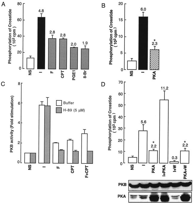
We have previously demonstrated that PKB and its PH domain-truncated form (ΔPH-PKB) can be activated by cAMP-elevating agents such as forskolin, chlorophenylthio (CPT)-cAMP, prostaglandin E1, and 8-bromo-cAMP (Fig. 1A). Although the major effect of cAMP elevation in the cell is to activate PKA, this nucleotide appears to have some PKA-independent actions as it is thought to act directly on ion channels (30, 40). Therefore, to establish that PKB activation in response to drugs augmenting intracellular cAMP was mediated by PKA, we tested the effect of cotransfection of the catalytic subunit of PKA on the activity of PKB overexpressed in 293 cells. As can be seen in Fig. 1B, coexpression of a catalytically active PKA led to a significant increase (2.3-fold) in PKB kinase activity. This effect is of a magnitude similar to that seen with cAMP-elevating agents such as forskolin (2.8-fold stimulation) (Fig. 1A), suggesting that PKA may be responsible for the PKB activation observed in response to these drugs. To further establish that PKA is involved in PKB activation, we used a cell-permeable and selective inhibitor of PKA, H-89 (dihydrochloride; Ki = 48 nM) (38). Incubation of H-89 (5 μM) together with forskolin or CPT-cAMP resulted in a 80 to 90% reduction in PKB activation compared to results with cells treated only with cAMP-elevating agents (Fig. 1C). We also found that cotransfection of PKA with the cDNA coding for PKI results in inhibition of PKA-induced activation of PKB (data not shown). Note that 5 μM H-89 inhibited by 95% the PKA activity in a cell-free assay (data not shown). Together, these data confirm that PKA itself is responsible for PKB activation by forskolin.
FIG. 1.
Effect of cAMP-elevating agents, the PKA catalytic subunit, and wortmannin on PKB activity in 293 cells. (A) Effect of cAMP-elevating agents on ΔPH-PKB. Serum-starved 293 EBNA cells overexpressing ΔPH-PKB were incubated for 15 min with 1 μM insulin (I), 10 μM forskolin (F), 1 mM CPT-cAMP (CPT), 2.5 μM prostaglandin E1 (PGE1), or 1 mM 8-bromo-cAMP (8-Br). After the incubation, cells were lysed and ΔPH-PKB was immunoprecipitated and its kinase activity was determined with Crosstide as described in Materials and Methods. ΔPH-PKB activity is expressed as counts per minute of 32P incorporated into Crosstide, and numbers above the bars indicate how many times above the basal level of stimulation PKB was stimulated. (B) Effect of the transfected PKA catalytic subunit. 293 EBNA cells were transfected with PKB and were either stimulated by insulin or cotransfected with the catalytic subunit of PKA. PKB was immunoprecipitated, and its activity was determined as described previously. (C) Effect of H-89 incubation on PKB activity. 293 EBNA cells were transfected with PKB and incubated or not incubated with H-89 (5 μM) and/or forskolin (10 μM; F), CPT-cAMP (1 mM; CPT), and insulin (1 μM; I). PKB was immunoprecipitated, and its activity was determined as described in Materials and Methods. (D) Effect of preincubation with wortmannin on PKA-induced activation of PKB. 293 EBNA cells were transfected with PKB in the presence or absence of PKA without preincubation or prior to preincubation for 30 min with 100 nM wortmannin (W). Subsequently, the cells were not treated or were treated with 1 μM insulin (I) for 5 min. The cells were lysed, PKB was immunoprecipitated, and kinase assays were performed as described in Materials and Methods. Results are expressed as counts per minute of 32P incorporated into Crosstide. The levels of the expressed enzymes measured by immunoblotting are shown below the graph. The results shown in panels A to D are means ± standard errors of results from a representative of two separate experiments performed in triplicate. Values significantly different from basal values (P < 0.05, Student’s t test) are denoted by ∗. NS, not stimulated.
We next wanted to determine if this PKA-mediated activation of PKB was independent of PI3-kinase as was previously demonstrated for drugs affecting cAMP levels (48). Therefore, the effect of wortmannin (an inhibitor of PI3-kinase) on PKA-induced PKB activation was examined. As expected, PKB activation by insulin was completely inhibited by pretreatment of the cells with wortmannin (Fig. 1D). However, pretreatment with wortmannin (100 nM) had no effect on PKB activity in PKA-transfected cells, since a 2.2-fold increase in the kinase activity was seen both with and without wortmannin. As shown in Fig. 1D, these differences were not due to variations in the level of protein expression.
Taken together, our results show that overexpression of the catalytic subunit of PKA increases PKB activity in 293 cells, suggesting that this effect is likely to be due to PKA activity itself. In addition, this effect appears to be independent of PI3-kinase.
PKA-induced activation of PKB in 293 cells depends on PKB phosphorylation and is indirect.
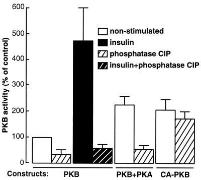
PKB activation in response to growth factors correlates with phosphorylation of the kinase on threonine 308 and serine 473. We therefore wanted to determine if the activation of PKB via PKA was also mediated by a change in the level of PKB phosphorylation. To do this, prior to the kinase reaction, PKB immunoprecipitates from transfected 293 cells were treated with CIP to dephosphorylate the wild-type and mutated PKB forms. To monitor that the phosphatase was indeed having an effect on PKB phosphorylation and not inhibiting the subsequent kinase reaction, immunoprecipitates of a constitutively active form of PKB (CA-PKB) were also treated with CIP. This construct contains acidic residues at position 308 and 473 of PKB and thus mimicks the effect of phosphorylation. The activity of this mutant should be resistant to phosphatase treatment. Therefore, if there is an effect of CIP on the activity of wild-type PKB, but not on CA-PKB, this would suggest that the phosphatase affects the phosphorylation state of PKB. As shown in Fig. 2, both insulin- and PKA-stimulated kinase activities of PKB were prevented by treating the immunoprecipitates with CIP. Exposure to the phosphatase had no effect on CA-PKB, confirming that CIP modifies the level of PKB phosphorylation.
FIG. 2.
Effect of phosphatase treatment on PKA-induced PKB activation in 293 cells. PKB and CA-PKB corresponding to mutations of both T308 and S473 to aspartate were expressed in 293 cells in the presence or absence of the catalytic subunit of PKA. Cell lysates were prepared and subjected to immunoprecipitation with an antibody to PKB. Immunoprecipitates were treated with CIP (20 U) for 30 min prior to the kinase assay. The results are the mean values ± standard errors of results from two experiments, each of which was performed in triplicate.
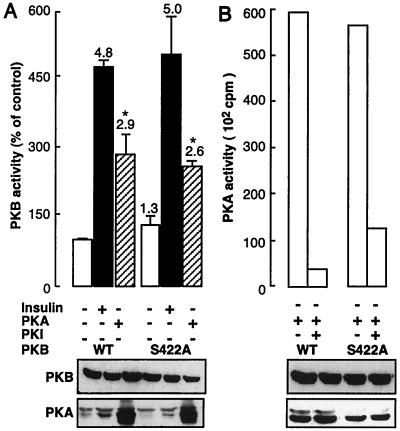
Next we were interested in determining whether activation of PKB by PKA was direct. We have previously shown that a recombinant glutathione S-transferase–PKB fusion protein is not efficiently phosphorylated in vitro by the catalytic subunit of PKA (48). However, since glutathione S-transferase–PKB was phosphorylated to a minor extent in vitro, we wanted to further explore the possibility that it was a direct substrate in vivo. As PKB possesses only a single PKA consensus phosphorylation site (KKLS422), we mutated S422 to alanine and analyzed the activity of this construct in 293 cells cotransfected or not cotransfected with the catalytic subunit of PKA. As can be seen in Fig. 3A, there was no effect of the S422 mutation on either insulin or PKA activation of PKB, since the levels of stimulation were identical for the wild-type and S422A forms of PKB. Figure 3B shows results with a control indicating that the PKA catalytic subunit transfected in 293 cells was active and that this activity was, as expected, abolished by the specific PKA inhibitor PKI. In conclusion, these data do not support the idea that PKB is a direct substrate of PKA.
FIG. 3.
Effect of the PKB S422A mutation on PKB’s activation by PKA. (A) The PKA phosphorylation consensus site was mutated by changing S422 to alanine (S422A). Then wild-type (WT) or mutant PKB was used to transfect 293 cells and the resulting kinase activity was determined as described in Materials and Methods. Results are expressed as a percentages of the value for the control and are the mean values ± standard errors of three experiments, each of which was performed in triplicate. (B) A PKA kinase assay was performed with Kemptide as a substrate in the presence or absence of PKI as described in Materials and Methods. Statistical significance, according to Student’s t test, is indicated as follows: ∗ indicates a P of <0.01 and ∗∗ indicates a P of <0.005 versus the value for the appropriate control.
PKB S473 is not required for PKA-induced PKB activation.
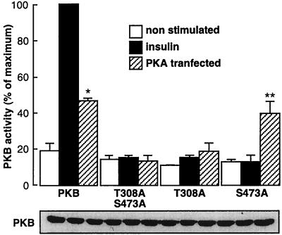
As phosphorylation of both T308 and S473 is necessary for the full activation of PKB by growth factors, we wanted to determine if these two residues were also important for PKB activation by PKA. Since this PKA action is independent of PI3-kinase, theoretically, this PKB activation process requires phosphorylation of other residues. To test this hypothesis, either T308, S473, or both residues together were mutated to alanine and these constructs were used to study the PKA effect. Cotransfection of the catalytically active PKA failed to stimulate the kinase activity of either the construct with the double mutation (T308A, S473A) or that with the single mutation (T308A) (Fig. 4). In contrast, a significant activation of the S473A mutant construct (twofold stimulation) was seen in response to PKA. However, the magnitude of this stimulation was not equivalent to that observed for the wild-type construct (2.5-fold stimulation). Taken together, these results demonstrate the following: (i) phosphorylation of T308 may be involved in PKB activation by PKA and (ii) phosphorylation of S473 does not appear to be necessary for PKB activation by PKA. Western blot analysis of PKB confirmed that the differences seen were not due to variation in the level of protein expression (Fig. 4, lower panel).
FIG. 4.
Effect of T308A and S473A on PKB activation by PKA in 293 cells. Single or double mutations of T308 and S472 to alanine were produced in PKB, and the constructs obtained were transfected in 293 cells. Immunoprecipitation was performed following stimulation with forskolin (10 μM), and the PKB activity was determined as described in Materials and Methods. A Western blot with antibody to PKB illustrates expression of PKB below the graph. Values shown are expressed as percentages of the maximal values and are the mean values ± standard errors of results from three separate experiments performed in triplicate. Values significantly different from basal values are denoted by ∗ and ∗∗ (∗ = P < 0.05 and ∗∗ = P < 0.005, Student’s t test).
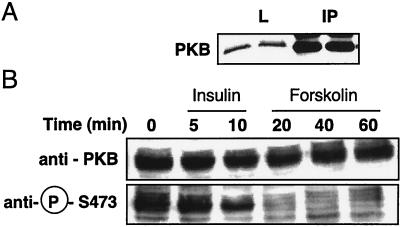
As all the above-described experiments were done with an overexpression system, we wanted to determine whether cAMP-elevating agents could lead to the activation of endogenous PKB. COS cells were found to be appropriate cells with which to perform these experiments as they express adequate amounts of PKB to efficiently detect subsequent kinase activity (Fig. 5A). To determine whether endogenous PKB is phosphorylated on S473 after stimulation by insulin or forskolin, we took advantage of an antibody which specifically recognizes phospho-S473. As can be seen in Fig. 5B, although PKB was significantly phosphorylated on S473 in response to insulin, phosphorylation of this residue was not seen in response to forskolin at any time point during the incubation. This result supports observations made for 293 cells with the S473A mutant PKB and indicates that phosphorylation of S473 is not important for the activation of PKB by PKA.
FIG. 5.
Effect of forskolin on PKB S473 phosphorylation. (A) Western blot showing endogenous expression of PKB in COS cells either after immunoprecipitation with anti-PKB (IP) or directly in the lysate (L). (B) COS cells were stimulated for the indicated times with insulin or forskolin prior to lysis. Cell extracts were subjected to SDS-PAGE (10% polyacrylamide) followed by Western blot analysis with antibody to PKB or to phospho-S473 PKB. Results shown are representative of at least two experiments.
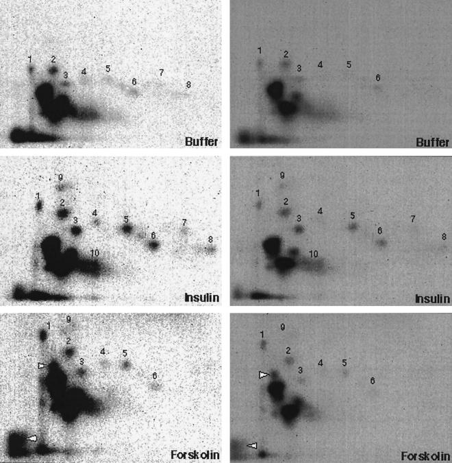
To confirm that residue S473 is not required for PKA-induced PKB activation and to determine which other residues may be involved, we analyzed PKB phosphorylation using phosphopeptide mapping of metabolically labeled COS cells transfected with PKB-HA. The 32P-labeled COS cells were treated with insulin for 5 min or forskolin or buffer for 30 min. Under these conditions, both insulin and forskolin stimulation resulted in an increase in PKB phosphorylation compared to the level in the nonstimulated cells (data not shown). Figure 6 represents the phosphopeptide maps of PKB-HA after stimulation by either insulin or forskolin. Compared to PKB obtained from nonstimulated cells, PKB obtained from insulin-stimulated cells was more intensely labeled at at least four phosphopeptides (i.e., spots 2, 3, 5, and 6) and showed two new phosphopeptides (i.e., spots 9 and 10), which very likely correspond to T308- and S473-containing peptides. While most phosphopeptides were found to be common, PKB from forskolin-treated cells showed at least two different phosphopeptides compared to PKB obtained from cells exposed to insulin, as indicated in Fig. 6. This indicates that the mechanism of PKB activation by forskolin is not identical to that used by insulin. Together, these results support the idea that PKA-induced activation of PKB is independent of S473 and very likely involves additional regulatory phosphorylated residues.
FIG. 6.
Phosphopeptide maps of endogenous PKB obtained from COS cells. Results of a two-dimensional peptide map analysis of 32P-labeled PKB-HA isolated from metabolically labeled COS cells are shown. COS cells were grown in medium containing 1 mCi of [32P]orthophosphate per ml for 4 h. Untreated, insulin (1 μM)-treated, or forskolin (10 μM)-treated cells were lysed, and equal amounts of lysate protein were immunoprecipitated with antibodies to HA. The immunoprecipitates were resolved by SDS-PAGE. The 32P-labeled band corresponding to PKB was excised from the gel, eluted, and digested with trypsin (20 μg). Peptides were separated on cellulose thin-layer plates by electrophoresis from left to right (anode on the right), followed by thin-layer chromatography from bottom to top. Autoradiograms of the thin-layer plates are shown.
Endogenous GSK-3 is inhibited by cAMP-elevating agents in COS cells.
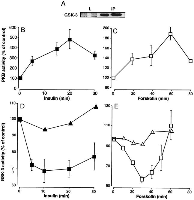
Next, we wanted to determine if cAMP-induced PKB activation has an effect on downstream events, such as inhibition of GSK-3. Experiments were performed with COS cells, as they were found to express measurable GSK-3 (Fig. 7A). First we compared time courses of PKB (Fig. 7B and C) and GSK-3 activities (Fig. 7D and E). The results showed that, in contrast with PKB activation by insulin, which is rapid and reaches a maximum (4.8-fold) within 20 min, PKB activation by forskolin was slow and resulted in a 2-fold increase in activity only after 60 min. With insulin stimulation, a significant decrease in GSK-3 activity was seen when PKB was highly active (between 5 and 20 min), with a progressive return to basal activity after 30 min. When cells were stimulated by forskolin, a decrease in GSK-3 activity corresponding to the highest PKB activity was also seen and basal levels were obtained when PKB activity declined. As for the results obtained for insulin, exposure to forskolin led to PKB stimulation and GSK-3 inhibition, illustrating that forskolin affects events downstream of PKB. To confirm that the GSK-3 inhibition was due to PKB, we transfected COS cells together with a dominant negative form of PKB mutated in three residues (T308A, S473A, and K179A). PKB with this triple mutation has been shown by others to function as a dominant negative PKB in neuroblastoma cells (61), and we found it to act in a similar fashion in our 293 or COS cells (data not shown). When this mutant PKB was overexpressed in COS cells, inhibition of GSK-3 in response to the presence of either insulin or forskolin was no longer seen, implicating PKB directly in this GSK-3 inhibition induced by either insulin or forskolin.
FIG. 7.
Time courses of the effects of forskolin and insulin on PKB and GSK-3 activity in COS cells (A) Western blot showing endogenous expression of GSK-3 in COS cells either after immunoprecipitation with anti-GSK3 (IP) or directly in the lysate (L). (B to E) Time courses of PKB and GSK-3 activities. COS cells were treated or not treated for the times indicated with insulin (1 μM) or forskolin (10 μM) prior to extraction. Cell lysates were immunoprecipitated with antibodies either to PKB or to GSK-3; thereafter, activities in the immune complexes were determined. The results are expressed as percentages of the control value at time zero and are mean values ± standard errors of results from two experiments. ▴ and ▵ represent the GSK-3 activities in the presence of overexpressed dominant negative PKB.
In response to insulin and forskolin, increased GSK-3 phosphorylation correlates with a decrease in enzymatic activity.
To determine whether the decrease in GSK-3 activity seen in response to the addition of insulin or forskolin was linked to its phosphorylation state, the ability of GSK-3 to serve as a substrate for insulin- or cAMP-activated PKB in vitro was examined. To do this, PKB was immunoprecipitated from either insulin- or forskolin-stimulated cells and GSK-3 was immunopurified from unstimulated cells. PKB and GSK-3 immunoprecipitates were then mixed together in the presence of [γ-32P]ATP. As can be seen in Fig. 8, GSK-3 was only weakly phosphorylated in its basal state. In response to insulin, the enzyme became rapidly phosphorylated (within 5 or 10 min) and thereafter decreased to basal levels within 30 min. The increase in phosphorylation correlated with the decrease in activity seen in Fig. 7D in response to insulin. The phosphorylation of GSK-3 by PKB in response to forskolin, as was seen for insulin, showed a pronounced increase in the level of GSK-3 phosphorylation with a parallel decrease in its activity. This result points to the existence of a direct link between the inhibition of GSK-3 activity in cells and its phosphorylation by PKB. This correlation was found regardless of whether PKB was activated by insulin or by forskolin.
FIG. 8.
In vitro phosphorylation of GSK-3. COS cells were treated with either insulin (1 μM) or forskolin (10 μM) for the indicated times, and PKB was immunoprecipitated. At the same time, GSK-3 was immunoprecipitated from nonstimulated cells. PKB and GSK-3 immunoprecipitates were mixed in the presence of [γ-32P]ATP, and the samples were subjected to SDS-PAGE under reducing conditions followed by autoradiography.
GFP-PKB translocation to the cell membrane in response to forskolin can be abolished by a high concentration (300 nM) of wortmannin.
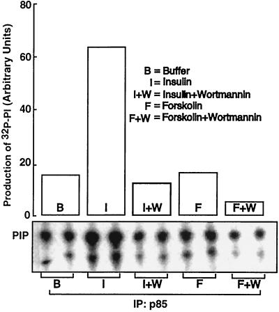
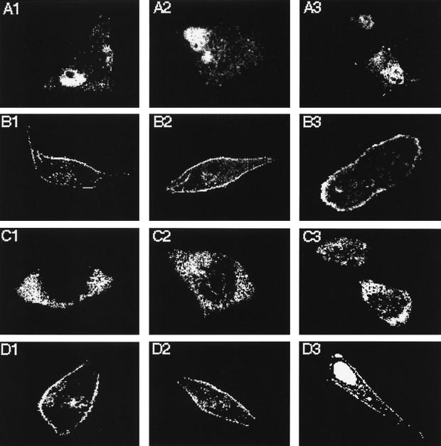
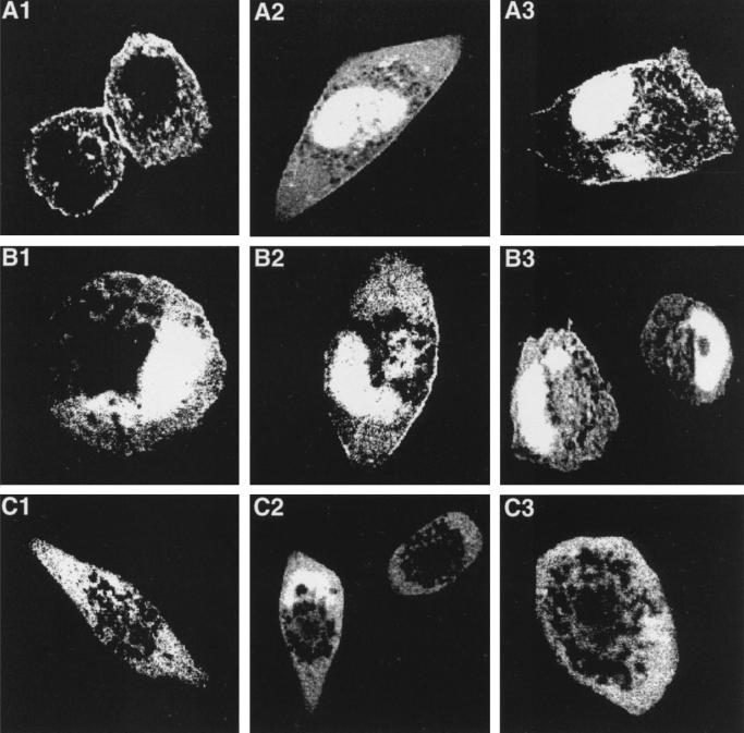
To further clarify the mechanism of PKB activation by cAMP-elevating agents, the effect of forskolin (or CPT-cAMP) on PKB localization was determined by confocal microscopy with GFP-tagged PKB. This construct was shown to be fully active when it was transfected in 293 or HeLa cells (data not shown). For these studies HeLa cells were used instead of 293 cells, since we found that 293 cells are difficult cells on which to perform immunohistochemistry. While preliminary data indicated that the distribution of PKB in 293 cells was similar to that seen in HeLa cells, these data were not conclusive. GFP-PKB plasmid was transfected into HeLa cells, and its subcellular localization was examined by confocal microscopy. In Fig. 9A1 to A3, which reflect results with nonstimulated cells, GFP-PKB is diffusely located throughout the cell. Translocation of GFP-PKB to the plasma membrane is clearly seen in response to insulin as illustrated by green fluorescence at the cell membrane (Fig. 9B1 to 9B3, where green fluorescence corresponds to white). Pretreatment with wortmannin (100 nM) prevented GFP-PKB from translocating to the plasma membrane (Fig. 9C1 to C3). Similar to the response to insulin, treatment with forskolin allowed translocation of PKB to the plasma membrane (Fig. 9D1 to D3 and 10A1 to A3). In contrast to that induced with insulin, the forskolin-induced translocation of PKB was not inhibited by pretreatment with 100 nM wortmannin (Fig. 10B1 to B3). However, at a higher concentration (300 nM) of wortmannin complete inhibition of PKB translocation could be observed (Fig. 10C1 to C3). Control experiments performed with cells transfected with an empty GFP vector showed no localization in the cell membrane in response to insulin or forskolin (data not shown). In Fig. 11 we show that cAMP did not activate PI3-kinase associated with the p85 regulatory subunit, indicating the involvement of a different kinase sensitive to the high concentration of wortmannin.
FIG. 11.
Effect of cAMP-increasing agents on PI3-kinase activity immunoprecipitated with antibody to p85. 293 cells were preincubated for 20 min with 100 nM wortmannin prior to stimulation for 5 min with 1 μM insulin or for 30 min with 10 μM forskolin. Total cellular protein lysates were immunoprecipitated with anti-p85 antibody, and associated phospholipid kinase activities were estimated in vitro with PtdIns or PtdIns-4-P plus PtdIns-4,5-P2 as the substrate. The lower panel shows a representative thin-layer chromatogram of products of PI3-kinase in the presence or absence of wortmannin. The position of 32P-labeled PI (PIP) is indicated. The graph shown in the upper panel corresponds to the radioactivity associated with each spot as quantified by phosphorimaging. B represents the nontreated cells. IP, immunoprecipitates.
DISCUSSION
In this work we investigated the mechanism of PKB activation induced by PKA, which appears to occur by a PI3-kinase-independent pathway. This is not the first demonstration that PKB can be activated independently of PI3-kinase. Indeed, Konishi et al. have shown that cellular stress, such as heat shock and hyperosmolarity, is able to stimulate PKB activity (37). Furthermore, Moule et al. have demonstrated that PKB can be activated by isoproterenol via β-adrenergic receptors, resulting in GSK-3 inhibition, although this could not be mimicked by elevation of cAMP (41), with the CPT-cAMP analog, in this cell type. Finally, we have previously shown that agents that can increase intracellular levels of cAMP can activate PKB in transfected 293 cells (48). In the present paper we have extended these findings and demonstrate that the kinase responsible for this activation is PKA. This is not surprising, since virtually all the effects of cAMP are mediated by PKA. However, in our experiments PKB activation did not appear to be due to direct phosphorylation of PKB by PKA. Data supporting this view are that mutation of S422, which is part of the sole consensus PKA phosphorylation site on PKB, does not abolish activation of PKB by PKA.
We next examined the mechanism of PKB stimulation in response to PKA. First we showed that this activation, as with growth factor receptor-mediated stimulation of PKB, is dependent upon PKB phosphorylation. This dependence was demonstrated by (i) treatment of immunopurified PKB with phosphoserine phosphatase, which abolished both insulin and PKA activation of PKB, and (ii) using a nonphosphorylatable mutant PKB which was resistant to activation by PKA. This led us to conclude that PKA-mediated activation of PKB requires PKB phosphorylation. This may be due to PKA activation of one or more PKBs and/or inactivation of a phosphatase(s) responsible for dephosphorylation and consequent inactivation of PKB regulatory sites. The next step was to determine if the two residues required for PKB activation by growth factors, threonine 308 and serine 473, were also important for PKA activation of PKB. In order to do this we used a panel of mutant PKBs in which positions 308 and 473 contained either alanine residues, preventing phosphorylation, or glutamic acid residues, mimicking phosphorylation. When T308 was mutated to alanine, either singly or in conjunction with S473A, the resulting PKB was resistant to activation by either insulin or PKA. Importantly, the S473A mutant PKB was still activated in response to forskolin. This result indicates that T308 is indispensable for PKB stimulation by PKA but that S473 is not. However, other residues different from those involved in growth factor-mediated activation of PKB may contribute to PKA-induced stimulation. Compatible with this view is the recent report by Yano et al. (61) which demonstrates that the Ca2+- and calmodulin-dependent protein kinase kinase activates PKB by direct phosphorylation of T308 independently of PI3-kinase.
To more thoroughly examine the mechanism of PKB stimulation by PKA, we looked at activation of endogenous PKB in response to forskolin in COS cells. These cells have high levels of endogenous PKB, and therefore the need to transfect is circumvented. For these experiments, an antibody which recognizes PKB phosphorylated on S473 was used. We found that this antibody did not recognize PKB activated by forskolin but that it did bind to insulin-stimulated PKB, consistent with the idea that S473 is not phosphorylated in response to activation by PKA. A comparable situation has been reported by Bellacosa et al., who showed that S473 phosphorylation is not required for PKB activation by PDGF (8). Finally, a phosphopeptide map analysis was performed with COS cells. The phosphopeptide maps obtained with PKB from forskolin-treated cells were at least partly different from those obtained from insulin-treated cells. This also supports the hypothesis that forskolin activation of PKB is mechanistically distinct from the pathway utilized by growth factors.
In addition to phosphorylation, regulation of PKB activity is dependent on subcellular localization. There is now evidence that PKBα is located in the cytosol of unstimulated cells but that it rapidly translocates to the plasma membrane following stimulation with IGF-1 (5), interleukin-2 (1), peroxovanadate (59), and, as was more recently shown, insulin (47). This translocation is prevented by inhibitors of PI3-kinase such as wortmannin (5) or by deletion of the PH domain of PKB (47). In this study, we find that forskolin stimulation also leads to PKB translocation to the plasma membrane. At first sight this result appears inconsistent with data showing that activation of PKB by PKA is PI3-kinase independent and hence phospholipid independent. However, PKA-induced PKB translocation is abolished after pretreatment of the cells with high concentrations (300 nM) of wortmannin, implying that translocation to the cell surface may require the generation of phospholipids. Furthermore, forskolin-induced PKB stimulation is maintained after a 300 nM wortmannin pretreatment (data not shown), demonstrating that the translocation process is not necessary for PKB activation by PKA. No translocation to the plasma membrane was seen with a mutant PKB with the PH domain deleted, suggesting a role for phospholipids in this translocation process (data not shown). Endogenous PI3-kinase is not activated by cAMP analogs in 293, HeLa, or COS cells, and PKB is still stimulated by forskolin when it is cotransfected with a dominant negative PI3-kinase in 293 cells (data not shown). Some class 2 PI3-kinases are relatively resistant to this inhibitor at concentrations greater than 100 nM (23, 57), and PI4-kinases have also been reported not to be inhibited by wortmannin, at concentrations up to 1 μM (55). It will thus be of interest to reveal which PI kinase, if implicated in this process, is responsible for this cAMP-induced translocation.
Next, we wanted to determine if PKB activation by PKA could have an impact upon events downstream of PKB. To date, only a few substrates have been discovered for PKB. There is evidence that some of the metabolic effects of insulin on glycogen synthesis are mediated by PKB, which can phosphorylate and inactivate GSK-3 in vitro and in vivo (56). The activities of both isoforms of GSK-3 have been shown to decrease by about 40% when rat L6 myotubes or rat skeletal muscle in situ is stimulated with insulin (18). GSK-3 may play a role in controlling glycogen metabolism since glycogen synthase is phosphorylated by GSK-3 and hence becomes inactive. However, it is unclear whether this is the major pathway regulating glycogen synthesis in all cells. For example, in 3T3-L1 adipocytes there is no effect of overexpression of constitutively active PKB on glycogen synthase activity. Consistent with this result is the recent notion that the glycogen-bound form of type 1 protein phosphatase is thought to play the dominant role in insulin regulation of glycogen metabolism in 3T3-L1 adipocytes (9).
In our study, we show that there is a decrease in GSK-3 activity following stimulation of COS cells with forskolin. At first glance this result may seem at variance with the general view that the actions of insulin and cAMP are opposing with regard to metabolism. However, there are several potential explanations for this apparent inconsistency. First, at the physiological level the predominant tissues for glycogen metabolism are the liver and the skeletal muscle. Therefore, since we used kidney cells, this decrease in GSK-3 activity may be a cell-specific effect. Second, the time courses of inactivation of GSK-3 by insulin and by cAMP were different; most notably, the inhibition by cAMP occurred later than that seen with insulin. Finally, GSK-3 has been implicated in cell functions very distinct from metabolism, like regulation of cell fate in Dictyostelium discoideum (31). Recently, a great deal of interest has been expressed about the Wnt→zw3→en signaling pathway, which is required for Drosophila and Xenopus development and also for growth regulation in mammalian cells. GSK-3 is involved in this pathway since it is the mammalian homolog of the zeste-white 3 (zw3, also known as shaggy) protein found in Drosophila melanogaster (16, 50). In addition, GSK-3 has been reported to phosphorylate the translation initiation factor eIF2B (58) and several transcription factors, including c-jun (42), that are reported to be involved in cell growth and apoptosis. It has also been shown more recently that GSK-3 may play a critical role in the regulation of apoptosis in the PI3-kinase/Akt cell survival circuitry (43).
In the past year it has become evident that one of the major functions of PKB is protection of cells from programmed death. This protection has been demonstrated for several cell types, including COS cells (39), fibroblasts (34), and neuronal cells (24, 45), and for several receptors, including the insulin or IGF-1 receptor (39). This may in part be due to the ability of PKB to phosphorylate and thereby inhibit the proapoptotic Bcl family member BAD. It has also been described that cAMP and PKA are antiapoptotic in certain cell types, including neutrophils (44). The mechanism of this antiapoptotic effect is not known, but from our work here, it is tempting to speculate that it is mediated via PKB. At this time there is much effort being expended in the study of regulation of apoptosis in mammalian cells. An interesting challenge is to unravel the physiological importance of the cross talk between insulin or IGF-1 signaling and cAMP effects and the possible convergence of this cross talk on the PKB pathway.
FIG. 9.
Localization of GFP-PKB in response to insulin or forskolin in transfected HeLa cells. HeLa cells were transfected with GFP-PKB as described in Materials and Methods. Forty-eight hours later, HeLa cells were preincubated (C1 to C3) or not preincubated (B1 to B3) for 20 min with wortmannin (100 nM) prior to stimulation for 10 min with insulin (1 μM) (B1 to B3) or for 30 min with forskolin (10 μM) (D1 to D3). Cells were washed and fixed with paraformaldehyde (4%). Slides were mounted and analyzed by confocal microscopy. Images represent the center section of the X-Y plane. (A1 to A3) Nonstimulated cells.
FIG. 10.
Effect of increasing concentrations of wortmannin on GFP-PKB localization in response to forskolin. HeLa cells were transfected with GFP-PKB as described in Materials and Methods. Forty-eight hours later, HeLa cells were preincubated for 20 min with wortmannin (100 nM [B1 to B3] or 300 nM [C1 to C3]) or buffer (A1 to A3) prior to stimulation for 30 min with forskolin (10 μM). Cells were mounted and analyzed by confocal microscopy. Images represent the center section of the X-Y plane.
ACKNOWLEDGMENTS
Our research was supported by the Institut National de la Santé et de la Recherche Médicale, the Association pour la Recherche sur le Cancer, the Université de Nice-Sophia Antipolis, la Ligue contre le Cancer, Groupe LIPHA-Merck (Lyon, France), and Sankyo (Dusseldörf, Germany, and Tokyo, Japan). C.L.S. is a recipient of a Poste Vert from INSERM.
REFERENCES
- 1.Ahmed N N, Grimes H L, Bellacosa A, Chan T O, Tsichlis P N. Transduction of interleukin-2 antiapoptotic and proliferative signals via Akt protein kinase. Proc Natl Acad Sci USA. 1997;94:3627–3632. doi: 10.1073/pnas.94.8.3627. [DOI] [PMC free article] [PubMed] [Google Scholar]
- 2.Alessi D, Cohen P. Mechanism of activation and function of protein kinase B. Curr Opin Genet Dev. 1998;8:55–62. doi: 10.1016/s0959-437x(98)80062-2. [DOI] [PubMed] [Google Scholar]
- 3.Alessi D, Kozlowski M T, Weng Q-P, Morrice N, Avruch J. 3-Phosphoinositide-dependent protein kinase 1 (PDK1) phosphorylates and activates the p70 S6 kinase in vivo and in vitro. Curr Biol. 1997;8:69–81. doi: 10.1016/s0960-9822(98)70037-5. [DOI] [PubMed] [Google Scholar]
- 4.Alessi D R, James S R, Downes C P, Holmes A B, Gaffney P R J, Reese C B, Cohen P. Characterization of a 3-phosphoinositide-dependent kinase which phosphorylates and activates protein kinase Bα. Curr Biol. 1997;7:261–269. doi: 10.1016/s0960-9822(06)00122-9. [DOI] [PubMed] [Google Scholar]
- 5.Andjelkovic M, Alessi D, Meier R, Fernandez A, Lamb N, Frech M, Cron P, Cohen P, Lucocq J, Hemmings B A. Role of translocation in the activation and function of protein kinase B. J Biol Chem. 1997;272:31515–31524. doi: 10.1074/jbc.272.50.31515. [DOI] [PubMed] [Google Scholar]
- 6.Andjelkovic M, Jakubowicz T, Cron P, Ming X-F, Han J-W, Hemmings B A. Activation and phosphorylation of a pleckstrin holology domain containing protein kinase (RAC-PK/PKB) promoted by serum and protein phosphatase inhibitors. Proc Natl Acad Sci USA. 1996;93:5699–5704. doi: 10.1073/pnas.93.12.5699. [DOI] [PMC free article] [PubMed] [Google Scholar]
- 7.Bellacosa A, Testa J R, Staal S P, Tsichlis P N. A retroviral oncogene, akt, encoding a serine-threonine kinase containing an SH2-like region. Science. 1991;254:274–277. doi: 10.1126/science.254.5029.274. [DOI] [PubMed] [Google Scholar]
- 8.Bellacosa A, Tung O, Naheed N, Datta K, Malstrom S, Stokoe D, McCormick F, Feng J, Tsichlis P. Akt activation by growth factors is a multiple-step process: the role of the PH domain. Oncogene. 1998;17:313–325. doi: 10.1038/sj.onc.1201947. [DOI] [PubMed] [Google Scholar]
- 9.Brady M, Bourbonais F, Saltiel A. The activation of glycogen synthase by insulin switches from kinase inhibition to phosphatase activation during adipogenesis in 3T3-L1 cells. J Biol Chem. 1998;273:14063–14066. doi: 10.1074/jbc.273.23.14063. [DOI] [PubMed] [Google Scholar]
- 10.Burgering B M T, Pronk G J, van Weeren P C, Chardin P, Bos J L. cAMP antagonizes p21ras-directed activation of extracellular signal-regulated kinase-2 and phosphorylation of mSos nucleotide exchange factor. EMBO J. 1993;12:4211–4220. doi: 10.1002/j.1460-2075.1993.tb06105.x. [DOI] [PMC free article] [PubMed] [Google Scholar]
- 11.Burgering T, Boudewijn M, Coffer P J. Protein kinase B (c-Akt) in phosphotidylinositol-3-OH kinase signal transduction. Nature. 1995;376:599–602. doi: 10.1038/376599a0. [DOI] [PubMed] [Google Scholar]
- 12.Cheng J Q, Godwin A K, Bellacosa A, Taguchi T, Franke T F, Hamilton T C, Tsichlis P N, Testa J R. AKT2, a putative oncogene encoding a member of a subfamily of protein-serine/threonine kinases is amplified in human ovarian carcinomas. Proc Natl Acad Sci USA. 1992;89:9267–9271. doi: 10.1073/pnas.89.19.9267. [DOI] [PMC free article] [PubMed] [Google Scholar]
- 13.Cheng J Q, Ruggeri B, Klein W M, Sonoda G, Altomare D A, Watson D K, Testa J R. Amplification of AKT2 in human pancreatic-cancer cells and inhibition of AKT2 expression and tumorigenicity by antisense RNA. Proc Natl Acad Sci USA. 1996;93:3636–3641. doi: 10.1073/pnas.93.8.3636. [DOI] [PMC free article] [PubMed] [Google Scholar]
- 14.Coffer P, Woodgett J. Molecular cloning and characterization of a novel protein-serine-threonine kinase related to the cAMP-dependent and protein kinase C families. Eur J Biochem. 1991;201:475–481. doi: 10.1111/j.1432-1033.1991.tb16305.x. [DOI] [PubMed] [Google Scholar]
- 15.Cong L-N, Chen H, Li Y, Zhou L, McGibbon M, Taylor S, Quon M. Physiological role of Akt in insulin-stimulated translocation of glut4 in transfected rat adipose cells. Mol Endocrinol. 1997;11:1881–1890. doi: 10.1210/mend.11.13.0027. [DOI] [PubMed] [Google Scholar]
- 16.Cook D, Fry M J, Hughes K, Sumathipala R, Woodgett J R, Dale T C. Wingless inactivats glycogen synthase kinase-3 via an intracellular signalling pathway which involves a protein kinase C. EMBO J. 1996;15:4526–4536. [PMC free article] [PubMed] [Google Scholar]
- 17.Cook S J, McCormick F. Inhibition by cAMP of Ras-dependent activation of Raf. Science. 1993;262:1069–1072. doi: 10.1126/science.7694367. [DOI] [PubMed] [Google Scholar]
- 18.Cross D A E, Alessi D R, Cohen P, Andjelkovich M, Hemmings B A. Inhibition of glycogen synthetase kinase-3 by insulin mediated by protein kinase B. Nature. 1995;378:785–789. doi: 10.1038/378785a0. [DOI] [PubMed] [Google Scholar]
- 19.Datta K, Bellacosa A, Chan T O, Tsichlis P N. Akt is a direct target of the phosphatidylinositol 3-kinase. J Biol Chem. 1996;271:599–602. doi: 10.1074/jbc.271.48.30835. [DOI] [PubMed] [Google Scholar]
- 20.Datta K, Franke T F, Chan T O, Makris A, Yang S-I, Kaplan D R, Morrison D K, Golemis E A, Tsichlis P N. AH/PH domain-mediated interaction between Akt molecules and its potential role in Akt regulation. Mol Cell Biol. 1995;15:2304–2310. doi: 10.1128/mcb.15.4.2304. [DOI] [PMC free article] [PubMed] [Google Scholar]
- 21.Delcommenne M, Tan C, Gray V, Rue L, Woodgett J, Dedhar S. Phosphoinositide-3-OH kinase-dependent regulation of glycogen synthase kinase 3 and protein kinase B/AKT by the integrin-linked kinase. Proc Natl Acad Sci USA. 1998;95:11211–11216. doi: 10.1073/pnas.95.19.11211. [DOI] [PMC free article] [PubMed] [Google Scholar]
- 22.Deprez J, Vertommen D, Alessi D R, Hue L, Rider M H. Phosphorylation and activation of heart 6-phosphofructo-2-kinase by protein kinase B and other protein kinases of the insulin signaling cascades. J Biol Chem. 1997;272:17269–17275. doi: 10.1074/jbc.272.28.17269. [DOI] [PubMed] [Google Scholar]
- 23.Domin J, Waterfield M D. Using structure to define the function of phosphoinositide 3-kinase family members. FEBS Lett. 1997;410:91–95. doi: 10.1016/s0014-5793(97)00617-0. [DOI] [PubMed] [Google Scholar]
- 24.Dudek H, Datta S, Franke T, Birnbarun M, Yao R, Cooper G. Regulation of neuronal survival by the serine-threonine protein kinase Akt. Science. 1997;275:661–665. doi: 10.1126/science.275.5300.661. [DOI] [PubMed] [Google Scholar]
- 25.Franke T F, Yang S-I, Chan T O, Datta K, Kazlauskas A, Morrison D K, Kaplan D R, Tsichlis P N. The protein kinase encoded by the Akt proto-oncogene is a target of the PDGF-activated phosphatidylinosital 3-kinase. Cell. 1995;81:727–736. doi: 10.1016/0092-8674(95)90534-0. [DOI] [PubMed] [Google Scholar]
- 26.Frech M, Andjelkovic M, Ingley E, Reddy K K, Falck J R, Hemmings B A. High affinity binding of inositol phosphates and phosphoinositides to the pleckstrin homology domain of RAC/protein kinase B and their influence on kinase activity. J Biol Chem. 1997;272:8474–8481. doi: 10.1074/jbc.272.13.8474. [DOI] [PubMed] [Google Scholar]
- 27.Göransson O, Wijkander J, Manganiello V, Degerman E. Insulin-induced translocation of protein kinase B to the plasma membrane in rat adipocytes. BBRC. 1998;246:249–254. doi: 10.1006/bbrc.1998.8602. [DOI] [PubMed] [Google Scholar]
- 28.Graves L M, Bornfield K E, Raines E W, Potts B C, Macdonald S G, Ross R, Krebs E G. Protein kinase antagonizes platelet-derived growth factor-induced signaling by mitogen-activated protein kinase in human arterial smooth muscle cells. Proc Natl Acad Sci USA. 1993;90:10300–10304. doi: 10.1073/pnas.90.21.10300. [DOI] [PMC free article] [PubMed] [Google Scholar]
- 29.Graves L M, Lawrence J C. Insulin, growth factors, and cAMP, antagonism in the signal transduction pathways. Trends Endocrinol Metab. 1996;7:43–50. doi: 10.1016/1043-2760(95)00204-9. [DOI] [PubMed] [Google Scholar]
- 30.Gray P C, Scott J D, Catteral W A. Regulation of ion channels by cAMP-dependent protein kinase and A-kinase anchoring proteins. Curr Biol. 1998;8:330–334. doi: 10.1016/s0959-4388(98)80057-3. [DOI] [PubMed] [Google Scholar]
- 31.Harwood A, Plyte S, Woodgett J, Strutt H, Kay R. Glycogen synthase kinase 3 regulates cell fate in Dictyostelium. Cell. 1995;80:139–148. doi: 10.1016/0092-8674(95)90458-1. [DOI] [PubMed] [Google Scholar]
- 32.Hordijk P L, Verlaan I, Jalink K, van Corvan E J, Moolenaar W H. cAMP abrogates the p21ras-mitogen-activated protein kinase pathway in fibroblasts. J Biol Chem. 1994;269:3534–3538. [PubMed] [Google Scholar]
- 33.Jones P F, Jakubowicz T, Pitossi F J, Maurer F, Hemmings B A. Molecular cloning and identification of a serine/threonine protein kinase of the second messenger family. Proc Natl Acad Sci USA. 1991;88:4171–4175. doi: 10.1073/pnas.88.10.4171. [DOI] [PMC free article] [PubMed] [Google Scholar]
- 34.Kauffman-Zeh A, Rodriguez-Viciana P, Ulrich E, Coffer P, Downward J, Evan G. Suppression of c-myc-induced apoptosis by ras signalling through PI(3)K and PKB. Nature. 1997;385:544–548. doi: 10.1038/385544a0. [DOI] [PubMed] [Google Scholar]
- 35.Kohn A D, Kovacina K S, Roth R A. Insulin stimulates the kinase activity of RAC-PK, a pleckstrin homology domain containing Ser/Thr kinase. EMBO J. 1995;14:4288–4295. doi: 10.1002/j.1460-2075.1995.tb00103.x. [DOI] [PMC free article] [PubMed] [Google Scholar]
- 36.Kohn A D, Summers S A, Birnbaum M, Roth R A. Expression of a constitutively active akt Ser:Thr kinase in 3T3-L1 stimulates glucose uptake and glucose transporter 4 translocation. J Biol Chem. 1996;271:31372–31378. doi: 10.1074/jbc.271.49.31372. [DOI] [PubMed] [Google Scholar]
- 37.Konishi H, Matsuzaki H, Tanaka M, Takemura Y, Kuroda S, Ono Y, Kikkawa U. Activation of protein kinase B (Akt/RAC-protein kinase) by cellular stress and its association with heat shock protein Hsp27. FEBS Lett. 1997;410:493–498. doi: 10.1016/s0014-5793(97)00541-3. [DOI] [PubMed] [Google Scholar]
- 38.Krueger K A, Bhatt H, Landt M, Easom R A. Calcium-stimulated phosphorylation of MAP-2 in pancreatic betaTC3-cells is mediated by Ca2+/calmodulin-dependent kinase II. J Biol Chem. 1997;272:27464–27469. doi: 10.1074/jbc.272.43.27464. [DOI] [PubMed] [Google Scholar]
- 39.Kulik G, Klippel A, Weber M J. Antiapoptotic signaling by the insulin-like growth factor I receptor, phosphatidylinositol 3-kinase, and Akt. Mol Cell Biol. 1997;17:1595–1606. doi: 10.1128/mcb.17.3.1595. [DOI] [PMC free article] [PubMed] [Google Scholar]
- 40.Matthews G. Ion channels that are directly activated by cyclic nucleotides. Trends Pharmacol Sci. 1991;12:245–247. doi: 10.1016/0165-6147(91)90563-8. [DOI] [PubMed] [Google Scholar]
- 41.Moule S K, Welsh G I, Edgel N J, Foulstone E J, Proud C G, Denton R M. Regulation of protein kinase B and glycogen synthase kinase-3 by insulin and β-adrenergic agonists in rat epididymal fat cells. J Biol Chem. 1997;272:7713–7719. doi: 10.1074/jbc.272.12.7713. [DOI] [PubMed] [Google Scholar]
- 42.Nikolakaki E, Coffer P J, Hemelsoet R, Woodgett J R, Defize L H. Glycogen synthase kinase 3 phosphorylates Jun family members in vitro and negatively regulates their transactivating potential in intact cells. Oncogene. 1993;8:833–840. [PubMed] [Google Scholar]
- 43.Pap M, Cooper G. Role of glycogen synthase kinase-3 in the phophatidylinositol 3-kinase/Akt cell survival pathway. J Biol Chem. 1998;273:19929–19932. doi: 10.1074/jbc.273.32.19929. [DOI] [PubMed] [Google Scholar]
- 44.Parvathenani L, Buescher S, Chacon-Cruz E, Beebe S. Type I cAMP-dependent protein kinase delays apoptosis in human neutrophils at site upstream of caspase-3. J Biol Chem. 1998;273:6736–6743. doi: 10.1074/jbc.273.12.6736. [DOI] [PubMed] [Google Scholar]
- 45.Philpott K L, McCarthy M J, Klippel A, Rubin L L. Activated phosphatidylinositol 3-kinase and Akt kinase promote survival of superior cervical neurons. J Cell Biol. 1997;139:809–815. doi: 10.1083/jcb.139.3.809. [DOI] [PMC free article] [PubMed] [Google Scholar]
- 46.Roskoski R. Assays of protein kinase. Methods Enzymol. 1983;99:3–6. doi: 10.1016/0076-6879(83)99034-1. [DOI] [PubMed] [Google Scholar]
- 47.Sable C L, Filippa N, Filloux C, Hemmings B A, Van Obberghen E. Involvement of the pleckstrin homology domain in the insulin-stimulated activation of protein kinase B. J Biol Chem. 1998;273:29600–29606. doi: 10.1074/jbc.273.45.29600. [DOI] [PubMed] [Google Scholar]
- 48.Sable C L, Filippa N, Filloux C, Hemmings B A, Van Obberghen E. cAMP stimulates protein kinase B in a wortmannin-insensitive manner. FEBS Lett. 1997;409:253–257. doi: 10.1016/s0014-5793(97)00518-8. [DOI] [PubMed] [Google Scholar]
- 49.Sevetson B R, Kong X, Lawrence J C J. Increasing cAMP attenuates activation of mitogen-activated protein kinase. Proc Natl Acad Sci USA. 1993;90:10305–10309. doi: 10.1073/pnas.90.21.10305. [DOI] [PMC free article] [PubMed] [Google Scholar]
- 50.Siegfried E, Chou T-B, Perrimon N. wingless signaling acts through zeste-white 3, the Drosophila homolog of glycogen synthase kinase-3, to regulate engrailed and establish cell fate. Cell. 1992;71:1167–1179. doi: 10.1016/s0092-8674(05)80065-0. [DOI] [PubMed] [Google Scholar]
- 51.Staal S, Hartley J, Rowe W. AKT-8, an acute transforming retrovirus isolated from a rodent T-cell lymphoma. Proc Natl Acad Sci USA. 1977;74:3065–3070. doi: 10.1073/pnas.74.7.3065. [DOI] [PMC free article] [PubMed] [Google Scholar]
- 52.Stephens L, Anderson K, Stokoe D, Erdjument-Bromage H, Painter G, Holmes A, Gaffney P, Reese C, McCormick F, Tempst P, Coadwell J, Hawkins P. Protein kinase B kinases that mediate phosphatidylinositol 3,4,5-trisphosphate-dependent activation of protein kinase B. Science. 1998;279:710–714. doi: 10.1126/science.279.5351.710. [DOI] [PubMed] [Google Scholar]
- 53.Sutherland C, Leighton I A, Cohen P. Inactivation of glycogen synthase kinase-3β by phosphorylation: new kinase connections in insulin and growth-factor signalling. Biochem J. 1993;296:15–19. doi: 10.1042/bj2960015. [DOI] [PMC free article] [PubMed] [Google Scholar]
- 54.Tanti J F, Grillo S, Grémaux T, Coffer P J, Van Obberghen E, Lemarchand-Brustel Y. Potential role of protein kinase B in glucose transporter 4 translocation in adipocytes. Endocrinology. 1997;138:2005–2009. doi: 10.1210/endo.138.5.5136. [DOI] [PubMed] [Google Scholar]
- 55.Ui M, Okada T, Hakezi K, Hakezi O. Wortmannin as a unique probe for an intracellular signalling protein, phosphoinositide 3-kinase. Trends Biochem Sci. 1995;20:303–307. doi: 10.1016/s0968-0004(00)89056-8. [DOI] [PubMed] [Google Scholar]
- 56.van Weeren P, de Bruyn K, de Vries-Smits A, van Lint J, Burgering B. Essential role for protein kinase B (PKB) in insulin-induced glycogen synthase kinase 3 inactivation. J Biol Chem. 1998;273:13150–13156. doi: 10.1074/jbc.273.21.13150. [DOI] [PubMed] [Google Scholar]
- 57.Virbasius J V, Guilhermes A, Czech M. Mouse p179 is a novel phosphatidyl 3-kinase containing a C2 domain. J Biol Chem. 1996;271:13304–13307. doi: 10.1074/jbc.271.23.13304. [DOI] [PubMed] [Google Scholar]
- 58.Welsh G, Stokes C, Wang X, Sakaue H, Ogawa W, Kasuga M, Proud C. Activation of translation initiation factor eIF2B by insulin requires phosphatidyl inositol 3-kinase. FEBS Lett. 1997;410:418–422. doi: 10.1016/s0014-5793(97)00579-6. [DOI] [PubMed] [Google Scholar]
- 59.Wijkander J, Stenson-Holst L, Rahn T, Resjo S, Castan I, Manganiello V, Belfrage P, Degerman E. Regulation of protein kinase B in rat adipocytes by insulin, vanadate, and peroxyvanadate. J Biol Chem. 1997;272:21520–21526. doi: 10.1074/jbc.272.34.21520. [DOI] [PubMed] [Google Scholar]
- 60.Wu J, Dent P, Jelinek T, Wolfman A, Weber M J, Sturgill T W. Inhibition of the EGF-activated MAP kinase signaling pathway by adenosine 3′, 5′-monophosphate. Science. 1993;262:1065–1069. doi: 10.1126/science.7694366. [DOI] [PubMed] [Google Scholar]
- 61.Yano S, Tokumitsu H, Soderling T R. Calcium promotes cell survival through CaM-K kinase activation of the protein-kinase-B pathway. Nature. 1998;396:584–587. doi: 10.1038/25147. [DOI] [PubMed] [Google Scholar]


