Abstract
Background
One of the major challenges in precision medicine is accurate prediction of individual patient’s response to drugs. A great number of computational methods have been developed to predict compounds activity using genomic profiles or chemical structures, but more exploration is yet to be done to combine genetic mutation, gene expression, and cheminformatics in one machine learning model.
Results
We presented here a novel deep-learning model that integrates gene expression, genetic mutation, and chemical structure of compounds in a multi-task convolutional architecture. We applied our model to the Genomics of Drug Sensitivity in Cancer (GDSC) and Cancer Cell Line Encyclopedia (CCLE) datasets. We selected relevant cancer-related genes based on oncology genetics database and L1000 landmark genes, and used their expression and mutations as genomic features in model training. We obtain the cheminformatics features for compounds from PubChem or ChEMBL. Our finding is that combining gene expression, genetic mutation, and cheminformatics features greatly enhances the predictive performance.
Conclusion
We implemented an extended Graph Neural Network for molecular graphs and Convolutional Neural Network for gene features. With the employment of multi-tasking and self-attention functions to monitor the similarity between compounds, our model outperforms recently published methods using the same training and testing datasets.
Supplementary Information
The online version contains supplementary material available at 10.1186/s12859-021-04352-9.
Keywords: Deep learning, Genomics, Cheminformatics, Drug response
Background
In the past decade, the concept of personalized medicine has been widely accepted with the advance of molecular medicine and genomics, seeking to underpin the association between individuals’ biological background and drugs response. Identification of molecular biomarkers is now a common practice in clinical studies, especially in the field of cancer therapy. The availability of genomics data and therapeutic agents make cancer an ideal field for the study of precision medicine that aims to match patient’s molecular background with the selection of drugs. In addition, understanding the interplay between biology and drugs chemical properties is also a key to the practice of drug repositioning. Research in the field of cancer has gained deep insights into its molecular features [1–3]. While clinical studies of drug response is expensive, a comprehensive catalogue of stable cancer cell lines that captures patient’s molecular features has been used for large-scale in vitro screening of anticancer drugs [4–6]. Data from these studies provide rich sources for investigating the nature of drug–gene interaction, and can provide meaningful guidance to clinical practice.
Since large-scale screening datasets have become publicly available, the computational community has made great efforts in developing predictive models that link gene-level signatures directly with drug response [7]. A variety of machine learning techniques [8] have been evaluated, including elastic net, regularized matrix factorization, sparse-linear/non-linear regressions, kernel methods, network-based methods, and ensemble models. Valuable insights were gained from these studies, such as the predictive power of different genomic data types, the performance of different algorithms and the benefit of incorporating prior knowledge [9–14]. Chemical structures can also be used to predict drug activity in a comparable fashion [15]. Some studies have tried to integrate genomic and chemical signatures in a predictive model. Several previously published methods showed that combining chemical structural information and cell lines’ molecular profile could improve prediction accuracy [16–22].
We have developed a novel deep-learning predictive model, self-attention gene weight layer network (SWnet), that aims to leverage the current advancement of machine learning methods and integrate genomics and cheminformatics. Our method has unique characteristics compared to existing methods: (1) with the application of Graph Neural Network (GNN), we converted the 2D representations of chemicals into continuous vectors in the latent space; (2) we used the gene weight layer to combine the information of gene mutation and gene expression, and then used the multi-task model to extract the interaction information of chemical structure and genetics to improve the prediction accuracy; (3) we applied self-attention mechanism [23] to incorporate the structural similarity between compounds into model training.
SWnet was trained and validated using the Genomics of Drug Sensitivity in Cancer (GDSC) dataset [5] and Cancer Cell Line Encyclopedia (CCLE) dataset [24]. We compared our result to a few recent studies that represent the most advanced methods in the field and showed that our predictive model outperformed other methods trained on the same dataset.
Methods
Training and testing data
Genomics of Drug Sensitivity in Cancer (GDSC) is the largest public resource of anticancer drug sensitivity screening using over 1000 human cancer cell lines. Drugs in GDSC are comprised of approved drugs, experimental drugs in clinical trials, and tool compounds. The Cancer Cell Line Encyclopedia (CCLE) is another commonly used resource with genomic profiling of cancer cell lines, which allows predictive modelling of anticancer drug sensitivity [1, 24]. The structures of compounds in GDSC and CCLE were obtained from PubChem or ChEMBL [25, 26]. The Simplified Molecular Input Line Entry System (SMILES) was converted to Morgan fingerprints using RDKit [27] and the similarity between compounds was determined by Tanimoto distance. The structures of compounds from GDSC belong to 209 Bemis-Murcko scaffold and can be further grouped into 56 clusters based on chemical fingerprints (with linkage type of UPGMA, and distance of 0.6 by Tanimoto distance), showing a high level of chemical diversity.
Sensitivity is measured by the natural log-transformed half maximal inhibitory concentration (). Cell lines have been extensively characterized at molecular level. We used gene expression and genetic mutation as biological features in our model training. The original expression matrix (RMA value from Array Express) was normalized to z-score per cell line. The binary mutation matrix was produced by collapsing all the somatic nonsynonymous mutations for each gene, regardless of the genomic location.
The high dimension of the genomic feature matrices is likely to cause overfitting. Subsets of genes based on biological functions could be selected to reduce the dimension, as proposed by other studies [12, 28]. We selected genes based on the following criteria: (1) relevance to cancer based on the Catalogue of Somatic Mutations in Cancer (COSMIC) database [3]; (2) gene expression showing non-redundancy, which means that a subset of genes are selected to represent the whole expression profile of the transcriptome. According to Broad L1000 project [6], this set of genes has been shown to be sufficient to predict the transcriptome change upon drug treatment. We used the gene set to reduce the possibility of overfitting. Finally, we merged the two gene lists and obtained 1478 genes across 1018 cell lines, whose expression and mutation represent the genomic features in model training.
Model architecture
Dual convergence model architecture
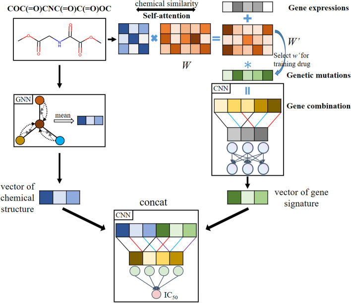
Our deep-learning model adopted a dual convergence architecture as shown in Fig. 1, which means the mutations and expression of genes and chemical structures of drugs information were modelled separately since they contained different types of information. The two were later merged into one prediction model. Genomic signatures and molecular graphs were processed in parallel through GNN and Convolutional Neural Network (CNN) layers to extract independent features, which were then concatenated. The gene branch in SWnet used 3 convolutional layers and 1 fully connected layer to generate gene embeddings. Then the embeddings of molecule and gene were concatenated and input into a prediction subnetwork, which consists of convolutional layer, pooling layer and batch normalization layer, to extract high-level features. Finally, the high-level features were input to fully connected layer to predict . It is noted that we used 1D CNNs in gene branch and prediction subnetwork to decrease the number of trainable parameters, therefore reducing the complexity of our model to avoid the risk of overfitting and improve the model performance. Based on dual convergence architecture, we also integrated multi-task learning and self-attention mechanism to further improve the performance. We described the architecture in more details in the following sections.
Fig. 1.
The overview of model architecture. Chemical structure, gene expression and genetic mutation serve as input of the model. Graph neural network encoded the drugs and convolutional neural network extracted gene expression and genetic mutation features simultaneously. Through self-attention, we incorporated the chemical similarity into the input to train gene weight layer . Gene weight layer combined gene expression, genetic mutation and drug similarity. Then the model concatenated drug vector and genetic vector to predict the
Parsing of compound chemical structures using graph method
There are many representations of chemical structures in the field of machine learning, such as molecular fingerprints, text-based representations (SMILES/INCHI), graph-based and 3D structure & surface. There are two main methods to encode molecules using continuous embedding vectors. One method is to use RNNs in variants autoencoder deep-learning network to generate continuous embedding vectors from SMILES [29, 30]. The other method is to learn features from graph-structured inputs [31–33].
In our study, we implemented an extended Graph Neural Network (GNN) [34] for molecular graphs. Normally, we represent a graph as , where V is the set of nodes and E is the set of edges. For a molecule, represents the ith atom and represents the chemical bond between ith atom and jth atom. The GNN takes a graph G as input, then produces a graph-level representation . In our extended GNN, we used r-radius subgraphs [9] to solve the issue that representation learning was ineffective due to the low model complexity. The r-radius subgraphs were induced by the neighboring nodes and edges within radius r from a node. Different from that in normal GNNs, in the extended GNN, we randomly initialized embedding for r-radius node and r-radius edge, and then updated the representation by backpropagation.
The process of GNN for molecular graphs can be described as a transition function and an output function. Figure 2 illustrates the general GNN architecture for molecular graphs. The transition function iteratively updates the node information by combining its neighboring nodes and edges, and the output function maps the node-level feature vectors to graph-level representation. The extended GNN has transition function for nodes and edges respectively, and updates the current node embedding through leveraging previous node and edge embeddings. For edge transition, we updated the edge embedding through both end node embeddings and as follows:
| 1 |
| 2 |
where is the element-wise sigmoid function and f is a non-linear activation function like ReLU, and are the trainable parameters and bias vector respectively, d is the dimension of edge embedding vector.
Fig. 2.
The overview of GNN architecture. a The process of updating node embedding through neighboring nodes. b The process of updating embedding through side node embedding. c The final graph after the transition function. d The graph-level representation through arithmetic mean of all node embeddings
We used similar iterative procedure for node transition, and we update the ith node embedding at time step through the following transition function:
| 3 |
| 4 |
where and are the trainable parameter matrix and bias vector respectively, and N(i) is the set of neighboring indices of i. The extended GNN computes the ith node embedding through combining the edge embedding and the neighboring node embedding iteratively, and the node gradually obtains global information on the graph.
The next operation takes the graph-level representation as input which is from the output function. Our output function is an arithmetic mean of all node embedding from transition function as follows:
| 5 |
where N is the number of nodes in the graph. While this is a simple operation to obtain the graph-level representation, it works well in practice.
The graph-level representation extracted by GNN embedded the 2D structure information of molecules into the feature vector, compared with the binary fingerprints used by other methods [15, 35], it effectively avoids the problem of sparse features. We think the graph-level representation extracted by GNN can benefit our task.
Weight vector/matrix to evaluate the significance of genetic mutation
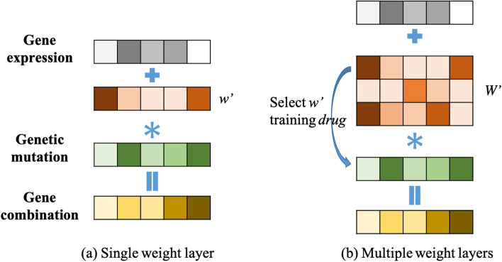
Most published research use only gene expression data or only genetic mutation data to train models [18]. We wanted to integrate these two data types, so we added a weight layer to the deep learning model. In this way, the mutation status of each gene is fed into the model with a weight that can be trained. As shown in Fig. 3, a combined feature for gene is calculated as:
| 6 |
where geneCom is the combination of expression (geneExp) and mutation (geneVar). represents the weight for genetic mutation. represents the vector dot product. The feature of each gene is the gene expression value plus the product of genetic mutation value and weight by Eq.(6). We have two kinds of weights: one is single weight vector whose dimension is w (1, 1478), and the other is multi-weight vector whose dimension is W (N, 1478). N is the number of anticancer drugs. Thus, for each compound, we trained for a vector w (1, 1478) that represented the significance of mutant genes. In the later section, we will show how to train the multi-layer weight matrix.
Fig. 3.
Gene weight layer. Gene feature was combined as the sum of normalized gene expression value and a weight adjusted genetic mutation value. Two ways of generating the mutation component were tested. a Single weight layer: a common weight vector was applied to all genetic mutations without considering the heterogeneity of gene-drug relationship. b Multiple weight layers: a weight matrix was applied to account for the heterogeneity of gene-drug relationship so that each drug would have its own weight vector for genetic mutation
Multi-task learning
Multi-task learning (MTL) problem is defined as multiple supervised learning tasks considered together [12]. In the previous section, we described a multi-layer weight matrix to measure the interaction between individual compound and genetic mutation. Each layer (vector of size ) is thus independent, and the simplest approach would be to solve these tasks independently through separating the dataset by compounds. However, it has been shown in previous studies that MTL can improve the performance [12, 36]. Since each task has the same model structure, we trained the 221 tasks all at once, using a common MSE loss for all drugs.
Self-attention
For the multi-task model described in previous section, each drug has its own gene weight parameters, which are supposed to be relevant to the drug itself. But we believe that similar drugs may have similar gene weight parameters. To take into consideration the similarity between drugs’ chemical structures, we applied a self-attention method, which uses drug similarity matrix to update the parameters of gene weight layer.
An attention function can be described as a query and a set of key-value pairs to an outputs, where the query, keys, values and output are all vectors. The output is computed as a weighted sum of the values, where the weight assigned to each value is computed by compatibility of the query with the corresponding key [23].
| 7 |
Q, K represent SMILES of compounds. V represents weight parameters of gene weight layer. a(Q, K) is an alignment function [20] which gives scores how well the inputs and the outputs match, and we normalized the scores by softmax function. The softmax function is a normalized exponential function as follows:
| 8 |
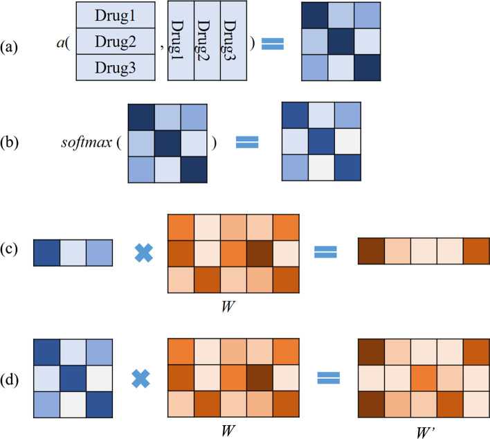
In our model, the calculation process is shown in Fig. 4. First, we used RDKit Tools to calculate the similarity of Morgan fingerprints between compounds to obtain a(Q, K). Then we performed matrix multiplication with the weight matrix. a(Q, K) represents the similarity matrix between all compounds. The output has the same dimension as W, but contains drug similarity information. Figure 1 illustrates the self-attention mechanism for prediction.
Fig. 4.
Self-attention calculation process. a Function a calculates the similarity between drugs. b The softmax function calculates the proportion of similarity score. c For one compound, through matrix multiplication, the weight layer will have compound similarity features. d The layer will be updated during the training process and finally reflects the contribution of genetic mutation to each drug’s sensitivity. By multiplying the W layer with the softmax matrix, drug–drug similarity is taken into account
For the drug response prediction of a new drug, its corresponding w could be calculated from the trained . The calculation process is described in Fig. 4a–c. This way, the drug response prediction for new drugs will be more accurate.
Other methods for comparison
We selected four previously published methods for comparison. We used Mean Square Error (MSE) and to evaluate the performance of models. The MSE metric measures the average squared difference between the predicted values and the actual values , it is calculated as follows:
| 9 |
where the N represents the number of samples. The quantifies the degree of any linear correlation between the predicted values and the actual values , it is calculated as follows:
| 10 |
These methods were all trained and tested on GDSC and CCLE datasets.
Kernelized Bayesian multi-task learning (KBMTL). This method formulated a novel Bayesian algorithm that combines kernel-based non-linear dimensionality reduction and binary classification or regression [11]. The joint Bayesian formulation projects data points into a shared subspace and learns predictive models for all drugs in this subspace. In our paper, we used the Gaussian kernel with kernel width to calculate similarity between cell lines. The parameters were (, , , , , R) = (1,1,1,1,0.1,1,10) and we used 200 iterations in all the experiments for KBMTL.
Similarity-regularized matrix factorization (SRMF). This method predicted anticancer drug responses of cell lines by using chemical structures of drugs and baseline gene expression levels in cell lines [14]. Specifically, chemical structural similarity of drugs and gene expression profile similarity of cell lines were considered as regularization terms, which were incorporated to the drug response matrix factorization model. We used Morgan fingerprint to calculate the drug similarity matrix in our paper. And the regularization parameters in SRMF were (K, , , ) = (45, , 0, ). Finally, we used 50 iterations in all the experiments for SRMF.
Weighted Graph Regularized Matrix Factorization (WGRMF). This method constructed a p-nearest neighbor graph to sparsify drug similarity matrix and cell line similarity matrix, respectively [37]. Using the sparse matrices in the graph regularization terms, it performed matrix factorization to generate the latent matrices for drugs and cell lines in anticancer drug response prediction task. We used Morgan fingerprint to calculate the drug similarity matrix in our paper. The regularization parameters used in our paper were (k, p, , , ) = (182, 20, 1, 2, ) for GDSC dataset and (k, p , , ) = (24, 10, 1, 2, 1) for CCLE dataset. Finally, we used 50 iterations in all the experiments for WGRMF.
Cancer Drug Response profile scan (CDRscan). This paper employed a deep learning dual convergence architecture, where the genomic mutational fingerprints of cell lines and the molecular fingerprints of drugs were processed individually, and then merged by ‘virtual docking’, an in silico modelling of drug treatment [35]. By inputting molecular fingerprints of drugs, they achieved better prediction result.
Graph convolutional network for drug response prediction (GraphDRP). In GraphDRP [22], drugs were represented in molecular graphs directly capturing the bonds among atoms. Meanwhile, cell lines were depicted as binary vectors of genomic aberrations. The features of drugs and cell lines were learned by the convolutional layers, and then the combination of drug and cell line features were used to predict value. In our paper, we used GIN graph model in GrapDRP to learn the representation of the drug in GDSC and CCLE datasets.
Results
In this section, we showed the performance of our method on GDSC and CCLE datasets. Meanwhile, we studied the effect of gene weight layer, multi-task, self-attention and different r-radius GNN for our model on GDSC dataset. We designed three experiments. The first is the initial model with single matrix of gene weight layer. The second is MTL model with multiple matrices of gene weight layer. The third is self-attention model base on MTL but with increased similarity feature of drugs. The three experiments are with different r-radius (r = 1, 2, 3). As shown in Table 3, after introducing the self-attention mechanism, the performance has been significantly improved. In addition, the trained gene weight layer helped to identify genes whose mutation status may strongly influence the prediction of drug ecacy.
Table 3.
Performance of model training with different weight layers, self-attention, or r-radius
| Weight layer | Self-attention | r-radius | MSE | |
|---|---|---|---|---|
| Single | No | 1 | 1.0848 | 0.8477 |
| Single | No | 2 | 0.9804 | 0.8623 |
| Single | No | 3 | 1.0694 | 0.8498 |
| Multi | No | 1 | 1.0654 | 0.8504 |
| Multi | No | 2 | 1.0078 | 0.8585 |
| Multi | No | 3 | 0.9767 | 0.8628 |
| Multi | Yes | 1 | 1.0724 | 0.8495 |
| Multi | Yes | 2 | 0.9785 | 0.8626 |
| Multi | Yes | 3 | 0.9384 | 0.8683 |
| Multi | Yes | 4 | 0.9917 | 0.8608 |
| Multi | Yes | 5 | 1.2083 | 0.8304 |
Bold values represents the result is the best performance among the models participating in the comparison
Performance on GDSC and CCLE datasets
GDSC data was pre-processed according to Methods Section Training and testing data. The final GDSC dataset consists of 983 cell lines with 1478 gene-level signatures and 221 drugs. A total of 177,128 instances of “chemical structure + gene signature vs. drug sensitivity” were in the final input for training the deep learning models (removing missing values where drug sensitivity is not available). The CCLE dataset consists of 24 compounds, covering 469 distinct cell lines for a total of 10853 instances.
In order to train and test the model, we randomly divided the GDSC dataset into the training (159415 instances) and testing (17,713 instances) sets, and split the CCLE dataset into the training (9767 instances) and testing (1086 instances) sets, which corresponded to 90% and 10% of the total instances, respectively.
Compared with previously published methods, our deep learning model shows superior predictive performance (Table 1). Other models extracted features in all gene expression to achieve good predictive performance, but it is very difficult to extract really effective features from so many gene expression datasets. Different from other papers, CDRscan used mutation position to train model. They selected sequence variation information at 28328 positions from 567 genes in CGC [38].
Table 1.
Comparison of performances with previously published methods
| Dataset | Model | MSE | |
|---|---|---|---|
| GDSC | KBMTL | 1.2642 | 0.8225 |
| SRMF | 0.9874 | 0.8614 | |
| WGRMF | 0.9844 | 0.8618 | |
| GraphDRP | 1.2586 | 0.8229 | |
| CDRscan | 2.1525 | 0.6978 | |
| SWnet | 0.9384 | 0.8683 | |
| CCLE | KBMTL | 1.9480 | 0.5439 |
| SRMF | 1.2565 | 0.7058 | |
| WGRMF | 1.3026 | 0.6950 | |
| GraphDRP | 1.3121 | 0.6928 | |
| CDRscan | 1.1960 | 0.7200 | |
| SWnet | 1.1604 | 0.7283 |
Bold values represents the result is the best performance among the models participating in the comparison
Single weight layer
In this section, we discussed the effectiveness of genetic mutation features and gene weight layer. To study the influence of genetic mutation on model performance, we trained SWnet without gene weight layer. In Table 2, we input two data types into SWnet gene branch: (1) gene expression only; (2) gene expression plus genetic mutation. As shown in Table 2, the best MSE is 0.9727 when the gene expression features as input and for radius. Meanwhile, the best MSE is 0.9853 when the gene expression and genetic mutation as input and for radius. From the results we could see that the introduction of genetic mutation through simple addition would change the distribution of gene expression data and lead to the deterioration of training results.
Table 2.
Performance of model training without gene weight layer in different r-radius
| Features | r-radius | MSE | |
|---|---|---|---|
| Gene expression | 1 | 1.0765 | 0.8489 |
| 2 | 0.9727 | 0.8634 | |
| 3 | 1.0763 | 0.8489 | |
| Gene expression + genetic mutation | 1 | 1.0663 | 0.8503 |
| 2 | 0.9853 | 0.8616 | |
| 3 | 1.1061 | 0.8447 |
Bold values represents the result is the best performance among the models participating in the comparison
To study the significance of gene weight layer on model, we used the gene weight layer in Table 3 to combine the gene expression and genetic mutation. We tried applying the gene weight layer to the following: (1) genetic mutation only; (2) gene expression only; (3) genetic mutation and gene expression. Our model performed the best () when we applied the gene weight layer to genetic mutation only. Based on this experimental result, we finally decided to apply the gene weight layer to the genetic mutation only. In Table 3, when radius, single weight layer without self-attention, the MSE is 0.9804. It is smaller than 0.9853, illustrating that through gene weight layer the two genetic traits are dynamically combined, achieving better performance than simple addition strategy. However, Table 3 shows that in single gene weight layer, , the MSE is 0.9804, bigger than 0.9727 in Table 2. This means the single weight layer has little side effects on model training. Taking genetic mutation data as input means more features are introduced into the model. Meanwhile, GNN is a deeper model, harder to train than CNN. This leads to the fact that during SWnet training, back propagation is more inclined to GNN network training, so as to obtain better MSE results. On the other hand, although our CNN gene branch added single weight layer, it has little effect on model training.
In fact, before training GNN as drug feature extract model, we have trained a model of SWnet with a similar structure. Its drug feature extraction is realized through the convolutional neural network and the input feature is fingerprints. When we did simple addition of the gene features, MSE was 1.0223. If we combined the gene features through gene weight layer, MSE was 0.9912. This indicates that the convolutional layer can make the genetic mutation participate in the training of the model well and improve the performance of the model. It is worth mentioning that although single weight layer has little effect on SWnet when drug branch is GNN, the MSE is 0.9804, better than CNN drug branch whose MSE is 0.9912.
Multi-task and self-attention
In section “Single weight layer”, we have solved the problem of how to input both gene expression and genetic mutations through single gene weight layer. In this section, we discussed how to design the gene weight layer to balance the complexity between GNN and CNN. First, we introduced multi-task learning. The single gene weight layer was upgraded to multiple gene weight layer. Each drug corresponded to a single gene weight layer at a specific location. However, there are too many weight layers to train, resulting in the model’s predictive performance degradation. As shown in Table 3 when using multi-weight layers and without self-attention, the MSE is 0.9911.
Then we introduced self-attention mechanism to improve gene branch of the model. With self-attention structure, drug similarity enhanced the training of multi-weight layers, and then gene expression and genetic mutation features can be better fused together. As shown in Table 3, when and using multi-weight layers and self-attention, the and .
Different r-radius
In section “Single weight layer” and section “Multi-task and self-attention” we have learned that the relative complexity of GNN and CNN determines the predictive ability of the model. With different r-radius subgraphs in GNN, we can adjust the complexity of the GNN branch. So we designed all experiments with different r-radius.
As shown in Table 3, single gene weight SWnet achieved the best performance when radius, self-attention SWnet achieved the best performance when radius. Compared the two model, since self-attention SWnet contained multiple matrix W and self-attention mechanism, it has more ability to extract molecule feature, therefore the GNN branch can be more complicated. Self-attention SWnet became overfitting when radius. Meanwhile, single gene layer SWnet model is simpler than self-attention SWnet, it means that the model would become overfitting faster when radius.
Relationship between drug targets and genetic mutation status revealed by the gene weight layer
The trained gene weight layer reflected the normalized contribution of the binary genetic mutation status to the prediction of drug efficacy. To explore biological relevance of the model, genes with have been identified for each drug, which represents the genetic mutation with the strongest predictive power. Proteins encoded by these genes might interact among themselves or interact with the drug targets. We identified these connections from protein–protein interaction database [39]. The protein–protein interactions were illustrated for BRAF inhibitors and BCL2 inhibitors (Fig. 5). Connections within 2 degrees from the target were shown in the network. For BRAF, two of the drugs (AZ628 and Dabrafenib) have their strongest predictive genes directly interacting with BRAF. For the other 3 BRAF inhibitors, there is at least one strongest predictive gene connected with BRAF within the second degree. The same was observed for BCL2 inhibitors. Interestingly, TP53 serves as a connection hub in the BCL2 case and SRC in the BRAF case. It is known that BCL2 and TP53 are two important nodes in the apoptosis signaling pathway and it has been reported that combining TP53 activation and BCL2 inhibition could result in synthetic lethality in AML [40]. Similarly, BRAF V600E mutation and SRC mutations have been found to be mutually exclusive in colorectal cancer patients, and both could serve as molecular markers for prognosis [41]. Lists of genes with for each drug are available in Additional file 1: Table S1. These protein–protein interactions indicated the relevance of the gene weights in our model.
Fig. 5.
Physical interactions often occur among drug target proteins and proteins encoded by genes with the highest weight (). a Five BRAF inhibitors were included in the training data. Genes with were extracted from the gene weight layer after model training, and their interactions with BRAF were analyzed in the String database. Connections within 2 degrees from BRAF were shown, grouped by drug. b Three BCL2 inhibitors was included in the training data. Analysis procedure was the same as above. The network showed connections within 2 degrees from BCL2
Split training data with cell lines
To further explore the generalization ability of the model, we split the data set by cell lines, so that part of the genomic information never participated in the model training. We randomly divided training data with cell lines and trained GNN with different r-radius subgraphs, resulting in 159913 instances (90%) for training and 17215 instances (10%) for testing. As shown in Table 4, the MSE is 2.2793 when single weight layer with radius. Since the cell lines are of different tissue lineages, the performance of the model is not as good as that when all the cell lines appeared in the training dataset. Meanwhile, when and using multi-weight layers and self-attention, the . This result is close to single layer SWnet. Due to weak cell line similarity, the different r-radius subgraphs have little effects of model performance.
Table 4.
Performance of SWnet in terms of MSE in random split dataset by cell lines with different r-radius subgraphs
| Weight layer | Self-attention | r-radius | MSE | |
|---|---|---|---|---|
| Single | No | 1 | 2.3090 | 0.6756 |
| Single | No | 2 | 2.3102 | 0.6755 |
| Single | No | 3 | 2.2793 | 0.6799 |
| Multi | Yes | 1 | 2.2975 | 0.6773 |
| Multi | Yes | 2 | 2.2840 | 0.6792 |
| Multi | Yes | 3 | 2.3304 | 0.6726 |
Bold values represents the result is the best performance among the models participating in the comparison
Conclusion
In this paper, we proposed a new end-to-end deep learning model called SWnet. With the gene weight layer, we input the gene expression and genetic mutation at once. We used GNN to encode chemical structures of drugs with different r. We predicted the based on gene signatures and molecular graphs and achieved better predictive performance than methods reported in previous literatures. We used the self-attention mechanism for the first time to explore the interactions between genetics and chemical structure of drugs.
SWnet combined genomic signatures and molecular graphs for drug efficacy prediction. We applied SWnet to GDSC and CCLE dataset, and showed that SWnet outperformed other models. With more datasets becoming available in the public domain, we envision training SWnet with more datasets and eventually using SWnet for in silico drug screenings. This model holds great promise for cancer therapy and precision medicine.
It is worth noting that the molecular structure information extracted by GNN is important for drug response prediction, where we applied the extend GNN to extract graph-level representation for molecule in this work. However, we used a simple arithmetic mean to get the graph-level representation from node-level representations. We believe that there will be more effective ways to extract the graph-level representation. We will improve the current GNN for more effective node information fusion in molecular graph.
The complex deep learning model SWnet can hardly be well-trained when it deals with a small dataset. In the future, we will design a GNN with a simpler structure to encode the drug structure to fit small datasets. We will try to fix the weight parameters for common atoms when updating node features in molecule. This may make GNN focus more on the structure of the molecule and the special atoms in it, and it will make it easier to train GNN with small datasets.
Supplementary Information
Additional file 1. Drug and genes information for efficacy prediction.
Acknowledgements
We thank Novartis team for its sponsorship and discussion, and with special thanks to Xinyi Yang and Yangjunyi Li for prior work that links to the ideation of this study.
Abbreviations
- GDSC
Genomics of drug sensitivity in cancer
- CCLE
Cancer cell line encyclopedia
- GNN
Graph neural network
- CNN
Convolutional neural network
- SWnet
Self-attention gene weight layer network
- SMILES
Simplified molecular input line entry system
Half maximal inhibitory concentration
- COSMIC
Catalogue of somatic mutations in cancer
- MTL
Multi-task learning
- MSE
Mean square error
- KBMTL
Kernelized Bayesian multi-task learning
- SRMF
Similarity-regularized matrix factorization
- WGRMF
Weighted graph regularized matrix factorization
- CDRscan
Cancer drug response profile scan
- GraphDRP
Graph convolutional network for drug response prediction
Author contributions
ZZ, HG, and DQ contributed to the study design; XC, LT data acquisition and data analysis; ZZ and PW model construction, code validation, data analysis, ZZ, PW and XC interpreted the results. ZZ, PW and XC prepared the figures and drafted the manuscript. ZZ, XC, PW, LT, HG and DQ edited and revised the manuscript. All authors read and approved the final manuscript.
Funding
This work has been supported in part by National Natural Science Foundation of China Grant 81974276 and by Novartis sponsorship. The funding bodies played no role in the design of the study and collection, analysis, and interpretation of data and in writing the manuscript.
Availability of data and materials
Source code can be downloaded from https://github.com/zuozhaorui/SWnet.git.
Declarations
Ethics approval and consent to participate
Not applicable.
Consent for publication
Not applicable.
Competing interests
The authors declare that they have no competing interests.
Footnotes
Publisher's Note
Springer Nature remains neutral with regard to jurisdictional claims in published maps and institutional affiliations.
Zhaorui Zuo and Penglei Wang have contributed equally to this work
Contributor Information
Hui Ge, Email: hui.ge@novartis.com.
Dahong Qian, Email: dahong.qian@sjtu.edu.cn.
References
- 1.Barretina J, Caponigro G, Stransky N, Venkatesan K, Margolin AA, Kim S, Wilson CJ, Lehár J, Kryukov GV, Sonkin D, et al. The cancer cell line encyclopedia enables predictive modelling of anticancer drug sensitivity. Nature. 2012;483(7391):603–607. doi: 10.1038/nature11003. [DOI] [PMC free article] [PubMed] [Google Scholar]
- 2.Weinstein JN, Collisson EA, Mills GB, Shaw KRM, Ozenberger BA, Ellrott K, Shmulevich I, Sander C, Stuart JM. The cancer genome atlas pan-cancer analysis project. Nat Genet. 2013;45(10):1113–1120. doi: 10.1038/ng.2764. [DOI] [PMC free article] [PubMed] [Google Scholar]
- 3.Tate JG, Bamford S, Jubb HC, Sondka Z, Beare DM, Bindal N, Boutselakis H, Cole CG, Creatore C, Dawson E, et al. Cosmic: the catalogue of somatic mutations in cancer. Nucleic Acids Res. 2019;47(D1):941–947. doi: 10.1093/nar/gky1015. [DOI] [PMC free article] [PubMed] [Google Scholar]
- 4.Shoemaker RH. The nci60 human tumour cell line anticancer drug screen. Nat Rev Cancer. 2006;6(10):813–823. doi: 10.1038/nrc1951. [DOI] [PubMed] [Google Scholar]
- 5.Yang W, Soares J, Greninger P, Edelman EJ, Lightfoot H, Forbes S, Bindal N, Beare D, Smith JA, Thompson IR, et al. Genomics of drug sensitivity in cancer (GDSC): a resource for therapeutic biomarker discovery in cancer cells. Nucleic Acids Res. 2012;41(D1):955–961. doi: 10.1093/nar/gks1111. [DOI] [PMC free article] [PubMed] [Google Scholar]
- 6.Subramanian A, Narayan R, Corsello SM, Peck DD, Natoli TE, Lu X, Gould J, Davis JF, Tubelli AA, Asiedu JK, et al. A next generation connectivity map: L1000 platform and the first 1,000,000 profiles. Cell. 2017;171(6):1437–1452. doi: 10.1016/j.cell.2017.10.049. [DOI] [PMC free article] [PubMed] [Google Scholar]
- 7.Costello JC, Heiser LM, Georgii E, Gönen M, Menden MP, Wang NJ, Bansal M, Hintsanen P, Khan SA, Mpindi J-P, et al. A community effort to assess and improve drug sensitivity prediction algorithms. Nat Biotechnol. 2014;32(12):1202–1212. doi: 10.1038/nbt.2877. [DOI] [PMC free article] [PubMed] [Google Scholar]
- 8.Chen J, Zhang L. A survey and systematic assessment of computational methods for drug response prediction. Brief Bioinform. 2020;22(1):232–246. doi: 10.1093/bib/bbz164. [DOI] [PubMed] [Google Scholar]
- 9.Costa F, De Grave K. Fast neighborhood subgraph pairwise distance kernel. In: ICML;2010.
- 10.Rappaport N, Nativ N, Stelzer G, Twik M, Guan-Golan Y, Iny Stein T, Bahir I, Belinky F, Morrey CP, Safran M et al. Malacards: an integrated compendium for diseases and their annotation. Database. 2013;2013. [DOI] [PMC free article] [PubMed]
- 11.Gönen M, Margolin AA. Drug susceptibility prediction against a panel of drugs using kernelized Bayesian multitask learning. Bioinformatics. 2014;30(17):556–563. doi: 10.1093/bioinformatics/btu464. [DOI] [PMC free article] [PubMed] [Google Scholar]
- 12.Tan M. Prediction of anti-cancer drug response by kernelized multi-task learning. Artif Intell Med. 2016;73:70–77. doi: 10.1016/j.artmed.2016.09.004. [DOI] [PubMed] [Google Scholar]
- 13.Ammad-Ud-Din M, Khan SA, Wennerberg K, Aittokallio T. Systematic identification of feature combinations for predicting drug response with Bayesian multi-view multi-task linear regression. Bioinformatics. 2017;33(14):359–368. doi: 10.1093/bioinformatics/btx266. [DOI] [PMC free article] [PubMed] [Google Scholar]
- 14.Wang L, Li X, Zhang L, Gao Q. Improved anticancer drug response prediction in cell lines using matrix factorization with similarity regularization. BMC Cancer. 2017;17(1):1–12. doi: 10.1186/s12885-016-3022-6. [DOI] [PMC free article] [PubMed] [Google Scholar]
- 15.Martin EJ, Polyakov VR, Tian L, Perez RC. Profile-QSAR 2.0: kinase virtual screening accuracy comparable to four-concentration ic50s for realistically novel compounds. J Chem Inf Model. 2017;57(8):2077–2088. doi: 10.1021/acs.jcim.7b00166. [DOI] [PubMed] [Google Scholar]
- 16.Ammad-Ud-Din M, Georgii E, Gonen M, Laitinen T, Kallioniemi O, Wennerberg K, Poso A, Kaski S. Integrative and personalized QSAR analysis in cancer by kernelized Bayesian matrix factorization. J Chem Inf Model. 2014;54(8):2347–2359. doi: 10.1021/ci500152b. [DOI] [PubMed] [Google Scholar]
- 17.Zhang F, Wang M, Xi J, Yang J, Li A. A novel heterogeneous network-based method for drug response prediction in cancer cell lines. Sci Rep. 2018;8(1):1–9. doi: 10.1038/s41598-018-21622-4. [DOI] [PMC free article] [PubMed] [Google Scholar]
- 18.Manica M, Oskooei A, Born J, Subramanian V, Sáez-Rodríguez J, Rodríguez Martínez M. Toward explainable anticancer compound sensitivity prediction via multimodal attention-based convolutional encoders. Mol Pharm. 2019;16(12):4797–4806. doi: 10.1021/acs.molpharmaceut.9b00520. [DOI] [PubMed] [Google Scholar]
- 19.Baptista D, Ferreira PG, Rocha M. Deep learning for drug response prediction in cancer. Brief Bioinform. 2021;22(1):360–379. doi: 10.1093/bib/bbz171. [DOI] [PubMed] [Google Scholar]
- 20.Bahdanau D, Cho K, Bengio Y. Neural machine translation by jointly learning to align and translate. 2014. arXiv:1409.0473.
- 21.Liu Q, Hu Z, Jiang R, Zhou M. Deepcdr: a hybrid graph convolutional network for predicting cancer drug response. Bioinformatics. 2020;36:i911–i918. doi: 10.1093/bioinformatics/btaa822. [DOI] [PubMed] [Google Scholar]
- 22.Nguyen T-T, Nguyen GTT, Nguyen T, Le D-H. Graph convolutional networks for drug response prediction. IEEE/ACM Trans Comput Biol Bioinform. 2021 doi: 10.1109/TCBB.2021.3060430. [DOI] [PubMed] [Google Scholar]
- 23.Vaswani A, Shazeer N, Parmar N, Uszkoreit J, Jones L, Gomez AN, Kaiser L, Polosukhin I. Attention is all you need. 2017. arXiv:1706.03762.
- 24.Ghandi M, Huang FW, Jané-Valbuena J, Kryukov GV, Lo CC, McDonald ER, Barretina J, Gelfand ET, Bielski CM, Li H, et al. Next-generation characterization of the cancer cell line encyclopedia. Nature. 2019;569(7757):503–508. doi: 10.1038/s41586-019-1186-3. [DOI] [PMC free article] [PubMed] [Google Scholar]
- 25.Gaulton A, Bellis LJ, Bento AP, Chambers J, Davies M, Hersey A, Light Y, McGlinchey S, Michalovich D, Al-Lazikani B, et al. Chembl: a large-scale bioactivity database for drug discovery. Nucleic Acids Res. 2012;40(D1):1100–1107. doi: 10.1093/nar/gkr777. [DOI] [PMC free article] [PubMed] [Google Scholar]
- 26.Kim S, Chen J, Cheng T, Gindulyte A, He J, He S, Li Q, Shoemaker BA, Thiessen PA, Yu B, et al. Pubchem 2019 update: improved access to chemical data. Nucleic Acids Res. 2019;47(D1):1102–1109. doi: 10.1093/nar/gky1033. [DOI] [PMC free article] [PubMed] [Google Scholar]
- 27.Landrum G. RDKit: open-source cheminformatics software. Berlin: Springer; 2016. [Google Scholar]
- 28.Tan M, Özgül OF, Bardak B, Ekşioğlu I, Sabuncuoğlu S. Drug response prediction by ensemble learning and drug-induced gene expression signatures. Genomics. 2019;111(5):1078–1088. doi: 10.1016/j.ygeno.2018.07.002. [DOI] [PubMed] [Google Scholar]
- 29.Kusner MJ, Paige B, Hernández-Lobato JM. Grammar variational autoencoder. In: International conference on machine learning, pp. 1945–1954;2017.
- 30.Gómez-Bombarelli R, Wei JN, Duvenaud D, Hernández-Lobato JM, Sánchez-Lengeling B, Sheberla D, Aguilera-Iparraguirre J, Hirzel TD, Adams RP, Aspuru-Guzik A. Automatic chemical design using a data-driven continuous representation of molecules. ACS Cent Sci. 2018;4(2):268–276. doi: 10.1021/acscentsci.7b00572. [DOI] [PMC free article] [PubMed] [Google Scholar]
- 31.Scarselli F, Gori M, Tsoi AC, Hagenbuchner M, Monfardini G. The graph neural network model. IEEE Trans Neural Networks. 2008;20(1):61–80. doi: 10.1109/TNN.2008.2005605. [DOI] [PubMed] [Google Scholar]
- 32.Li Y, Tarlow D, Brockschmidt M, Zemel R. Gated graph sequence neural networks. 2015. arXiv:1511.05493.
- 33.Veličković P, Cucurull G, Casanova A, Romero A, Lio P, Bengio Y. Graph attention networks.2017. arXiv:1710.10903.
- 34.Tsubaki M, Tomii K, Sese J. Compound-protein interaction prediction with end-to-end learning of neural networks for graphs and sequences. Bioinformatics. 2019;35(2):309–318. doi: 10.1093/bioinformatics/bty535. [DOI] [PubMed] [Google Scholar]
- 35.Chang Y, Park H, Yang H-J, Lee S, Lee K-Y, Kim TS, Jung J, Shin J-M. Cancer drug response profile scan (CDRscan): a deep learning model that predicts drug effectiveness from cancer genomic signature. Sci Rep. 2018;8(1):1–11. doi: 10.1038/s41598-018-27214-6. [DOI] [PMC free article] [PubMed] [Google Scholar]
- 36.Caruana R. Multitask learning. Mach Learn. 1997;28(1):41–75. doi: 10.1023/A:1007379606734. [DOI] [Google Scholar]
- 37.Guan N-N, Zhao Y, Wang C-C, Li J-Q, Chen X, Piao X. Anticancer drug response prediction in cell lines using weighted graph regularized matrix factorization. Mol Therapy-Nucleic Acids. 2019;17:164–174. doi: 10.1016/j.omtn.2019.05.017. [DOI] [PMC free article] [PubMed] [Google Scholar]
- 38.Futreal PA, Coin L, Marshall M, Down T, Hubbard T, Wooster R, Rahman N, Stratton MR. A census of human cancer genes. Nat Rev Cancer. 2004;4(3):177–183. doi: 10.1038/nrc1299. [DOI] [PMC free article] [PubMed] [Google Scholar]
- 39.Szklarczyk D, Gable AL, Lyon D, Junge A, Wyder S, Huerta-Cepas J, Simonovic M, Doncheva NT, Morris JH, Bork P, et al. String v11: protein-protein association networks with increased coverage, supporting functional discovery in genome-wide experimental datasets. Nucleic Acids Res. 2019;47(D1):607–613. doi: 10.1093/nar/gky1131. [DOI] [PMC free article] [PubMed] [Google Scholar]
- 40.Pan R, Ruvolo V, Mu H, Leverson JD, Nichols G, Reed JC, Konopleva M, Andreeff M. Synthetic lethality of combined Bcl-2 inhibition and p53 activation in AML: mechanisms and superior antileukemic efficacy. Cancer Cell. 2017;32(6):748–760. doi: 10.1016/j.ccell.2017.11.003. [DOI] [PMC free article] [PubMed] [Google Scholar]
- 41.Shimada Y, Muneoka Y, Nagahashi M, Ichikawa H, Tajima Y, Hirose Y, Ando T, Nakano M, Sakata J, Kameyama H, et al. BRAF v600e and SRC mutations as molecular markers for predicting prognosis and conversion surgery in stage iv colorectal cancer. Sci Rep. 2019;9(1):1–10. doi: 10.1038/s41598-019-39328-6. [DOI] [PMC free article] [PubMed] [Google Scholar]
Associated Data
This section collects any data citations, data availability statements, or supplementary materials included in this article.
Supplementary Materials
Additional file 1. Drug and genes information for efficacy prediction.
Data Availability Statement
Source code can be downloaded from https://github.com/zuozhaorui/SWnet.git.


