Abstract
Itaconic acid is produced by immune responsive gene 1 (IRG1)-coded enzyme in activated macrophages and known to play an important role in metabolism and immunity. In this study, mechanism of itaconic acid functioning as an anti-inflammatory metabolite was investigated with molecular biology and immunology techniques, by employing IRG1-null (prepared with CRISPR) and wild-type macrophages. Experimental results showed that itaconic acid significantly promoted the pentose phosphate pathway (PPP), which subsequently led to significantly higher NADPH oxidase activity and more reactive oxygen species (ROS) production. ROS production increased the expression of anti-inflammatory gene A20, which in turn decreased the production of inflammatory cytokines IL-6, IL-1β and TNF-α. NF-κB, which can up-regulate A20, was also vital in controlling IRG1 and itaconic acid involved immune-modulatory responses in LPS-stimulated macrophage in this study. In addition, itaconic acid inhibited the growth of Salmonella typhimurium in cell through increasing ROS production from NADPH oxidase and the hatching of Schistosoma japonicum eggs in vitro. In short, this study revealed an alternative mechanism by which itaconic acid acts as an anti-inflammatory metabolite and confirmed the inhibition of bacterial pathogens with itaconic acid via ROS in cell. These findings provide the basic knowledge for future biological applications of itaconic acid in anti-inflammation and related pathogens control.
Subject terms: Bacteria, Bacterial host response, Adaptive immunity, Antimicrobial responses, Cytokines, Infection, Inflammation, Immunology, Microbiology
Introduction
Itaconic acid is an important mammalian metabolite which mediates the crosstalk between infection, immunity and metabolism. It is produced by immune responsive gene 1 (IRG1)-coded enzyme which catalyzes cis-aconitic acid in activated macrophages under conditions of infection or pro-inflammation1–4. The increased production of itaconic acid has been found in spleens of BALB/c mice infected with Salmonella typhimurium5. The Schistosoma japonicum worm and egg burden of mice co-infected with both S. japonicum and S. typhimurium were significantly reduced, and the elevated level of itaconic acid caused by S. typhimurium coinfection may play an important role6. In other bacterial infection studies, increased levels of itaconic acid in target organ or tissue have been found7,8. The itaconic acid has been shown to have an antimicrobial effect through inhibiting isocitrate lyase, the key enzyme of an essential pathway (glyoxylate shunt) for bacterial growth in vivo9,10. Itaconic acid can also inhibit bacterial and viral infections via modulating the production of reactive oxygen species (ROS)11,12, the function which is believed to belong to some of the antibiotics13.
Itaconic acid has demonstrated its anti-inflammatory functions in vitro and in vivo through the mechanisms which have been proposed in several investigations14–18. ROS play an important role in the anti-inflammatory mechanism of itaconic acid15,16, in which ROS has been shown to be generated from the fatty acid oxidation mediated by oxidative phosphorylation (OXPHOS)11,16,19,20. Endogenous itaconic acid may cause elimination of mitochondrial substrate-level phosphorylation in LPS-activated murine macrophage lineage21. Itaconic acid can inhibit succinate dehydrogenase, which may lead to alterations of tricarboxylic acid cycle (TCA) and modulate intracellular succinate level14,22. Itaconic acid also can resemble phenylpyruvate to suppress glycolysis by decreasing the level of fructose 2,6-bisphosphate (F26BP)23. Hence, more attention has been focused on glycolysis, TCA circle and OXPHOS when considering mechanisms of anti-inflammatory effects of itaconic acid19,20,24. There have been fewer studies on the pentose phosphate pathway (PPP), which is another glucose metabolic pathway in parallel to glycolysis25. PPP generates the pentose precursor for the synthesis of nucleotides and NADPH that may be catalyzed by NADPH oxidase to produce superoxide and other ROS26,27.
In the current investigation, an alternative mechanism of anti-inflammatory effects of itaconic acid with PPP involved was explored. IRG1 in monocyte/macrophage-like cell line RAW264.7 was knocked out with CRISPR-Cas9 technique to prepare IRG1-null macrophage28, molecular biology and immune methods were employed to study how IRG1 regulates macrophage responses to related stimuli through PPP pathway using this cell model. Experimental results demonstrated that itaconate can promote the pentose phosphate pathway to produce more NADPH, which was used by NADPH oxidase to produce more ROS. ROS also induced expression of gene A20 (TNFAIP3, tumor necrosis factor alpha-induced protein 3) and contributed to anti-inflammatory effects in RAW264.7. Significantly elevated ROS level inhibited the propagation of S. typhimurium in RAW264.7, and may be also the cause for the low hatching rate of S. japonicum eggs with presence of itaconic acid. Our research provided understanding into new mechanisms of the anti-inflammatory effects of itaconic acid/IRG1 and shed new light on the anti-inflammatory functions of itaconate.
Results
Single cell selection of RAW264.7-IRG1KO
IRG1 is the gene responsible for producing itaconic acid. In order to evaluate the function of itaconic acid, CRISPR-Cas9 was employed to delete the IRG1 gene in RAW264.7 cell line. Twenty puromycin-selected positive single-cell clones were evaluated with Cruiser Enzyme kit. Gel-electrophoresis showed that single-cell clones (lane 2, 4, 7, 11) have the desired base mutations (Supplementary information: Fig. S1). In order to further confirm successful knock-out of IRG1, levels of itaconic acid in cells and cell culture medium was measured with NMR spectrometer. No itaconic acid was observed from the knock-out cell line and their spent medium irrespective of whether the cells were treated with lipopolysaccharide (LPS, 10 ng/mL) or not. However, the wild-type RAW264.7 was able to produce itaconic acid with or without LPS stimulation and more itaconic acid was produced with LPS treatment (Supplementary information: Fig. S2). This observation further confirmed successful deletion of the IRG1 gene. The IRG1 knocked out cell line was named as RAW264.7-IRG1KO.
Endogenous itaconic acid attenuated LPS-induced inflammatory cytokines
IL-1β, IL-6 and TNF-α are important pro-inflammatory cytokines which can be produced by activated macrophages. In this study, LPS significantly induced elevation in levels of IL-1β, IL-6 and TNF-α in both cell mediums of wild-type RAW264.7 and RAW264.7-IRG1KO cultured for 24 h when compared with control (Fig. 1A–C). Interestingly, immune response of IRG1-null cells to LPS treatment were more pronounced compared to its wild-type RAW264.7 cells (Fig. 1A–C), suggesting that endogenous itaconic acid was capable of diminishing LPS-induced inflammatory cytokines.
Figure 1.
Different responses of RAW264.7 and RAW264.7-IRG1KO to LPS stimulation. ELISA kit test results of cytokines IL-1β (A), IL-6 (B) and TNF-α (C) in RAW264.7 and RAW264.7-IRG1KO cell culture medium when exposed to LPS (24 h). ITA, itaconic acid; LPS, lipopolysaccharide. White bar, RAW264.7; black bar, RAW264.7-IRG1KO. Values represent the mean ± S.E.M. *, p < 0.05; **, p < 0.01.
Itaconic acid promoted the production of ROS that plays a role in diminishing LPS-induced inflammation
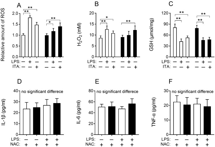
The levels of total ROS in cell, H2O2 in the cell culture medium and glutathione (GSH) in cell lysate were measured for cells treated with LPS or itaconic acid for 24 h. Experimental results showed that total ROS and H2O2 production in cell RAW264.7 subjected to LPS or itaconic acid was significantly increased compared to the corresponding untreated control (Fig. 2A, B). While the RAW264.7-IRG1KO cells produced significantly higher levels of H2O2 only when incubating with itaconic acid, and LPS treated RAW264.7-IRG1KO cells did not produce significantly increased level of H2O2 (Fig. 2B). These observations suggested that both endogenous and exogenous itaconic acid promoted the production of ROS and that the IRG1 deletion could reduce the production of ROS. For RAW264.7-IRG1KO cells, treatment with itaconic acid also depleted level of GSH compared to untreated control cells, confirming that itaconic acid promoted the production of ROS (Fig. 2C). However, depleted levels of GSH were also observed in LPS treated RAW264.7-IRG1KO cells (Fig. 2C). IRG1 can promote endotoxin tolerance via ROS in macrophage16. Therefore, ROS scavenger N-acetyl-L-cysteine (NAC, 2.5 mM) was added when treating RAW264.7 and RAW264.7-IRG1KO with LPS, and the results showed that no significant changes in the levels of IL-1β, IL-6 or TNF-α (Fig. 2D–F). This observation suggested that ROS scavenge could reduce LPS-induced inflammatory cytokines in both wild-type and RAW264.7-IRG1KO cells and eliminate the immune response difference between wild-type and RAW264.7-IRG1KO cells when treated with LPS. Therefore, knocking out of IRG1 diminished the production of ROS in murine macrophage RAW264.7, and resulted in more pronounced pro-inflammatory cytokines production to LPS treatment when compared to wild-type RAW264.7 cells (Fig. 1A–C).
Figure 2.
Results of ROS level in cell (A), H2O2 in cell medium (B) and GSH level (C) in cell lysate. The levels of IL-1β (D), IL-6(E), TNF-α (F) in cell culture medium from RAW264.7 and RAW264.7-IRG1KO with different treatments (24 h). White bar, RAW264.7; black bar, RAW264.7-IRG1KO. LPS, lipopolysaccharide; ITA, itaconic acid; NAC, N-acetyl-L-cysteine. Relative ROS amount in cells with different treatments was normalized to RAW264.7 control and represents the fold change. Values represent the mean ± S.E.M. *, p < 0.05; **, p < 0.01.
Itaconic acid promoted the pentose phosphate pathway (PPP) and NADPH oxidase activity
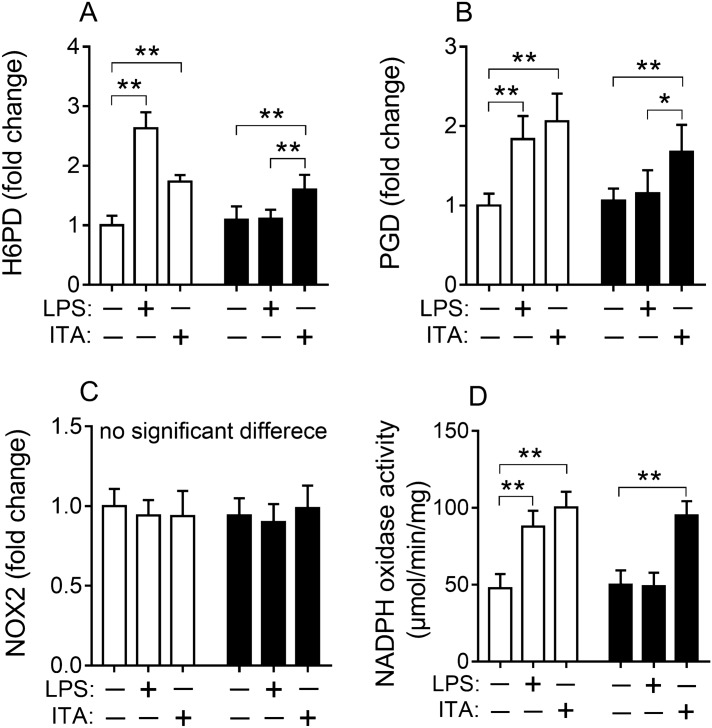
It is known that the first step of PPP pathway is the oxidative phase where NADPH is generated and can be used to produce ROS29. Hence, we hypothesized that higher production of ROS in the LPS treated wild-type RAW264.7 cells (compared with LPS treated RAW264.7-IRG1KO cells) may be generated from the PPP pathway. In order to test this hypothesis, the expression level of two key genes Glucose-6-phosphate dehydrogenase (H6PD) and 6-phosphogluconate dehydrogenase (PGD) in this pathway was tested. The expression levels of H6PD and PGD were significantly increased in the wild-type RAW264.7 cells treated with LPS for 24 h when compared to the control (Fig. 3A, B). However, this observation was not made for RAW264.7-IRG1KO cells with the same LPS treatment, given that there were no changes in the expression levels of H6PD and PGD was observed for RAW264.7-IRG1KO cells (Fig. 3A, B). Interestingly, the exogenous itaconic acid was also able to up-regulate the expression levels of H6PD and PGD for both wild-type and RAW264.7-IRG1KO cells (Fig. 3A, B), further confirming that itaconic acid stimulates PPP pathway. Gene (NOX2) expression level of NADPH oxidase was also tested, since this enzyme catalyzes the production of ROS using NADPH as substrate. However, no significant changes of NOX2 expression was detected for both LPS and itaconic acid treated RAW264.7 and RAW264.7-IRG1KO cells (Fig. 3C). Then the enzymatic activity of NADPH oxidase in different treated cells was tested, and both LPS and itaconic acid treatments can upregulate the activity of NADPH oxidase in RAW264.7 cells (Fig. 3D), whilst only exogenous itaconic acid was able to promote the activity of NADPH oxidase in IRG1-null cells (Fig. 3D). Overall, these results suggested that itaconic acid promoted the PPP pathway, and up-regulated NADPH oxidase to produce ROS. While in the absent of itaconic acid, these effects were diminished.
Figure 3.
Relative expression of gene H6PD (A), PGD (B), NOX2 (C) and NADPH oxidase activity (D) in RAW264.7 and RAW264.7-IRG1KO cells with different treatments (24 h). White bar, RAW264.7; black bar, RAW264.7-IRG1KO. LPS, lipopolysaccharide; ITA, itaconic acid. H6PD, glucose 6-phosphate dehydrogenase; PGD, 6-phosphogluconate dehydrogenase; NOX2, NADPH oxidase 2. Relative gene expression level of cells with different treatments was normalized to RAW264.7 control and represents the fold change. Values represent the mean ± S.E.M. *, p < 0.05; **, p < 0.01.
LPS and itaconic acid increased ROS in macrophages improved expression of gene A20 that was regulated by NF-κB pathway
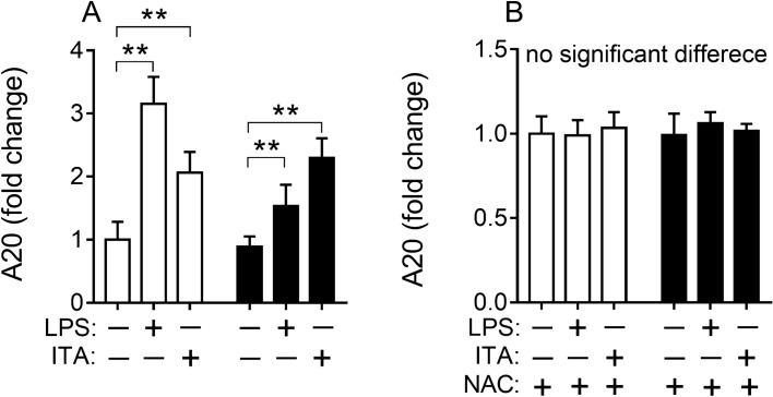
Gene A20 (TNFAIP3, tumor necrosis factor alpha-induced protein 3) has been shown to be critical in restraining inflammation induced by endotoxin- and TNF-α-caused NF-κB responses30,31. The expression of A20 is up-regulated by ROS15,16. Therefore, A20 expression was investigated in wild-type RAW264.7 and RAW264.7-IRG1KO cells which were treated with LPS or itaconic acid. Both LPS and itaconic acid treatments induced higher A20 expression in both cell lines and with higher levels of A20 being expressed with the presence of endogenous itaconic acid (Fig. 4A). This effect was eliminated by N-acetyl-L-cysteine (NAC) treatment, an antioxidant capable of removing ROS, which is independent of IRG1 regulation (Fig. 4B). NF-κB signaling pathway is considered a very important “rapid-acting” pro-inflammatory pathway32,33. Therefore, Bay11-7082 an inhibitor of NF-κB was employed to test the role of NF-κB in this study. The results showed that inhibiting NF-κB eliminated responses of A20 to LPS and itaconic acid treatments (Supplementary information: Fig. S3A). Additionally, when NF-κB was inhibited with Bay11-7082, the levels of IL-1β, IL-6 and TNF-α were unchanged when both wild-type RAW264.7 cells and RAW264.7-IRG1KO cells were treated with LPS and itaconic acid (Supplementary information: Fig. S3B–D). These observations confirmed that NF-κB, which can up-regulate A20, was vital in pro- and anti-inflammatory responses with IRG1 or itaconic acid involved in LPS-stimulated macrophage.
Figure 4.
Relative expression of gene A20 in RAW264.7 and RAW264.7-IRG1KO cells with different treatments (24 h). White bar, RAW264.7; black bar, RAW264.7-IRG1KO. LPS, lipopolysaccharide; ITA, itaconic acid; NAC, N-acetyl-L-cysteine. A20, TNFAIP3, TNF alpha induced protein 3. Relative gene expression level of cells with different treatments was normalized to RAW264.7 control and represents the fold change. Values represent the mean ± S.E.M. **, p < 0.01.
Endogenous and exogenous itaconic acid both can inhibit S. typhimurium replication in the macrophage
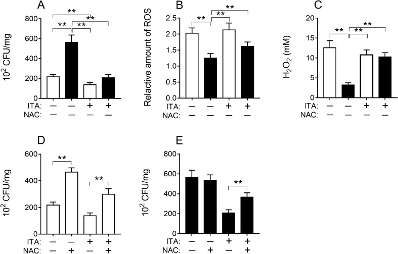
The macrophage plays an important role in the innate immune response to control infection34. Deletion of IRG1 diminished the capacity of RAW264.7 to produce itaconic acid, and subsequently these cells were unable to suppress the growth of S. typhimurium (Fig. 5A). However, this capability was regained by adding itaconic acid exogenously; exogenous itaconic acid appeared to improve the capability of wild-type RAW264.7 in eliminating S. typhimurium further (Fig. 5A). Consistent with this finding, ROS levels produced by IRG1-null cells reduced significantly and adding exogenous itaconic acid could remedy some of this effect (Fig. 5B, C). Moreover, the effects of the removal of ROS with NAC on the capability of these cells in eradicating the S. typhimurium were further evaluated (Fig. 5D, E). The results showed that incubating these cells with NAC reduced ROS and the capability of eradicating the S. typhimurium (Fig. 5D, E).
Figure 5.
Growth of S. typhimurium in cells (A, D, E) and levels of ROS (B), H2O2 (C) in S. typhimurium infected cells and culture medium under different treatment conditions (24 h). White bar, RAW264.7; black bar, RAW264.7-IRG1KO. ITA, itaconic acid; NAC, N-acetyl-L-cysteine. Relative ROS amount in cells with different treatments was normalized to RAW264.7 control (NAC) and represents the fold change. Values represent the mean ± S.E.M. **, p < 0.01.
Itaconic acid inhibited the hatching of S. japonicum eggs
Previously, we have demonstrated that co-infection of mice with both S. japonicum and S. typhimurium can significantly reduce S. japonicum worm and egg burden6. Concurrently, we also discovered increased levels of itaconic acid in the spleen and liver of co-infected mice5,6. To test effects of itaconic acid on S. japonicum eggs, matured S. japonicum eggs from the liver of BALB/c mice after 5 weeks of S. japonicum infection were collected and subjected to different concentrations of itaconic acid. The result showed that even at a very low concentration, itaconic acid completely inhibited the hatching of S. japonicum eggs (Supplementary information: Fig. S4).
Discussion
Itaconic acid has antimicrobial effect through inhibiting isocitrate lyase9 and has been considered not to be produced by mammalian cells for a long time7. However, this notion was subverted on the discovery that itaconic acid can be produced by immune-responsive gene 1 coded protein in LPS activated macrophages2,3. In addition, reactive oxygen species (ROS) has been known to play an important role in the anti-inflammation of itaconic acid15,16. Recently, the mechanism of the anti-inflammation of itaconic acid has been proposed through two ways: through IκBζ–ATF3 inflammatory axis regulated by electrophilic properties of itaconate and its derivatives17, or through activating Nrf2 via alkylation of KEAP18. These studies suggest that itaconic acid may be a potential anti-inflammatory drug35,36. In the current investigation, the mechanism of itaconic acid/IRG1 regulating the pentose phosphate pathway to produce ROS was assessed for its anti-inflammatory and anti-bacterial activities.
As the first step of the investigation, an IRG1-null (RAW264.7-IRG1KO) cell line was generated by employing CRISPR-Cas9 to delete IRG1 gene from the macrophage RAW264.7. Successful knockout of the IRG1 gene was evident in the DNA and further manifested by the absence of endogenous production of itaconic acid (Supplementary information: Figs. S1 and S2). The RAW264.7-IRG1KO cells in the absence of itaconic acid had significant inflammatory response, e.g. increasing levels of IL-1β, IL-6 and TNF-α, when exposed to LPS (Fig. 1). This observed anti-inflammatory function of itaconic acid is consistent with studies conducted previously by other groups37,38. Immune-responsive gene 1 has been shown to be associated with ROS production11,12,20, therefore the role of ROS production in the anti-inflammatory functions of itaconic acid was subsequently tested in this study. The measurement results of ROS in cells, H2O2 in cell culture medium and GSH in cell lysates confirmed that significantly increased levels of ROS and decreased levels of GSH was associated with the presence of endogenous itaconic acid (Fig. 2A–C). In addition, when ROS in cells was removed by anti-oxidant (N-acetyl-L-cysteine) treatment, the anti-inflammatory effects of itaconic acid diminished in macrophages (Fig. 2D–F), which illustrated that anti-inflammation of itaconic acid could be mediated by ROS. Moreover, exogenous addition of itaconic acid could aid in the production of ROS (Fig. 2A, B). All the above results confirmed that anti-inflammatory effects of itaconic acid are via promoting the production of ROS, which is consistent with previously reported results showing that ROS functions as a signaling molecule39 and possesses anti-microbial activity40–42.
In order to identify where excessive amounts of ROS were generated, key enzymes in the pentose phosphate pathway (PPP) were investigated and the results showed that PPP was stimulated by the presence of itaconic acid. Since NADPH is the intermediate metabolite of the PPP and can be used by NADPH oxidase as a substrate to produce ROS, the expression levels of NOX2 and NADPH oxidase were subsequently evaluated. And it was found that both endogenous and exogenous itaconic acid can promote the PPP pathway and the activity of NADPH oxidase (Fig. 3A, B, D). The ubiquitin-editing enzyme A20 has been shown to regulate immune responses, including IRG116,43. Thereafter the role of A20 in itaconic acid associated anti-inflammatory effects was subsequently assessed. Experimental results demonstrated that both exogenous and exogenous itaconic acid upregulated the expression of A20, and this upregulation could be eliminated by the antioxidant NAC (Fig. 4). Furthermore, inhibition of NF-κB by Bay11-7082 also can eliminate itaconic acid associated upregulation of A20 with concurrent eliminating anti-inflammatory effects of itaconic acid (Supplementary information: Fig. S3).
In this study, the experimental results showed that the anti-inflammatory effects of itaconic acid were motivated by its promotion of ROS production via PPP pathway, which is also regulated by A20 through the NF-κB pathway (Supplementary information: Fig. S3). And the increased levels of ROS induced by itaconic acid played an important role in inhibiting the growth of S. typhimurium in cell (Fig. 5). In addition, itaconic acid is found capable of suppressing parasitic growth, which is manifested by inhibiting hatching of S. japonicum eggs in vitro even at very low concentrations (Supplementary information: Fig. S4). These findings provided an rationale for our previous work, showing a reduction of schistosoma worm and egg burden in mice co-infected with S. japonicum and S. typhimurium, where the presence of itaconic acid was also concurrently discovered in the co-infected mice6.
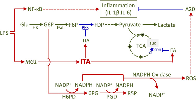
Itaconic acid, an under-studied small metabolite, has been found to play important functions in immune and metabolic regulation. The production of itaconic acid is controlled by the gene IRG1. The IRG1-null macrophage cell model, RAW264.7-IRG1KO, alongside its wild-type counterpart, RAW264.7, was employed to investigate the mechanism of anti-inflammatory effects of itaconic acid in this study. Itaconic acid promotes the production of ROS by stimulating the PPP-pathway. Intracellular ROS has the ability of eradicating cellular bacteria and can also induce the expression of anti-inflammatory gene A20 through NF-κB pathway, affecting the secretion of macrophage cytokines, and then exerting anti-inflammatory affects in vivo (Fig. 6). In addition, itaconic acid was showed having anti-parasitic effects. Our study has revealed an alternative mechanism on the anti-inflammatory functions of itaconic acid, in which metabolic pathway PPP involving ROS production play a necessary role.
Figure 6.
Schematic representation of the mechanism of anti-inflammatory effects of itaconic acid/IRG1 (Drew with Microsoft Office Powerpoint 2007). Glu, glucose; HK, Hexokinase; G6P, glucose-6-phosphate; PGI, glucosephosphate isomerase; F6P, fructose-6-phosphate; PFK, phosphofructo kinase; FDP, fructose-1,6-diphosphat; H6PD, hexose-6-phosphate dehydrogenase; 6PG, 6-phosphogluconate; PGD, phosphogluconate dehydrogenase; R5P, ribulose-5-phosphate; ROS, reactive oxygen species; A20, TNFAIP3, TNF alpha induced protein 3; TCA, tricarboxylic acid cycle; SUC, succinate; ITA, itaconic acid; SDH, succinate dehydrogenase.
Methods
IRG1 knocked out in RAW264.7
RAW264.7 was cultured in DMEM medium with 10% fetal bovine serum at 37 ℃ with 5% CO2. CRISPR-Cas9 was carried out to knock out IRG1 in RAW264.7 cells according to protocol published previously44. The plasmid, pSpCas9(BB)-2A-Puro (PX459)V2.0 (Addgene), was used to transfect RAW264.7 cells with FuGENE HD Transfection Reagent (Promega). The guide sequence was 5′-TGAGTGGCAGCGTTCGCTATGGG-3′, and the synthetic oligonucleotide sequence need to be inserted in plasmid PX459 were forward: 5′-CACCGTGAGTGGCAGCGTTCGCTAT-3′ and reverse: 5′-AAACATAGCGAACGCTGCCACTCAC-3′. Single-cell clones were selected at 24 h after transfection and followed with puromycin (3 μg/mL, Sigma) treatment for 48 h. Primers used to amplify the target IRG1 DNA fragment (~ 500 bp) from genome with possible indel were forward: 5′-TTGTCCTTCbTGGGCATGGATA-3′ and reverse: 5′-TCCAACAGCCAGATGTGAGAA-3′. This pair of primers was also used to amplify the target IRG1 DNA fragment from wild-type RAW264.7 genome. Purified PCR products (~ 500 bp) of target IRG1 DNA fragment from puromycin-selected single-cell clones and wild-type RAW264.7 cells were mixed together to produce hybrid-DNA fragments, and then the hybrid-DNA fragments were subjected to Cruiser Enzyme (Genloci Biotechnologies Inc.) validation. Cruiser Enzyme-digested hybrid DNA fragments were used to run DNA gel electrophoresis. PCR products from selected single-cell clones with indel in gene IRG1 will have unpaired base when to produce hybrid-DNA fragment, which can be digested by Cruiser Enzyme to produce two different DNA fragment. In our experiments, it should produce a ~ 200 bp DNA fragment and a ~ 300 bp fragment. PCR products amplified from wild-type RAW264.7 were used as negative controls. The IRG1 knocked out cell line was named as RAW264.7-IRG1KO, which was further confirmed for the absence of itaconic acid using nuclear magnetic resonance (NMR) spectrometer.
Cell culture for gene expression, cytokines, ROS, H2O2, GSH and NADPH oxidase activity detection
Both wild-type RAW264.7 and RAW264.7-IRG1KO were cultured in DMEM with 10% fetal bovine serum at 37 °C with 5% CO2. Itaconate (10 mM, Sigma), lipopolysaccharide (LPS, 10 ng/mL, Sigma), N-acetyl-L-cysteine (NAC, 2.5 mM, Beyotime) and Bay11-7082 (1.5 μM, Beyotime) were used to treat cells and six repeats of each treatment were performed. Total RNA was isolated with RNAiso plus (Takara). RT-qPCR was carried out with PrimeScript RT Reagent Kit with gDNA Eraser (Takara) and SYBR Green PCR Master Mix (ABI). Relative expressions of genes H6PD (glucose 6-phosphate dehydrogenase), PGD (6-phosphogluconate dehydrogenase), NOX2 (NADPH oxidase 2), A20 (TNFAIP3, TNF alpha induced protein) were measured using RT-qPCR and were normalized to expression of the housekeeping gene β-actin. Primers used for RT-qPCR analysis were listed in Table S1 (Supplementary information). IL-1β, IL-6 and TNF-α in cell culture medium were tested with ELISA Kits purchased from BOSTER (Wuhan, China). ROS in cell was tested with probe 2,7-dichlorofluorescin diacetate (DCFH-DA)-based Reactive Oxygen Species Assay Kit (E004-1–1, Nanjing Jiancheng Bioengineering Institute, China). H2O2 in cell culture medium (A064-1–1, Hydrogen Peroxide Assay Kit), glutathione (GSH) (A061-1–1, Glutathione Assay Kit) and NADPH oxidase activity (A127-1–1, NADPH Oxidase Assay Kit) in cell lysate were determined using respective kits purchased from Nanjing Jiancheng Bioengineering Institute (China).
Infection of RAW264.7 and RAW264.7-IRG1KO with S. typhimurium
Salmonella typhimurium strain 14028 (ATCC) was obtained from Prof. Guo (Wuhan University) and cultured aerobically at 37 °C in Luria–Bertani (LB) broth (Sigma) overnight. Cultured bacteria were recovered by centrifuging at 10,000 g for 30 s, then washed twice and re-suspended in sterile PBS (pH7.2) with a suitable concentration. Salmonella typhimurium (~ 100:1) was added into medium of cultured cells (RAW264.7 and RAW264.7-IRG1KO). After 30 min of co-culture, gentamicin was added at a final concentration of 100 ug/mL to clear bacteria in medium. One hour later, medium was replaced with fresh medium containing 16 ug/mL gentamicin and maintained with or without treatments (10 mM itaconic acid, 2.5 mM N-acetyl-L-cysteine) for 24 h. Then cells were collected, weighted, and lysed for 10 min with tritonX-100 (0.1%). Cell lysates with bacteria were diluted with PBS into desired concentration and the bacteria number was determined by plate counts on solid LB culture medium. Six repeats were conducted for each of the treatment.
Collection and itaconate treatment of S. japonicum eggs
Schistosoma japonicum cercariae were released from living oncomelanias6, which were purchased from Jiangsu Institute of Parasitic Diseases (China). Cercariae were counted in small dechlorinated water drops on cover slides under anatomical microscope. Six-week old SPF BALB/c mice (Hunan SJA Laboratory Animal Co., Ltd., China) were infected with approximate eighty S. japonicum cercariae each via abdominal skin. Forty five days after infection, mice were sacrificed and schistosome eggs were collected following the procedure: fresh liver tissues were cut into small pieces and homogenized and sequentially passed through nylon mesh sieves of 80, 120 and 300 mesh, washed with 0.9% saline to prevent eggs from hatching.
Schistosoma japonicum eggs were kept in 250 mL flask with dechlorinated water in artificial climate incubator at 26 ℃. Different concentrations of itaconic acid (0.00, 1.90, 3.80, 5.80, 7.70 μM) were used to treat S. japonicum eggs. Five repeats were conducted for each of the treatment. Then miracidia were counted under an anatomical microscope, 2, 4, 6 h after treatment.
Ethics statement
All animal experiments were approved by the Animal Welfare Committee of Wuhan Institute of Physics and Mathematics, Chinese Academy of Sciences (Permission No. S-051–10-04-OU). All animal experiment procedures were carried out in strict accordance with the National Guidelines for Experimental Animal Welfare (People’s Republic of China, 2006), and in compliance with the ARRIVE guidelines.
Statistical analysis
The data were subjected to either analysis of variance (ANOVA) followed by Tukey test for post hoc comparisons, and the software for statistical analysis used in this study is Prism 7.0 (GraphPad Software Inc., La Jolla, CA). Values represent the mean ± S.E.M.
Supplementary Information
Acknowledgements
Startup funding from Lee Kong Chian School of Medicine, Nanyang Technological University is acknowledged.
Author contributions
Conceptualization: X.Z. and Y.W.; Methodology: X.Z.; Investigation: X.Z., Y.G., Z.L., J.Y.; Writing—original, X.Z. and Y.L.; Writing—Review and Editing, Y.W., H.T., and X.Z.; Funding Acquisition: Y.W. and H.T.; Supervision: Y.W.
Data availability
We declare that the data supporting the findings of this study are available within the article and its supplemental material files or from the corresponding author on request.
Competing interests
The authors declare no competing interests.
Footnotes
Publisher's note
Springer Nature remains neutral with regard to jurisdictional claims in published maps and institutional affiliations.
Supplementary Information
The online version contains supplementary material available at 10.1038/s41598-021-97352-x.
References
- 1.Lee CGL, Jenkins NA, Gilbert DJ, Copeland NG, Obrien WE. Cloning and analysis of gene-regulation of a novel LPS-inducible cDNA. Immunogenetics. 1995;41:263–270. doi: 10.1007/BF00172150. [DOI] [PubMed] [Google Scholar]
- 2.Strelko CL, Lu WY, Dufort FJ, Seyfried TN, Chiles TC, et al. Itaconic acid is a mammalian metabolite induced during macrophage activation. J. Am. Chem. Soc. 2011;133:16386–16389. doi: 10.1021/ja2070889. [DOI] [PMC free article] [PubMed] [Google Scholar]
- 3.Michelucci A, Cordes T, Ghelfi J, Pailot A, Reiling N, et al. Immune-responsive gene 1 protein links metabolism to immunity by catalyzing itaconic acid production. Proc. Natl. Acad. Sci. U. S. A. 2013;110:7820–7825. doi: 10.1073/pnas.1218599110. [DOI] [PMC free article] [PubMed] [Google Scholar]
- 4.Naujoks J, Tabeling C, Dill BD, Hoffmann C, Brown AS, et al. IFNs modify the proteome of Legionella-containing vacuoles and restrict infection via IRG1-derived itaconic acid. PLoS Pathog. 2016;12:e1005408. doi: 10.1371/journal.ppat.1005408. [DOI] [PMC free article] [PubMed] [Google Scholar]
- 5.Zhu XY, Lei HH, Wu JF, Li JV, Tang HR, et al. Systemic responses of BALB/c mice to Salmonella typhimurium infection. J. Proteome Res. 2014;13:4436–4445. doi: 10.1021/pr500770x. [DOI] [PubMed] [Google Scholar]
- 6.Zhu XY, Chen L, Wu JF, Tang HR, Wang YL. Salmonella typhimurium infection reduces Schistosoma japonicum worm burden in mice. Sci. Rep. 2017;7:1349. doi: 10.1038/s41598-017-00992-1. [DOI] [PMC free article] [PubMed] [Google Scholar]
- 7.Shin JH, Yang JY, Jeon BY, Yoon YJ, Cho SN, et al. H-1 NMR-based metabolomic profiling in mice infected with Mycobacterium tuberculosis. J. Proteome Res. 2011;10:2238–2247. doi: 10.1021/pr101054m. [DOI] [PubMed] [Google Scholar]
- 8.Nguyen TV, Alfaro AC, Merien F, Young T, Grandiosa R. Metabolic and immunological responses of male and female new Zealand Greenshell mussels (Perna canaliculus) infected with Vibrio sp. J. Invertebr. Pathol. 2018;157:80–89. doi: 10.1016/j.jip.2018.08.008. [DOI] [PubMed] [Google Scholar]
- 9.Mcfadden BA, Purohit S. Itaconate, an isocitrate lyase-directed inhibitor in Pseudomonas indigofera. J. Bacteriol. 1977;131:136–144. doi: 10.1128/jb.131.1.136-144.1977. [DOI] [PMC free article] [PubMed] [Google Scholar]
- 10.Patel TR, Mcfadden BA. Caenorhabditis-elegans and Ascaris-suum—Inhibition of isocitrate lyase by itaconate. Exp. Parasitol. 1978;44:262–268. doi: 10.1016/0014-4894(78)90107-8. [DOI] [PubMed] [Google Scholar]
- 11.Hall CJ, Boyle RH, Astin JW, Flores MV, Oehlers SH, et al. Immunoresponsive gene 1 augments bactericidal activity of macrophage-lineage cells by regulating β-oxidation-dependent mitochondrial ROS production. Cell Metab. 2013;18:265–278. doi: 10.1016/j.cmet.2013.06.018. [DOI] [PubMed] [Google Scholar]
- 12.Ren K, Lv YZ, Zhuo YJ, Chen CM, Shi HF, et al. Suppression of IRG-1 reduces inflammatory cell infiltration and lung injury in respiratory syncytial virus infection by reducing production of reactive oxygen species. J. Virol. 2016;90:7313–7322. doi: 10.1128/JVI.00563-16. [DOI] [PMC free article] [PubMed] [Google Scholar]
- 13.Van Acker H, Coenye T. The role of reactive oxygen species in antibiotic-mediated killing of bacteria. Trends Microbiol. 2017;25:456–466. doi: 10.1016/j.tim.2016.12.008. [DOI] [PubMed] [Google Scholar]
- 14.Lampropoulou V, Sergushichev A, Bambouskova M, Nair S, Vincent EE, et al. Itaconate links inhibition of succinate dehydrogenase with macrophage metabolic remodeling and regulation of inflammation. Cell Metab. 2016;24:158–166. doi: 10.1016/j.cmet.2016.06.004. [DOI] [PMC free article] [PubMed] [Google Scholar]
- 15.Uddin MJ, Joe Y, Kim SK, Jeong SO, Ryter SW, et al. IRG1 induced by heme oxygenase-1/carbon monoxide inhibits LPS-mediated sepsis and pro-inflammatory cytokine production. Cell Mol. Immunol. 2016;13:170–179. doi: 10.1038/cmi.2015.02. [DOI] [PMC free article] [PubMed] [Google Scholar]
- 16.Li YK, Zhang P, Wang CC, Han CF, Meng J, et al. Immune responsive gene 1 (IRG1) promotes endotoxin tolerance by increasing A20 expression in macrophages through reactive oxygen species. J. Biol. Chem. 2013;288:16225–16234. doi: 10.1074/jbc.M113.454538. [DOI] [PMC free article] [PubMed] [Google Scholar]
- 17.Bambouskova M, Gorvel L, Lampropoulou V, Sergushichev A, Loginicheva E, et al. Electrophilic properties of itaconate and derivatives regulate the I kappa B zeta-ATF3 inflammatory axis. Nature. 2018;556:501–504. doi: 10.1038/s41586-018-0052-z. [DOI] [PMC free article] [PubMed] [Google Scholar]
- 18.Mills EL, Ryan DG, Prag HA, Dikovskaya D, Menon D, et al. Itaconate is an anti-inflammatory metabolite that activates Nrf2 via alkylation of KEAP1. Nature. 2018;556:113–117. doi: 10.1038/nature25986. [DOI] [PMC free article] [PubMed] [Google Scholar]
- 19.Weiss JM, Davies LC, Karwan M, Ileva L, Ozaki MK, et al. Itaconic acid mediates crosstalk between macrophage metabolism and peritoneal tumors. J. Clin. Invest. 2018;128:3794–3805. doi: 10.1172/JCI99169. [DOI] [PMC free article] [PubMed] [Google Scholar]
- 20.Hall CJ, Sanderson LE, Lawrence LM, et al. Blocking fatty acid-fueled mROS production within macrophages alleviates acute gouty inflammation. J. Clin. Invest. 2018;128:1752–1771. doi: 10.1172/JCI94584. [DOI] [PMC free article] [PubMed] [Google Scholar]
- 21.Nemeth B, Doczi J, Csete D, Kacso G, Ravasz D, et al. Abolition of mitochondrial substrate-level phosphorylation by itaconic acid produced by LPS-induced Irg1 expression in cells of murine macrophage lineage. FASEB J. 2016;30:286–300. doi: 10.1096/fj.15-279398. [DOI] [PubMed] [Google Scholar]
- 22.Cordes T, Wallace M, Michelucci A, Divakaruni AS, Sapcariu SC, et al. Immunoresponsive gene 1 and itaconate inhibit succinate dehydrogenase to modulate intracellular succinate levels. J. Biol. Chem. 2016;291:14274–14284. doi: 10.1074/jbc.M115.685792. [DOI] [PMC free article] [PubMed] [Google Scholar]
- 23.Sakai A, Kusumoto A, Kiso Y, Furuya E. Itaconate reduces visceral fat by inhibiting fructose 2,6-bisphosphate synthesis in rat liver. Nutrition. 2004;20:997–1002. doi: 10.1016/j.nut.2004.08.007. [DOI] [PubMed] [Google Scholar]
- 24.De Santa F, Vitiello L, Torcinaro A, Ferraro E. The role of metabolic remodeling in macrophage polarization and its effect on skeletal muscle regeneration. Antioxid. Redox Signal. 2019;30:1553–1598. doi: 10.1089/ars.2017.7420. [DOI] [PubMed] [Google Scholar]
- 25.Stincone A, Prigione A, Cramer T, Wamelink MM, et al. The return of metabolism: Biochemistry and physiology of the pentose phosphate pathway. Biol. Rev. Camb. Philos. Soc. 2015;90:927–963. doi: 10.1111/brv.12140. [DOI] [PMC free article] [PubMed] [Google Scholar]
- 26.Netti GS, Prattichizzo C, Montemurno E, Simone S, et al. Exposure to low- vs iso-osmolar contrast agents reduces NADPH-dependent reactive oxygen species generation in a cellular model of renal injury. Free Radic. Biol. Med. 2014;68:35–42. doi: 10.1016/j.freeradbiomed.2013.11.016. [DOI] [PubMed] [Google Scholar]
- 27.Lu J, Wu L, Jiang T, Wang Y, Zhao H, et al. Angiotensin AT2 receptor stimulation inhibits activation of NADPH oxidase and ameliorates oxidative stress in rotenone model of Parkinson's disease in CATH.a cells. Neurotoxicol. Teratol. 2015;47:16–24. doi: 10.1016/j.ntt.2014.11.004. [DOI] [PubMed] [Google Scholar]
- 28.Zhou DX, Huang C, Lin Z, Zhan SX, Kong LN, et al. Macrophage polarization and function with emphasis on the evolving roles of coordinated regulation of cellular signaling pathways. Cell Signal. 2014;26:192–197. doi: 10.1016/j.cellsig.2013.11.004. [DOI] [PubMed] [Google Scholar]
- 29.Munhoz AC, Riva P, Simoes D, Curi R, Carpinelli AR. Control of insulin secretion by production of reactive oxygen species: Study performed in pancreatic islets from fed and 48-hour fasted wistar rats. PLoS ONE. 2016;11:e0158166. doi: 10.1371/journal.pone.0158166. [DOI] [PMC free article] [PubMed] [Google Scholar]
- 30.Soni D, Wang DM, Regmi SC, Mittal M, Vogel SM, et al. Deubiquitinase function of A20 maintains and repairs endothelial barrier after lung vascular injury. Cell Death Discov. 2018;4:60. doi: 10.1038/s41420-018-0056-3. [DOI] [PMC free article] [PubMed] [Google Scholar]
- 31.Tiruppathi C, Soni D, Wang DM, Xue J, Singh V, et al. The transcription factor DREAM represses the deubiquitinase A20 and mediates inflammation. Nat. Immunol. 2014;15:239–247. doi: 10.1038/ni.2823. [DOI] [PMC free article] [PubMed] [Google Scholar]
- 32.Lawrence, T. The nuclear factor NF-kappaB pathway in inflammation. Cold Spring Harb. Perspect. Biol.1, a001651 (2009). PMID: 20457564. [DOI] [PMC free article] [PubMed]
- 33.Lee EG, Boone DL, Chai S, Libby SL, Chien M, et al. Failure to regulate TNF-induced NF-kappaB and cell death responses in A20-deficient mice. Science. 2000;289:2350–2354. doi: 10.1126/science.289.5488.2350. [DOI] [PMC free article] [PubMed] [Google Scholar]
- 34.Rosowski EE. Determining macrophage versus neutrophil contributions to innate immunity using larval zebrafish. Dis. Model Mech. 2020;13:dmm041889. doi: 10.1242/dmm.041889. [DOI] [PMC free article] [PubMed] [Google Scholar]
- 35.Otto G. Immunometabolism: Itaconate helps KEAP1's cool. Nat. Rev. Immunol. 2018;18:294–295. doi: 10.1038/nri.2018.27. [DOI] [PubMed] [Google Scholar]
- 36.Perico L, Wyatt CM, Benigni A. A new BEACON of hope for the treatment of inflammation? The endogenous metabolite itaconate as an alternative activator of the KEAP1-Nrf2 system. Kidney Int. 2018;94:646–649. doi: 10.1016/j.kint.2018.07.018. [DOI] [PubMed] [Google Scholar]
- 37.Zhao C, Jiang P, He Z, Yuan X, Guo J, et al. Dimethyl itaconate protects against lippolysacchride-induced mastitis in mice by activating MAPKs and Nrf2 and inhibiting NF-kappaB signaling pathways. Microb. Pathog. 2019;133:103541. doi: 10.1016/j.micpath.2019.05.024. [DOI] [PubMed] [Google Scholar]
- 38.Dominguez-Andres J, Novakovic B, Li Y, Scicluna BP, Gresnigt MS, et al. The itaconate pathway is a central regulatory node linking innate immune tolerance and trained immunity. Cell Metab. 2019;29:211–220. doi: 10.1016/j.cmet.2018.09.003. [DOI] [PubMed] [Google Scholar]
- 39.Thannickal VJ, Fanburg BL. Reactive oxygen species in cell signaling. Am. J. Physiol. Lung Cell Mol. Physiol. 2000;279:L1005–L1028. doi: 10.1152/ajplung.2000.279.6.L1005. [DOI] [PubMed] [Google Scholar]
- 40.Yen WC, Wu YH, Wu CC, Lin HR, Stern A, et al. Impaired inflammasome activation and bacterial clearance in G6PD deficiency due to defective NOX/p38 MAPK/AP-1 redox signaling. Redox Biol. 2019;28:101363. doi: 10.1016/j.redox.2019.101363. [DOI] [PMC free article] [PubMed] [Google Scholar]
- 41.Nguyen TV, Alfaro AC, Young T, Green S, Zarate E, et al. Itaconic acid inhibits growth of a pathogenic marine Vibrio strain: A metabolomics approach. Sci. Rep. 2019;9:5937. doi: 10.1038/s41598-019-42315-6. [DOI] [PMC free article] [PubMed] [Google Scholar]
- 42.Shastri MD, Shukla SD, Chong WC, Dua K, Peterson GM, et al. Role of oxidative stress in the pathology and management of human tuberculosis. Oxid. Med. Cell. Longev. 2018;2018:7695364. doi: 10.1155/2018/7695364. [DOI] [PMC free article] [PubMed] [Google Scholar]
- 43.Van Quickelberghe E, Martens A, Goeminne LJE, et al. Identification of immune-responsive gene 1 (IRG1) as a target of A20. J. Proteome Res. 2018;17:2182–2191. doi: 10.1021/acs.jproteome.8b00139. [DOI] [PubMed] [Google Scholar]
- 44.Ran FA, Hsu PD, Wright J, Agarwala V, Scott DA, et al. Genome engineering using the CRISPR-Cas9 system. Nat. Protoc. 2013;8:2281–2308. doi: 10.1038/nprot.2013.143. [DOI] [PMC free article] [PubMed] [Google Scholar]
Associated Data
This section collects any data citations, data availability statements, or supplementary materials included in this article.
Supplementary Materials
Data Availability Statement
We declare that the data supporting the findings of this study are available within the article and its supplemental material files or from the corresponding author on request.


