



Abstract
The human cerebellum contributes to both motor and non-motor processes. Within the cerebellum, different subregions support sensorimotor and broader cognitive functions, due to regional patterns in anatomical connectivity with the cerebral cortex and spinal and vestibular systems. We evaluated the effects of transcranial direct current stimulation (tDCS) targeting different cerebellar regions on language task performance and whole-brain functional activation patterns. Functional MRI data were acquired while 43 healthy young adults (15 males, 28 females; 23.3 ± 3.0 years) performed a sentence completion task before and after 20min of 1.5mA anodal tDCS. Participants received tDCS targeting either the anterior sensorimotor cerebellum (n=11; 3cm right of inion, over lobule V); the right posterolateral cerebellum (n=18; 1cm down and 4cm right of inion, over lobule VII); or sham tDCS (n=14). TDCS targeting the right posterolateral cerebellum improved task accuracy relative to the sham condition (p=0.04) and increased activation in left frontal and temporal cortices relevant to task performance (post-tDCS>pre-tDCS; T 3.17, FDR p<0.05 cluster correction). The regions of increased BOLD signal after right posterolateral cerebellar tDCS fell within the network showing functional connectivity with right cerebellar lobule VII, suggesting specific modulation of this network. In contrast, tDCS targeting the sensorimotor cerebellum did not impact task performance and increased BOLD signal only in one cluster extending into the precentral gyrus. These findings indicate that sensorimotor and cognitive functional cerebellar subregions differentially impact behavioral task performance and task-relevant activation patterns, further contributing to our understanding of the cerebellar modulation of motor and non-motor functions.
Keywords: neuromodulation, functional MRI, cognition, motor, language, cerebellum
Introduction
Containing over half of the neurons in the brain, the cerebellum is extensively interconnected with the cerebral cortex via multiple parallel circuits. These connections provide anatomical substrates for the cerebellar contribution to a wide range of behaviors, from motor adaptation to higher cognitive functions (for reviews, Ito 2008; Ramnani, 2014; Mariën et al., 2014; Schmahmann et al., 2019). Anatomical projections (Kelly and Strick, 2003) and functional activation and connectivity patterns (Buckner et al., 2011; King et al., 2019) provide evidence for a functional topography within the cerebellum, with different subregions supporting overt motor control (anterior lobe and medial lobule VI; regions in lobule VIII) and a range of cognitive functions (lateral regions of lobule VI; lobule VII, regions of VIII, and IX; see Figure 1, and see Stoodley and Schmahmann, 2009; Buckner et al., 2011; Stoodley et al., 2012; Kerren-Happuch et al., 2014; King et al., 2019).
Figure 1. Cerebellar anatomy.
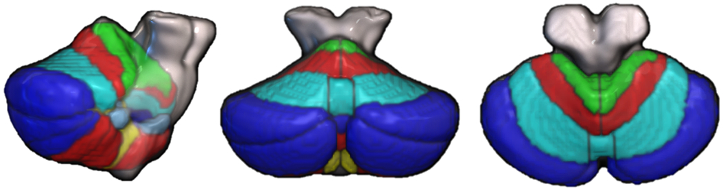
Lobules I-IV = green, V = red / superior, VI = cyan, VII = blue, VIII = red / inferior, IX = yellow, X = light blue (from the Spatially Unbiased Infratentorial [SUIT] atlas of the cerebellum; Diedrichsen et al., 2009). Left, lateral view; middle, posterior view; right, superior view.
Based on the relatively uniform cytoarchitecture of the cerebellum, it has been proposed that the cerebellum performs a common computation within these subregions, a “Universal Cerebellar Transform” (see Schmahmann, 2004; Schmahmann et al., 2019), though this theory has recently been challenged (Diedrichsen et al., 2019). Various hypotheses regarding this computation have emerged, with a common theme being that the cerebellum builds internal models of movement and “mental models” of thoughts (Ito, 2008), which are trained through feedback and, once optimized, enable outcome prediction (see Miall et al., 1993; Wolpert et al., 1998; Ito, 2008; Sokolov et al., 2017; Raymond and Medina, 2018). It has been argued that this broad predictive modeling (also known as the internal forward model) can be applied to a range of tasks, including movement kinematics, semantic prediction, social cognition, and cognitive control (see Van Overwalle et al., 2014; Lesage et al., 2017; Sokolov, 2018; Schmahmann et al., 2019; Van Overwalle et al., 2020; D’Mello et al., 2020). According to this theory, the cerebellum is able to automatize both basic and higher-level functions by rapidly detecting disruptions in action / thought sequences and utilizing prediction error to optimize performance (Popa et al., 2016; Heleven et al., 2019).
While the internal model hypothesis attempts to establish the specific cerebellar contribution to motor and cognitive control, there is also the question of how the cerebellum exerts its influence on such a range of functions. Recent evidence from animal models suggests that the cerebellum coordinates the timing of cortical neural oscillations in a site- and frequency-specific manner (McAfee et al., 2019; Lindeman et al., 2020). Human neuromodulation studies with transcranial direct current stimulation (tDCS) and transcranial magnetic stimulation (TMS) (see Ferrucci et al., 2015; Grimaldi et al., 2016; Miterko et al., 2019) offer the opportunity to investigate the impact of primary cerebellar modulation on behavioral performance and, when combined with neuroimaging, neural activation and functional connectivity patterns. Cerebellar neuromodulation impacts physiological measures of motor control (Galea et al., 2009; Hiraoka et al., 2009) and behavioral performance on a range of tasks, including working memory (Desmond et al., 2005; Pope and Miall, 2012; Sheu et al., 2019), predictive language processing (Lesage et al., 2012; D’Mello et al., 2017), and articulation and phonemic fluency (Turkeltaub et al., 2016; for review, see Grimaldi et al., 2016). In these studies, different cerebellar regions were targeted in order to impact motor control (anterior cerebellum) and cognitive task performance (posterior and lateral cerebellar regions). Likewise, our previous work has shown that sensorimotor and cognitive aspects of language are differentially affected with electrode positions targeting sensorimotor (lobule V) and cognitive (lobule VII) cerebellar regions, respectively (Turkeltaub et al. 2016).
Consistent with animal models, modulating the human cerebellum with tDCS has revealed location- and polarity-specific effects on broader cortical regions to which the cerebellum interconnects. For example, tDCS targeting a sensorimotor cerebellar region (lobule V) modulated cerebellar brain inhibition (CBI) of contralateral motor cortex, such that cathodal (inhibitory) tDCS decreased CBI and anodal (excitatory) tDCS increased it (Galea et al., 2009). In contrast, neuromodulation targeting posterolateral regions of the cerebellum impacted cerebral cortical networks relevant to cognitive functions. For instance, functional connectivity in the default mode network increased following intermittent theta-burst TMS targeting the lateral cerebellum (Halko et al., 2014). Anodal tDCS targeting right lobule VII (RVII) increased activation in RVII during semantic prediction and increased resting-state functional connectivity within reading and language networks, but not within sensorimotor networks (D’Mello et al., 2017) or from adjacent cerebellar lobules (Turkeltaub et al., 2016, Stoodley et al., 2017). These findings further suggest that it is possible to modulate different cerebellar regions and cerebro-cerebellar networks with tDCS depending on electrode placement.
It has been proposed that cerebellar tDCS may provide a potential therapeutic option for a number of clinical disorders (Grimaldi et al., 2016), particularly bipolar disorder (Minichino et al., 2015), aphasia (Turkeltaub et al., 2016; Sebastian et al., 2017), autism (Stoodley et al., 2017; Kelly et al., 2020), and schizophrenia (Escelsior et al., 2019). However, despite the potential therapeutic utility of cerebellar tDCS, its underlying mechanisms of action are still in question (Miterko et al., 2019). Given the different symptom patterns across these disorders, and the corresponding differences in atypical regional structural and functional neural findings, it makes sense to consider how well one can target a specific cerebellar region with tDCS. For example, anterior sensorimotor regions may modulate cerebral cortical sensorimotor networks relevant to motor recovery following stroke, while right posterolateral cerebellar hemisphere regions could be relevant to language recovery in aphasia due to their influence on left hemisphere language networks (see Turkeltaub et al., 2016; Sebastian et al., 2017; Marangolo et al., 2018; Sebastian et al., 2020).
Here, we aimed to further test the functional specificity and mechanism of action of cerebellar tDCS through determining (1) whether neuromodulation of cerebellar subregions differentially impacts behavioral task performance; and (2) whether different cerebral cortical regions are modulated following tDCS targeting sensorimotor vs. cognitive cerebellar subregions. To attempt to answer these questions, we compared the behavioral and neural impact of tDCS targeting sensorimotor and cognitive cerebellar regions in neurotypical young adults. We predicted that modulation of the sensorimotor cerebellar target would impact behavioral response times and sensorimotor regional activation patterns, whereas tDCS targeting the right posterolateral cerebellum would improve task accuracy and alter functional activation in left-hemisphere language regions associated with task performance.
Experimental Procedures
Participants.
Forty-eight participants provided written, informed consent to take part in the study. Thirty-four participants also took part in D’Mello et al. (2017). After exclusion due to scanner error (n=2), electrode shift (n=1), and behavioral data outliers (n=2), the final sample included 43 participants (15 males, 28 females; 23.3 ± 3.0 years old; 32 participants overlapped with D’Mello et al., 2017). All participants were compensated for their time. The study was approved by the Institutional Review Board of American University.
All participants were right-handed, native English speakers with no contraindications for tDCS and/or MRI (e.g. no metal implants). Participants had no history of neurological injury or any psychiatric or neurodevelopmental disorder and were not taking any medications that act on the CNS. Participants were randomly assigned to one of three groups that received either sham (n=14) or anodal tDCS targeting cerebellar right lobule VII (n=18) or right lobule V (n=11).
Study design.
Participants each completed one tDCS-neuroimaging session. Following informed consent and introduction to the language task, the tDCS electrodes were positioned prior to entering the scanner. After acquisition of the structural MRI, participants completed a resting-state scan and a language task inside the scanner as a measure of baseline functional connectivity, task activation patterns, and behavioral performance. Then, tDCS was administered inside the scanner for 20min while resting-state data were acquired. Following tDCS, participants completed another resting-state scan and another run of the language task.
Following the scanning session, participants completed a 26-item self-scored questionnaire to rate side-effects both during and after tDCS, such as tingling, itching, burning, attention, fatigue, and pain (see Kessler et al., 2012). Participants rated the extent to which they experienced these symptoms on a scale of 0 (not at all) to 10 (greatest imaginable).
Language task.
Participants performed a sentence completion task (see Figure 2 and D’Mello et al., 2017). The task design was motivated by the hypothesis that the cerebellum is critical to predictive language processing, and our previous work showed that anodal cerebellar tDCS over the right posterolateral cerebellum (cognitive target region) increased cerebellar activation during predictive sentences only (see D’Mello et al., 2017).
Figure 2. Sentence completion task.
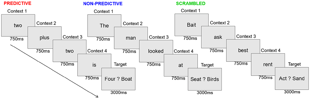
The target word was highly predictable for predictive trials, less predictable for non-predictive trials, and not predictable for scrambled trials (see D’Mello et al., 2017).
Participants viewed a sequence of four words (each presented for 750 ms) which established the context for the fifth target word (presented for 3000 ms). The target word was presented along with another word in a forced-choice paradigm, and participants were instructed to select the correct target word as quickly and accurately as possible using a button box with the first two fingers on their right hand. The cloze probability, or predictability of the final target word, was pre-determined by the context of the preceding word sequence, resulting in predictable (e.g. “Two plus two is …”) and non-predictable trials (e.g. “The man looked at …”) (for details, see D’Mello et al., 2017). In scrambled trials the word sequence did not form a sentence. There were 20 trials of each sentence condition (predictive, nonpredictive, scrambled) with a total task time of 6min 30sec. The task was presented and accuracy and response time data were collected using E-Prime software (Psychology Software Tools, Inc., Sharpsburg, PA).
Transcranial direct current stimulation.
TDCS involves running a small amount of direct current between two electrodes to increase (anodal) or decrease (cathodal) the chance of neuronal firing (Woods et al., 2016), although the specific impact of tDCS polarity on cerebellar circuits has yet to be established (see Ferrucci et al., 2016). Different configurations of the active and return electrodes on the scalp allow for targeting various regions of the cerebral cortex and cerebellum (see Ramaraju et al., 2018). TDCS was administered in the scanner using the MR-compatible NeuroConn MR Plus (Jali Medical, Inc, Waltham, MA). Anodal current was ramped up over 15s to 1.5mA, applied for 20min, and ramped down over 15s. The current amplitude was dictated by the impedance limits of the NeuroConn device inside the MR environment. In sham conditions, the current was increased over 15s and then ramped down, allowing participants to experience the initial tingling sensation associated with tDCS without receiving enough current to modulate neuronal excitability.
The right posterolateral cerebellum (RVII), which is associated with cognitive processes, was targeted by centering a 5x7 cm sponge-covered saline-soaked electrode 1cm inferior and 4cm lateral to the inion with the return electrode on the right clavicle (see Pope and Miall, 2012; Turkeltaub et al., 2016; D’Mello et al., 2017). The anterior sensorimotor cerebellum (RV) was targeted by centering a 5x5cm electrode 3cm lateral to the inion over the right cerebellum and the return electrode over the right clavicle (see Turkeltaub et al., 2016; Galea et al., 2009). The smaller electrode was used to target the anterior cerebellum in order to minimize the degree of overlap between the electric fields produced by the electrode montages. Participants in the sham condition had the electrode positioned 1cm inferior and 4cm lateral to the inion with the return electrode over the right clavicle. Participants were blinded to tDCS condition.
Neuroimaging protocols.
Neuroimaging was performed at the Center for Functional and Molecular Imaging at Georgetown University. Data were acquired on a Magnetom Trio 3T scanner (Siemens, Erlangen, Germany) with a 12-channel head coil. Structural T1-weighted (MP-RAGE) images were acquired in sagittal orientation with a TR of 1900 ms, TE of 2.52 ms, 9 degree flip angle, 1 mm slice thickness, 176 slices, and a voxel size of 1 mm3. During the two runs of the language task, data were acquired with a TR of 3000 ms, TE of 30 ms, 90 degree flip angle, 3 mm slice thickness, 3 mm slice spacing, 57 interleaved odd slices, 3.2 mm3 isotropic voxels, and 122 volumes.
Data Analyses.
Electric field modeling.
Estimated electric fields (EFs) produced by each tDCS montage were modeled using SimNIBS 2.1 (Thielscher et al., 2015). SimNIBS uses individual participant structural MR scans to model the EFs induced by noninvasive brain modulation, factoring in electrode size, position, polarity, and current intensity. Since the software does not enable modeling of reference electrodes below the neck, as we used in this study, we placed the return electrode along the right jaw bone in these models (posterior edge of electrode aligned with anterior edge of earlobe; see Figure 3). Head models were created for each subject using the headreco function and finite element methods were used to calculate electric field maps for each subject in native space. The electric field maps were then transformed into Montreal Neurological Institute (MNI) standard space using the superconvergent patch recovery interpolation method, whereby tissue boundaries are taken into consideration. Average MNI electric field maps were generated for the cognitive and sensorimotor groups using SPM12’s Imcalc function.
Figure 3. Electrode montage used for electric field modeling in SimNIBS.

Active electrode (anode) was positioned based on individual participant MRI scans and the return electrode (cathode) was positioned over the right jaw bone to approximate the right clavicle.
Behavioral data analyses.
Behavioral data were analyzed in RStudio (R version 3.6.1). The stats package was used for ANOVAs and t-tests and the lme4 package was used for linear mixed effects models. Language task performance was modeled with two separate response variables: median response time (ms) and mean percent accuracy. Outliers were assessed before and after normalization of response variables using a Tukey power transformation and modeling. Two outliers were identified in the sham group for low accuracy (<70%) and these two participants were removed from further analyses. In the linear mixed effects models, the random factor for participant was modeled in addition to the following fixed factors and their interactions: group (sham, cognitive, sensorimotor), sentence type (predictive, nonpredictive, scrambled), and time point with respect to tDCS (pre, post). Scores on the post-tDCS symptom questionnaires were compared between groups using a one-way ANOVA for each measure. Post-hoc analyses were conducted to assess directionality of significant effects. Statistical significance was assessed at p<0.05.
Preprocessing of neuroimaging data.
Task fMRI data were pre-processed using the CONN toolbox (Version 18b) implemented in MATLAB version 2018b (Whitfield-Gabrieli and Nieto-Castanon, 2012) (http://www.nitrc.org/projects/conn) according to procedures followed by D’Mello et al. (2017). The standard pipeline included slice-time correction, realignment, and unwarping, ART scrubbing for outlier detection using default intermediate thresholds, normalization, and smoothing (8 mm FWHM). Subject-specific variables for physiological noise (white matter and CSF signal), head motion parameters, and bandpass filtering were implemented to de-noise the data. A high-pass filter (0.008 – INF Hz) was applied to the task data to preserve task-related signal fluctuations. Motion outliers were assessed and participants with greater than 30% of volumes as outliers were removed. Within and between group differences in motion outliers were assessed using one-tailed paired t-tests (post-tDCS > pre-tDCS) and separate one-way ANOVAs (across groups) for each time point, respectively.
Analysis of language task activation patterns.
Activation patterns during the language task paradigm were modeled using SPM12. Because our primary question of interest was the impact of tDCS on task activation patterns, the whole task was modeled as a single, 6min 6sec block for each task run (pre- and post-tDCS) for each participant, with the six motion parameters as regressors. To determine the regions associated with task performance, activation prior to tDCS (pre-tDCS) across all participants was assessed using a peak threshold FWE-corrected p<0.05 with an FDR-corrected p<0.05 at the cluster level. To assess the effects of tDCS on task activation, the within-subject post > pre and pre > post contrast images were analyzed at the second level within each group (sham, cognitive, motor). Results were thresholded at a peak T=3.17 (p<0.005) and FDR-corrected cluster p<0.05.
Data visualization.
Behavioral data were visualized using RStudio (RStudio Team [2015] RStudio: Integrated Development for R. RStudio, Inc., Boston, MA; http://www.rstudio.com/). Imaging data were displayed using MRIcroGL (https://www.nitrc.org/projects/mricrogl).
Results
Visualization of electrode positions.
The active electrode was visible on the rendered MP-RAGE image for each participant and estimated electric field models were generated based on the active electrode position for each individual participant (see Figure 4). Visual inspection of the individual MP-RAGE images revealed one outlier in the cognitive group: the active electrode had shifted up to the right parietal cortex after the participant entered the scanner. Due to this misplacement, the participant was removed from the behavioral and neuroimaging analyses.
Figure 4. Active electrode placement and estimated electric fields for active tDCS groups.

Electrode positions for two representative participants with the electrode targeting the sensorimotor (top; red – centered 3 cm lateral to inion) and cognitive (bottom; blue – centered 4 cm lateral and 1 cm inferior to inion) cerebellum, and the position of the center of the electrode relative to the underlying brain anatomy. Estimated electric field maps (right, thresholded from −3.0 to −6.0) for the sensorimotor (red-yellow) and cognitive (blue-green) target groups reveal both overlapping and distinct electric fields.
Estimation of group average peak electric field.
We were unable to model the exact return electrode position due to the limited field of view in the MR scans, which did not include the clavicle. Therefore, these models are approximate and provided for illustrative purposes. Figure 4 shows the average electric field maps for the sensorimotor and cognitive active tDCS groups. While there was overlap between the estimated EF maps for the groups, the peak EFs differed between the two groups.
During- and post-tDCS symptoms questionnaires.
There were no significant differences in symptoms between the three groups during or after tDCS, with the exception of “itching” during tDCS (ANOVA p=0.025), which was significantly higher in the sensorimotor group (3.25 ± 2.22, on a scale of 1-10) compared with the sham tDCS group (0.77 ± 1.29, post-hoc paired t-test p=0.003). Supplemental Table 1 shows the scores for during- and post-tDCS symptoms. The highest intensity ratings were for “tingling” during tDCS (sham: 2.43 ± 2.20; cognitive tDCS: 3.6 ± 2.71; sensorimotor tDCS: 3.00 ± 2.17), though these did not differ between the groups. These findings suggest successful participant blinding of tDCS condition.
Motion during imaging.
There were no between-group differences in head motion outliers for the pre-tDCS scans (p=0.933) or post-tDCS scans (p=0.474). Additionally, there were no within-groups differences in motion pre- to post-tDCS (Cognitive, p=0.265; Sham, p=0.235; Sensorimotor p=0.072). No participants were removed from data analyses due to excessive motion.
Language task behavioral results.
Behavioral data were available for a subset of participants that received tDCS-fMRI (cognitive n=15, sham n=16, sensorimotor n=10). Mean accuracy scores are summarized by group (sham, sensorimotor, cognitive), sentence type, and time point (pre-, post-tDCS) in Table 1 and Figure 5. Accuracy is not reported for the scrambled sentences as there were no correct answers for those trials. As expected, participants completed the task with a high degree of accuracy.
Table 1. Accuracy across groups, sentence type, and time point.
Accuracy = Mean percentage accuracy, N = Number of participants, SD = Standard deviation, SE = Standard error.
| Group | N | Sentence type | Timepoint | Accuracy | SD | SE |
|---|---|---|---|---|---|---|
| Sham | 14 | Predictive | Pre | 0.959 | 0.025 | 0.007 |
| Post | 0.946 | 0.037 | 0.010 | |||
| Nonpredictive | Pre | 0.986 | 0.023 | 0.006 | ||
| Post | 0.989 | 0.021 | 0.006 | |||
| Cognitive | 15 | Predictive | Pre | 0.937 | 0.043 | 0.011 |
| Post | 0.962 | 0.037 | 0.002 | |||
| Nonpredictive | Pre | 0.977 | 0.037 | 0.005 | ||
| Post | 0.993 | 0.018 | 0.004 | |||
| Sensorimotor | 10 | Predictive | Pre | 0.962 | 0.020 | 0.006 |
| Post | 0.924 | 0.072 | 0.023 | |||
| Nonpredictive | Pre | 0.980 | 0.026 | 0.008 | ||
| Post | 0.980 | 0.063 | 0.020 |
Fig 5. Mean accuracy across groups, sentence type, and time point.

The violin plots show the distribution of the data, black dots represent individual participants, and white diamonds indicate mean accuracy.
After Tukey power transformation, mean percentage accuracy was entered as the response variable into a linear mixed effects model with group, sentence type, time point, and their interactions as fixed effects and participant as a random effect. After backward stepwise regression, the reduced model included the random effect of participant, main effects of group, condition, time point, and the interaction of group and time point (Supplemental Table 2). There was a significant main effect of sentence condition (p=1.47×10⁻¹¹), with higher accuracy on non-predictive sentences; this somewhat unexpected finding may be due to increased attention and/or deliberation during nonpredictive sentences, as indicated by the longer response times for nonpredictive relative to predictive sentences (see below). There was a significant group × time point interaction (p=0.048), suggesting different effects of tDCS on accuracy between the groups.
Post hoc analyses revealed that accuracy in the cognitive group improved more after tDCS relative to the sham group (p=0.041, cognitive group × time point interaction; Supplemental Table 3, Figure 6). Within the cognitive group, there was a significant effect of time point on accuracy (estimate 0.13, t value 2.56, p=0.013), but no sentence type × time point interaction, indicating that accuracy improved on both predictive and nonpredictive sentences post-tDCS. Within the sham group, there were no statistically significant changes in accuracy pre- to post-tDCS, and no sentence type × time point interaction. When the sensorimotor cerebellum was targeted, there were no statistically significant changes in accuracy pre- to post-tDCS and no sentence type × time point interaction.
Fig 6. Mean accuracy across groups and time point.

The violin plots show the distribution of the data, black dots represent individual participants, and white diamonds indicate mean accuracy.
Median response time (RT) data summarized by group, sentence type, and time point are shown in Figure 7 and Table 2. After Tukey transformation, median RT was entered as the dependent variable into a linear mixed effects model with group, sentence type, time point, and their interactions as fixed effects and participant as a random effect. After backward stepwise regression, the reduced model included the random effect of participant, main effects of group, sentence type, time point, and the interaction of sentence type × time point (Supplemental Tables 4 and 5). There was a significant sentence type × time point interaction, with both scrambled and nonpredictive sentences showing decreased RTs during the second task run, indicating a practice effect; this pattern was not evident for the predictive sentences. There was no significant group × time point interaction, indicating similar changes in RT pre- and post-tDCS across all groups.
Fig 7. Median response times (msec) across groups, sentence type, and time point.

The violin plots show the distribution of the data, black dots represent individual participants, and white diamonds indicate median response times.
Table 2. Median Response Time across groups, sentence type, and time point.
RT = Response time in msec, N = Number of participants, SD = Standard deviation, SE = Standard error
| Group | N | Sentence type | Time point | Median RT | SD | SE |
|---|---|---|---|---|---|---|
| Sham | 14 | Scrambled | Pre | 1003.25 | 361.37 | 96.58 |
| Post | 847.25 | 333.88 | 89.23 | |||
| Predictive | Pre | 559.75 | 123.27 | 32.95 | ||
| Post | 610.75 | 120.15 | 32.11 | |||
| Nonpredictive | Pre | 852.75 | 141.53 | 37.83 | ||
| Post | 685.00 | 104.75 | 28.00 | |||
| Cognitive | 15 | Scrambled | Pre | 1255.50 | 428.60 | 110.66 |
| Post | 980.50 | 318.02 | 82.11 | |||
| Predictive | Pre | 604.00 | 129.63 | 33.47 | ||
| Post | 588.00 | 102.64 | 26.50 | |||
| Nonpredictive | Pre | 838.50 | 168.99 | 43.63 | ||
| Post | 676.50 | 77.55 | 20.02 | |||
| Sensorimotor | 10 | Scrambled | Pre | 1072.25 | 299.53 | 94.72 |
| Post | 1119.25 | 299.89 | 94.83 | |||
| Predictive | Pre | 582.50 | 84.29 | 26.66 | ||
| Post | 688.50 | 130.29 | 41.20 | |||
| Nonpredictive | Pre | 925.50 | 114.55 | 36.22 | ||
| Post | 804.00 | 123.66 | 39.10 |
Taken together, the behavioral results show that electrode position modulates task performance accuracy following cerebellar tDCS. TDCS targeting the cognitive cerebellum improved performance on the sentence completion task, with a greater increase in accuracy relative to the sham condition. In contrast, tDCS targeting the sensorimotor cerebellum had no impact on task accuracy. To examine the potential neural correlates of these behavioral effects of tDCS, we analyzed the impact of regional cerebellar tDCS on task activation patterns.
Impact of cerebellar tDCS on task activation patterns
Pre-tDCS task activation patterns.
As expected, across all three groups pre-tDCS task performance engaged a network of regions involved in reading and language (Figure 9, Supplemental Table 6). In line with our previous findings with the same task paradigm (D’Mello et al., 2017), significant activation clusters were found in right-lateralized cerebellar lobules VI and VII and left-lateralized fusiform / inferior temporal, middle temporal, and inferior frontal cortical regions.
Fig 9. Pre-tDCS task activation across all groups.

Results thresholded at peak FWE p<0.05, FDR cluster p<0.05. IFG = inferior frontal gyrus; IOG = inferior occipital gyrus; medSFG = medial superior frontal gyrus; MOG = middle occipital gyrus; MTG = middle temporal gyrus; PostCG = postcentral gyrus; PreCG = precentral gyrus; SFG = superior frontal gyrus; SMG = supramarginal gyrus; STG = superior temporal gyrus
Post-tDCS changes within groups.
Given the moderate sample sizes, we assessed changes due to tDCS separately within groups (peak T=3.17 [p<0.005] with an FDR p<0.05 cluster correction; Table 3, Figure 10). In the sham group, there was increased activation during the second run of the task in left-lateralized cortical regions, including the inferior frontal gyrus (IFG), middle frontal gyrus (IFG), and superior parietal lobule (SPL). The sham group showed decreased engagement of right-lateralized frontal, cingulate, and inferior parietal regions during the second run of the task.
Table 3. Post-tDCS changes in BOLD signal in each group.
Cbllm = cerebellum; IFG = inferior frontal gyrus; IPL = inferior parietal lobule; medSFG = medial superior frontal gyrus; midCing = mid cingulate gyrus; MFG = middle frontal gyrus; MOG = middle occipital gyrus; MT = middle temporal gyrus; postCing = posterior cingulate gyrus; SFG = superior frontal gyrus; SMG = supramarginal gyrus; SPL = superior parietal lobule. X, y, z = MNI coordinates.
| Group | Cluster | Cluster p (FDR-corr) | Cluster k | Peak puncorr | Peak T | x | y | z | Location |
|---|---|---|---|---|---|---|---|---|---|
| Sham | |||||||||
| Post>Pre | 1 | 0.0158 | 193 | 2.75×10⁻⁸ | 11.06 | 36 | −32 | −22 | R fusiform |
| 0.0005 | 4.23 | 44 | −44 | −26 | R fusiform | ||||
| 2 | 4.46×10⁻⁷ | 877 | 1.01×10⁻⁵ | 6.50 | −28 | 36 | 52 | L MFG | |
| 2.74×10⁻⁵ | 5.87 | −26 | 30 | 58 | L MFG | ||||
| 3.77×10⁻⁵ | 5.68 | −54 | 34 | 4 | L IFG | ||||
| 3 | 0.015584 | 223 | 2.48×10⁻⁵ | 5.93 | −40 | −46 | −34 | L Cbllm Crus I | |
| 0.000352 | 4.41 | −20 | −36 | −16 | L fusiform | ||||
| 0.000492 | 4.23 | −28 | −38 | −18 | L fusiform | ||||
| 4 | 0.015584 | 206 | 0.000178 | 4.79 | −22 | −72 | 46 | L SPL | |
| 0.000688 | 4.05 | −32 | −74 | 50 | L IPL | ||||
| Pre>post | 1 | 2.86×10⁻⁶ | 700 | 3.52×10⁻⁶ | 7.19 | 60 | −44 | 48 | R IPL |
| 3.2×10⁻⁵ | 5.78 | 58 | −48 | 30 | R SMG | ||||
| 6.35×10⁻⁵ | 5.37 | 46 | −56 | 50 | R IPL | ||||
| 2 | 0.001874 | 302 | 6.72×10⁻⁶ | 6.76 | 12 | −46 | 32 | MidCing | |
| 2.62×10⁻⁵ | 5.90 | 10 | −44 | 42 | Precuneus | ||||
| 0.000657 | 4.07 | 0 | −46 | 30 | PostCing | ||||
| 3 | 3.14×10⁻⁷ | 898 | 2.49×10⁻⁵ | 5.93 | 48 | 40 | −4 | R IFG | |
| 3.37×10⁻⁵ | 5.75 | 16 | 50 | 24 | R SFG | ||||
| 3.45×10⁻⁵ | 5.73 | 48 | 14 | 0 | R IFG | ||||
| Cognitive | |||||||||
| Post>Pre | 1 | 6.05×10⁻¹⁰ | 1532 | 1.38×10⁻⁶ | 6.86 | −52 | 8 | 42 | L Precentral |
| 1.67×10⁻⁶ | 6.76 | −56 | 18 | 32 | L IFG | ||||
| 1.21×10⁻⁵ | 5.74 | −56 | 32 | 12 | L IFG | ||||
| 2 | 0.007017 | 257 | 1.23×10⁻⁵ | 5.73 | −28 | −62 | 40 | L MOG | |
| 0.000533 | 3.94 | −42 | −50 | 50 | L IPL | ||||
| 3 | 0.00606 | 279 | 3.47×10⁻⁵ | 5.22 | −28 | 18 | 56 | L MFG | |
| 5.03×10⁻⁵ | 5.04 | −40 | 0 | 56 | L Precentral | ||||
| 0.000238 | 4.31 | −44 | 10 | 54 | L MFG | ||||
| 4 | 0.000617 | 428 | 4.58×10⁻⁵ | 5.09 | −10 | 52 | 40 | L MedSFG | |
| 8.12×10⁻⁵ | 4.81 | −10 | 44 | 50 | L MedSFG | ||||
| 0.000111 | 4.67 | −14 | 20 | 62 | L SFG | ||||
| 5 | 0.045396 | 160 | 4.84×10⁻⁵ | 5.06 | −58 | −18 | −18 | L MT | |
| 8.39×10⁻⁵ | 4.80 | −54 | −22 | −8 | L MT | ||||
| Pre>post | N.S. | ||||||||
| Motor | |||||||||
| Post>pre | 1 | 0.035817 | 258 | 0.000151 | 5.40 | −32 | 4 | 64 | L MFG |
| 0.001498 | 3.89 | −42 | 10 | 56 | L MFG | ||||
| 0.004011 | 3.30 | −20 | −4 | 60 | L SFG | ||||
| Pre>post | 1 | 0.019825 | 329 | 4.05×10⁻⁶ | 8.35 | 54 | −42 | 44 | R IPL/SMG |
| 6.87×10⁻⁵ | 5.97 | 64 | −42 | 38 | R SMG | ||||
| 0.000183 | 5.26 | 54 | −54 | 52 | R IPL |
Figure 10.
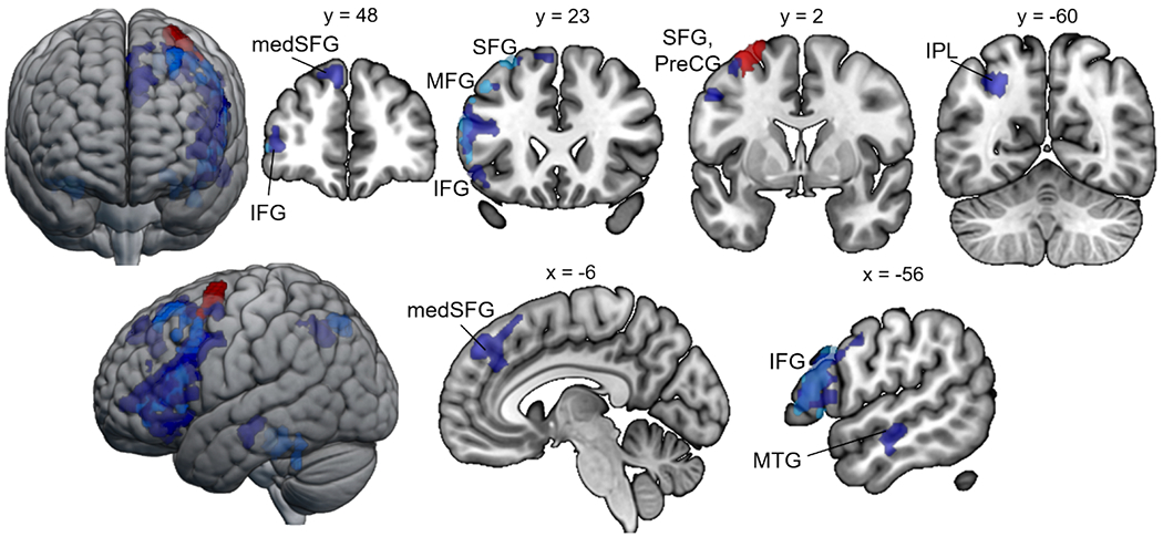
Increased activation after tDCS. Post > pre comparisons within groups. Light blue = sham post > pre; red = sensorimotor post > pre; blue = cognitive post > pre.
The cognitive tDCS group showed greater activation post-tDCS in left frontal regions, including the inferior frontal gyrus, middle frontal gyrus, and medial superior frontal gyrus, overlapping with the regions showing practice-associated changes in the sham group (Fig. 10). While the cognitive group showed more extensive activation of these regions, direct comparisons between groups for the Cognitivepost>pre vs. Shampost>pre were underpowered to meet the FDR-cluster corrected statistical threshold (see unthresholded maps in Supplemental Fig 1). The sham and cognitive groups also showed increased engagement of a similar region of the superior parietal lobule, and the cognitive group additionally showed increased inferior parietal and middle temporal activation post-tDCS. Notably, these clusters were all left-lateralized and overlapped with the task-associated regions identified pre-tDCS across the groups. There were no reductions in activation (pre-tDCS > post-tDCS) in the cognitive group. These findings suggest that neuromodulation of the posterolateral cerebellum might improve performance through increased engagement of the left-lateralized regions involved in the sentence completion task.
TDCS targeting the sensorimotor cerebellum also increased activation in the left MFG post-tDCS (Table 3, Fig. 10), although this cluster did not extend to the IFG as in the cognitive and sham groups. Moreover, this cluster was located posterior to the regions showing increases in the cognitive condition. Unthresholded maps (see Supplemental Figure 1) of the direct comparison between sensorimotor and sham groups (Sensorimotorpost>pre vs. Shampost>pre) indicate greater signal change in right frontal regions in the sensorimotor relative to the sham group. Post-tDCS decreases in the sensorimotor group were evident in the right IPL / supramarginal gyrus (SMG), which overlapped with the cluster showing decreased engagement in the sham group during the second run of the task (Table 3, Fig 11).
Figure 11.
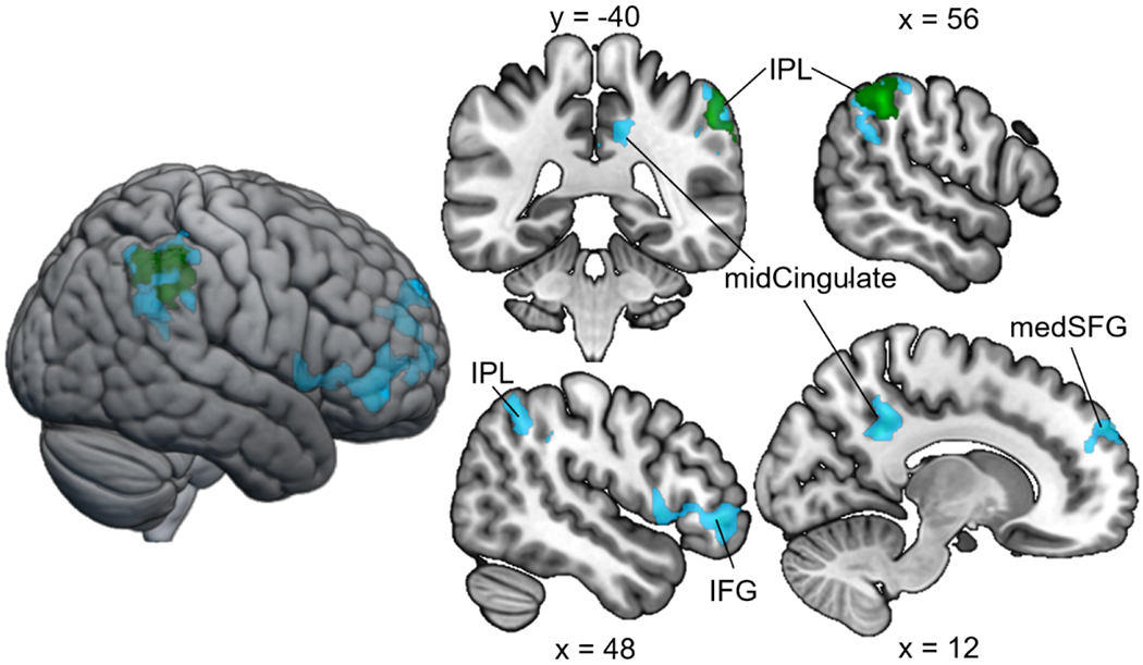
Decreased activation following tDCS. Pre > post comparisons within groups. Light blue = sham pre > post; green = sensorimotor pre > post.
Discussion
Our goal was to determine the impact of tDCS targeting different cerebellar regions on behavioral performance and activation patterns during a sentence completion task. TDCS targeting the right posterolateral cognitive cerebellum improved accuracy and increased activation in task-relevant cortical regions, whereas tDCS targeting the anterior sensorimotor cerebellum had no significant impact on task performance or the engagement of left cortical language networks. Our results are in line with the well-established functional topography framework of cerebellar function, whereby the anterior lobe plays a primary role in sensorimotor function and lateral regions of the posterior lobe in cognitive and affective processing (Stoodley & Schmahmann, 2009; King et al., 2019). These findings also add to the broader literature supporting a role for the right posterolateral cerebellum in language processing, including semantic prediction (Pope and Miall, 2012; Moberget et al., 2014; Mariën et al., 2014; D’Mello et al., 2017; Lesage et al., 2017).
These findings are also consistent with previous work reporting a dissociation between the effect of tDCS targeting the anterior cerebellum on articulation (impaired articulation rate) and right posterolateral cerebellar tDCS on verbal fluency (improved verbal fluency; Turkeltaub et al., 2016). Similarly, the present study showed differential effects of regional cerebellar tDCS on sentence completion, with tDCS targeting right lobule VII improving accuracy relative to the sham group, while tDCS targeting the anterior cerebellum had no effect on performance. Contrary to our prediction, there were no significant effects of tDCS on response time in either group.
TDCS targeting sensorimotor vs. cognitive cerebellar regions also differentially impacted task activation patterns. Following cognitive cerebellar tDCS, there was increased activation in task-relevant left-lateralized frontal, temporal, and parietal cortices, and there were no statistically significant decreases in activation from pre- to post-tDCS. In contrast, the group receiving tDCS targeting the sensorimotor cerebellum did not show a significant increase in activation in task-relevant brain regions following tDCS. The one cluster showing increased signal post-tDCS in the sensorimotor group was adjacent to the superior frontal regions showing increased activation in the cognitive group, but extended posteriorly into the precentral gyrus. This differential modulation of language and reading-related left-hemisphere cortical regions may underlie the observed behavioral dissociations on the sentence completion task between the active tDCS groups.
Cerebellar internal models.
It has been proposed that the cerebellum contributes to both motor and cognitive tasks via building internal models of information that enable prediction (e.g. Moberget and Ivry, 2016; Sokolov et al., 2017). Our results are consistent with previous studies showing that neuromodulation of the right posterolateral cerebellum impacts linguistic prediction (Lesage et al., 2012; Miall et al., 2016). However, there were no significant 3-way interactions between tDCS group, sentence type, and time point, indicating that the impact of tDCS on task performance was not specific to the predictive sentences. That said, it is important to note that “nonpredictive” sentences also require linguistic prediction. In imaging studies using this or a similar task, there is robust right cerebellar lobule VII activation associated with both sentence types (Moberget et al., 2014; D’Mello et al., 2017), with specific increase in signal in right lobule VII during strongly predictive sentences (D’Mello et al., 2017; Lesage et al., 2017). Further, previous work has shown that right posterolateral cerebellar tDCS specifically increased activation in right lobule VII during predictive sentence processing (D’Mello et al., 2017). Taken together, our findings that tDCS targeting right lobule VII improved performance on this linguistic prediction task are consistent with theoretical concepts that cerebellar internal models represent both motor and cognitive information (e.g. Ito, 2008).
Cerebellar modulation of broader brain networks.
Our results are also consistent with the concept that the cerebellum exerts its impact via modulation of cortical regions. The right posterolateral cerebellum is part of established language networks (see Price et al., 2012) and tDCS targeting right cerebellar lobule VII increased resting state functional connectivity within cortical language networks (Turkeltaub et al., 2016; D’Mello et al., 2017). We did not see any statistically significant BOLD signal changes within the cerebellum following tDCS, but there were changes in the engagement of cortical language regions in the cognitive tDCS group, including regions involved in semantic (e.g. Binder et al., 2009) and predictive language processing (e.g. Willems et al., 2016). Specifically, tDCS targeting the right posterolateral cerebellum altered task activation patterns in cerebral cortical areas that are functionally connected to right lobule VII, indicating cerebellar modulation of these cortical regions. With the exception of the left middle temporal cluster that showed increased signal in the cognitive group post-tDCS, all of the clusters of increased activation in the cognitive group fell within the resting state network for RVII (see Fig 12).
Fig 12. Cognitive post > pre-tDCS activation in the context of resting state connectivity of right cerebellar lobule VII.

Pre-tDCS resting state connectivity from right VII seed in all participants is shown in orange-yellow (p<0.001, FDR p<0.05 cluster correction; n=44 participants). Blue = Cognitive group post > pre-tDCS (t 3.17, FDR p<0.05 cluster correction).
The cerebellum also plays an important role in regulating attentional mechanisms of stimulus orientation and inhibitory control (Mannarelli et al., 2020). It is possible that the posterolateral regions of the cerebellum that are involved in dorsal and ventral attention networks were modulated with the RVII montage (see Buckner et al., 2011), and that attentional modulation may underlie the observed neural and behavioral effects.
Clinical implications.
The impact of right posterolateral cerebellar tDCS on language task performance and activation within language networks has potential implications for the treatment of language disorders. In particular, it has been proposed that cerebellar neuromodulation could be a potential therapeutic option for aphasia following left cortical hemisphere stroke (e.g. Turkeltaub et al., 2016), due to the structural and functional connectivity between the right cerebellum and left hemisphere language networks. Thus far, there have been mixed results in clinical studies, with some studies reporting improvements in spelling (Sebastian et al., 2017), naming (Sebastian et al., 2020), and verb generation (Marangolo et al., 2018), while others have not found significant treatment effects (DeMarco et al., in press). The impact of factors such as patient characteristics, electrode montage, duration, intensity and polarity of cerebellar tDCS need to be more thoroughly investigated before translation to the clinic.
The differential impact of the two electrode montages used here also has clinical implications, given the potential applications of cerebellar tDCS for a range of clinical conditions (e.g. see Ferrucci et al., 2016 for review). Our findings suggest that we were able to broadly impact different cerebellar regions with electrode montages that were based on prior studies reporting effects on motor (e.g. Galea et al., 2009) vs. cognitive (e.g. Pope and Miall, 2012) task performance. That said, we do not expect that we would be able to, for example, modulate specific subregions of lobule VII using 5x5cm electrodes. Such specific modulation of cerebro-cerebellar circuits might be advantageous in targeting, for example, the fronto-parietal and default mode networks for the treatment of different clinical conditions. The differential effects of our two electrode montages might also explain how small changes in electrode placement could lead to the sometimes variable effects of cerebellar tDCS on behavior that are reported in the literature (see Grimaldi et al., 2016; Moussa-Tooks et al., 2020).
Limitations.
The present study comes with several limitations. First, the sample size is relatively small and increases in sample size would likely increase statistical power to detect differences in the sensorimotor group relative to the sham group and to evaluate specific hypotheses about the role of the cerebellum in linguistic prediction. Second, although the electrode montages for the cognitive and sensorimotor positions yielded a dissociation in behavioral performance and neural activation patterns at the whole brain level, the size of the electrodes were not optimal for targeting specific cerebellar subregions. Future studies should utilize a combination of fMRI, electric field modeling, neuronavigation, HD-tDCS and/or concentric ring electrode montages to enable more precise anatomical and functional targeting of the cerebellum (Alam et al., 2016). Third, while our findings indicate that cerebellar modulation impacts both behavior and neural activation in functionally-connected regions of the cerebral cortex, this study does not provide information regarding the specific mechanism of action of cerebellar tDCS. Because we did not have a group receiving cathodal tDCS, we were unable to evaluate polarity-specific effects of cerebellar tDCS, although a recent meta-analysis found limited evidence for polarity-specific impacts of cerebellar tDCS on behavior (Oldratti and Schutter, 2018). Finally, the present study included a cognitive behavioral task, but did not include a sensorimotor task. Inclusion of both a language and motor task would allow for examination of a potential double dissociation of cerebellar tDCS on behavior, whereby the impact of tDCS targeting the sensorimotor cerebellum would be expected to influence the motor task, but have limited effects on the cognitive task (as shown here), and modulation of the cognitive cerebellar target would have the opposite effect on task performance.
Conclusions.
Here we show that tDCS targeting sensorimotor vs. cognitive cerebellar subregions differentially impacts behavioral task performance and functional activation patterns in healthy young adults. TDCS targeting the right posterolateral cerebellum improved accuracy on the sentence completion task and increased activation within task-relevant regions that are functionally connected with the cerebellar target. When the sensorimotor cerebellum was targeted, there were no effects on either task performance or increased engagement of task-relevant cortical language areas. Taken together, our findings indicate that the cerebellum modulates task performance through its long-range connectivity with specific cerebro-cerebellar networks.
Supplementary Material
Fig 8. Median response times (msec) across groups and time point.
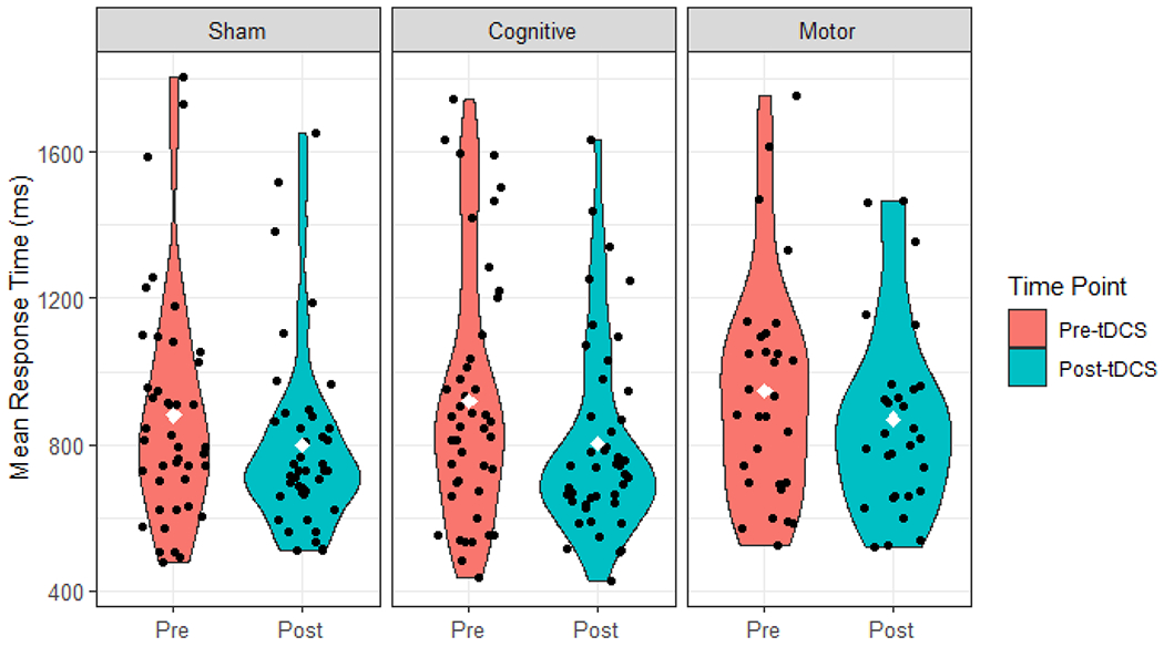
The violin plots show the distribution of the data, black dots represent individual participants, and white diamonds indicate median response times.
Highlights.
Different cerebellar subregions support motor and cognitive task performance
We tested behavioral and neural effects of transcranial direct current stimulation (tDCS) targeting cerebellar subregions
Posterolateral cerebellar tDCS improved language task performance and increased activation in left cortical regions
Sensorimotor cerebellar tDCS had no impact on task performance and disrupted task-relevant activation increases
Acknowledgments
This work was supported by the National Institutes of Health (R15MH106957 and R21DC014087) and doctoral student fellowships from American University to LCR and AMD. We would like to thank Stephanie Martin, Elizabeth Dvorak, Marissa Marko, Brianne Drury, and Caitlin Barrett for help with data collection, and Betty Malloy, Andrew DeMarco, and Peter Turkeltaub for helpful discussion.
Footnotes
Publisher's Disclaimer: This is a PDF file of an unedited manuscript that has been accepted for publication. As a service to our customers we are providing this early version of the manuscript. The manuscript will undergo copyediting, typesetting, and review of the resulting proof before it is published in its final form. Please note that during the production process errors may be discovered which could affect the content, and all legal disclaimers that apply to the journal pertain.
Declaration of interest: None
References
- Alam M, Truong DQ, Khadka N, Bikson M. Spatial and polarity precision of concentric high-definition transcranial direct current stimulation (HD-tDCS). Phys Med Biol. 2016June21;61(12):4506–21. [DOI] [PubMed] [Google Scholar]
- Binder JR, Desai RH, Graves WW, Conant LL. Where is the semantic system? A critical review and meta-analysis of 120 functional neuroimaging studies. Cereb Cortex. 2009. December;19(12):2767–96. [DOI] [PMC free article] [PubMed] [Google Scholar]
- Buckner RL, Krienen FM, Castellanos A, Diaz JC, Yeo BTT. The organization of the human cerebellum estimated by intrinsic functional connectivity. J Neurophysiol. 2011November;106(5):2322–45. [DOI] [PMC free article] [PubMed] [Google Scholar]
- DeMarco A, Dvorak E, Lacey E, Stoodley CJ, Turkeltaub PE. An exploratory study of cerebellar transcranial direct current stimulation in chronic stroke aphasia. In press, Cogn Behav Neurology. [DOI] [PMC free article] [PubMed] [Google Scholar]
- D’Mello AM, Gabrieli JDE, Nee DE. Evidence for hierarchical cognitive control in the human cerebellum. Curr Biol. 2020May18;30(10):1881–1892.e3. [DOI] [PMC free article] [PubMed] [Google Scholar]
- D’Mello AM, Turkeltaub PE, Stoodley CJ. Cerebellar tDCS Modulates Neural Circuits during Semantic Prediction: A Combined tDCS-fMRI Study. J Neurosci. 201708;37(6):1604–13. [DOI] [PMC free article] [PubMed] [Google Scholar]
- Desmond JE, Chen SHA, Shieh PB. Cerebellar transcranial magnetic stimulation impairs verbal working memory. Ann Neurol. 2005. October;58(4):553–60. [DOI] [PubMed] [Google Scholar]
- Diedrichsen J, King M, Hernandez-Castillo C, Sereno M, Ivry RB. Universal Transform or multiple functionality? Understanding the contribution of the human cerebellum across task domains. Neuron. 201905;102(5):918–28. [DOI] [PMC free article] [PubMed] [Google Scholar]
- Diedrichsen J, Balsters JH, Flavell J, Cussans E, Ramnani N. A probabilistic MR atlas of the human cerebellum. Neuroimage. 2009May15;46(1):39–46. [DOI] [PubMed] [Google Scholar]
- Escelsior A, Belvederi Murri M, Calcagno P, Cervetti A, Caruso R, Croce E, et al. Effectiveness of cerebellar circuitry modulation in Schizophrenia: A systematic review. J Nerv Ment Dis. 2019November;207(11):977–86. [DOI] [PubMed] [Google Scholar]
- Ferrucci R, Cortese F, Priori A. Cerebellar tDCS: How to do it. Cerebellum. 2015. February;14(1):27–30. [DOI] [PMC free article] [PubMed] [Google Scholar]
- Ferrucci R, Bocci T, Cortese F, Ruggiero F, Priori A. Cerebellar transcranial direct current stimulation in neurological disease. Cerebellum Ataxias. 2016. September; 3:16. [DOI] [PMC free article] [PubMed] [Google Scholar]
- Galea JM, Jayaram G, Ajagbe L, Celnik P. Modulation of cerebellar excitability by polarity-specific noninvasive direct current stimulation. J Neurosci. 2009. July 15;29(28):9115–22. [DOI] [PMC free article] [PubMed] [Google Scholar]
- Grimaldi G, Argyropoulos GP, Bastian A, Cortes M, Davis NJ, Edwards DJ, et al. Cerebellar transcranial Direct Current Stimulation (ctDCS): A novel approach to understanding cerebellar function in health and disease. Neuroscientist. 2016February;22(1):83–97. [DOI] [PMC free article] [PubMed] [Google Scholar]
- Halko MA, Farzan F, Eldaief MC, Schmahmann JD, Pascual-Leone A. Intermittent theta-burst stimulation of the lateral cerebellum increases functional connectivity of the default network. J Neurosci. 2014September3;34(36):12049–56. [DOI] [PMC free article] [PubMed] [Google Scholar]
- Heleven E, van Dun K, Van Overwalle F. The posterior cerebellum is involved in constructing social action sequences: An fMRI study. Sci Rep. 2019December;9(1):11110. [DOI] [PMC free article] [PubMed] [Google Scholar]
- Hiraoka K, Sugiyama K, Abe K. Effects of transcranial magnetic stimulation over the cerebellum on triphasic electromyographic pattern. Int J Neurosci. 2009;119(10):1523–37. [DOI] [PubMed] [Google Scholar]
- Ito M Control of mental activities by internal models in the cerebellum. Nat Rev Neurosci. 2008. April;9(4):304–13. [DOI] [PubMed] [Google Scholar]
- Kelly E, Meng F, Fujita H, Morgado F, Kazemi Y, Rice LC, et al. Regulation of autism-relevant behaviors by cerebellar-prefrontal cortical circuits. Nat Neurosci. 2020. September;23(9):1102–10. [DOI] [PMC free article] [PubMed] [Google Scholar]
- Kelly RM, Strick PL. Cerebellar loops with motor cortex and prefrontal cortex of a nonhuman primate. J Neurosci. 2003. September 10;23(23):8432–44. [DOI] [PMC free article] [PubMed] [Google Scholar]
- Kerren-Happuch E, Chen S-HA, Ho M-HR, Desmond JE. A meta-analysis of cerebellar contributions to higher cognition from PET and fMRI studies. Hum Brain Mapp. 2014February;35(2):593–615. [DOI] [PMC free article] [PubMed] [Google Scholar]
- Kessler SK, Turkeltaub PE, Benson JG, Hamilton RH. Differences in the experience of active and sham transcranial direct current stimulation. Brain Stimul. 2012April;5(2):155–62. [DOI] [PMC free article] [PubMed] [Google Scholar]
- King M, Hernandez-Castillo CR, Poldrack RA, Ivry RB, Diedrichsen J. Functional boundaries in the human cerebellum revealed by a multi-domain task battery. Nat Neurosci. 2019August;22(8):1371–8. [DOI] [PMC free article] [PubMed] [Google Scholar]
- Lesage E, Hansen PC, Miall RC. Right lateral cerebellum represents linguistic predictability. J Neurosci. 2017. June 28;37(26):6231–41. [DOI] [PMC free article] [PubMed] [Google Scholar]
- Lesage E, Morgan BE, Olson AC, Meyer AS, Miall RC. Cerebellar rTMS disrupts predictive language processing. Curr Biol. 2012September25;22(18):R794–795. [DOI] [PMC free article] [PubMed] [Google Scholar]
- Lindeman S, Kros L, Hong S, Mejias JF, Romano V, Negrello M, et al. Cerebellar Purkinje cells can differentially modulate coherence between sensory and motor cortex depending on region and behavior. Proc Natl Acad Sci USA. 2021. January 25; 118(2): e2015292118. [DOI] [PMC free article] [PubMed] [Google Scholar]
- Marangolo P, Fiori V, Caltagirone C, Pisano F, Priori A. Transcranial cerebellar direct current stimulation enhances verb generation but not verb naming in poststroke aphasia. J Cogn Neurosci. 2018February;30(2):188–99. [DOI] [PubMed] [Google Scholar]
- Mannarelli D, Pauletti C, Petritis A et al. Effects of cerebellar tDCS on inhibitory control: Evidence from a Go/NoGo task. Cerebellum. 2020July;14(19):788–798. [DOI] [PMC free article] [PubMed] [Google Scholar]
- Mariën P, Ackermann H, Adamaszek M, Barwood CHS, Beaton A, Desmond J, et al. Consensus paper: Language and the cerebellum: an ongoing enigma. Cerebellum. 2014June;13(3):386–410. [DOI] [PMC free article] [PubMed] [Google Scholar]
- McAfee SS, Liu Y, Sillitoe RV, Heck DH. Cerebellar lobulus simplex and Crus I differentially represent phase and phase difference of prefrontal cortical and hippocampal oscillations. Cell Rep. 2019May21;27(8):2328–2334.e3. [DOI] [PMC free article] [PubMed] [Google Scholar]
- Miall RC, Antony J, Goldsmith-Sumner A, Harding SR, McGovern C, Winter JL. Modulation of linguistic prediction by TDCS of the right lateral cerebellum. Neuropsychologia. 2016. June;86:103–9. [DOI] [PMC free article] [PubMed] [Google Scholar]
- Miall RC, Weir DJ, Wolpert DM, Stein JF. Is the cerebellum a Smith predictor? J Mot Behav. 1993September;25(3):203–16. [DOI] [PubMed] [Google Scholar]
- Minichino A, Bersani FS, Bernabei L, Spagnoli F, Vergnani L, Corrado A, et al. Prefronto-cerebellar transcranial direct current stimulation improves visuospatial memory, executive functions, and neurological soft signs in patients with euthymic bipolar disorder. Neuropsychiatr Dis Treat. 2015;11:2265–70. [DOI] [PMC free article] [PubMed] [Google Scholar]
- Miterko LN, Baker KB, Beckinghausen J, Bradnam LV, Cheng MY, Cooperrider J, et al. Consensus Paper: Experimental neurostimulation of the cerebellum. Cerebellum. 2019December;18(6):1064–97. [DOI] [PMC free article] [PubMed] [Google Scholar]
- Moberget T, Ivry RB. Cerebellar contributions to motor control and language comprehension: searching for common computational principles. Ann N Y Acad Sci. 2016April;1369(1):154–71. [DOI] [PMC free article] [PubMed] [Google Scholar]
- Moberget T, Gullesen EH, Andersson S, Ivry RB, Endestad T. Generalized role for the cerebellum in encoding internal models: evidence from semantic processing. J Neurosci. 2014February19;34(8):2871–8. [DOI] [PMC free article] [PubMed] [Google Scholar]
- Moussa-Tooks AB, Burroughs LP, Rejimon AC, Cheng H, Hetrick WP. Cerebellar tDCS consistency and metabolite changes: A recommendation to decrease barriers to replicability. Brain Stimul. 2020. December;13(6):1521–3. [DOI] [PMC free article] [PubMed] [Google Scholar]
- Oldrati V, Schutter DJLG. Targeting the human cerebellum with transcranial direct current stimulation to modulate behavior: A meta-analysis. Cerebellum. 2018. 17:228–236. [DOI] [PMC free article] [PubMed] [Google Scholar]
- Popa LS, Streng ML, Hewitt AL, Ebner TJ. The errors of our ways: Understanding error representations in cerebellar-dependent motor learning. Cerebellum. 2016. April;15(2):93–103. [DOI] [PMC free article] [PubMed] [Google Scholar]
- Pope PA, Miall RC. Task-specific facilitation of cognition by cathodal transcranial direct current stimulation of the cerebellum. Brain Stimulation. 2012. April;5(2):84–94. [DOI] [PMC free article] [PubMed] [Google Scholar]
- Price CJ. A review and synthesis of the first 20 years of PET and fMRI studies of heard speech, spoken language and reading. Neuroimage. 2012. August 15;62(2):816–47. [DOI] [PMC free article] [PubMed] [Google Scholar]
- Ramaraju S, Roula MA, McCarthy PW. Modelling the effect of electrode displacement on transcranial direct current stimulation (tDCS). J Neural Eng. 2018February1;15(1):016019. [DOI] [PubMed] [Google Scholar]
- Ramnani N. Automatic and controlled processing in the corticocerebellar system. Prog Brain Res. 2014;210:255–85. [DOI] [PubMed] [Google Scholar]
- Raymond JL, Medina JF. Computational principles of supervised learning in the cerebellum. Annu Rev Neurosci. 2018. July 8;41(1):233–53. [DOI] [PMC free article] [PubMed] [Google Scholar]
- Schmahmann JD. Disorders of the cerebellum: ataxia, dysmetria of thought, and the cerebellar cognitive affective syndrome. J Neuropsychiatry Clin Neurosci. 2004;16(3):367–78. [DOI] [PubMed] [Google Scholar]
- Schmahmann JD, Guell X, Stoodley CJ, Halko MA. The theory and neuroscience of cerebellar cognition. Annu Rev Neurosci. 2019. July 8;42:337–64. [DOI] [PubMed] [Google Scholar]
- Sebastian R, Kim JH, Brenowitz R, Tippett DC, Desmond JE, Celnik PA, et al. Cerebellar neuromodulation improves naming in post-stroke aphasia. Brain Commun. 2020;2(2):fcaa179. [DOI] [PMC free article] [PubMed] [Google Scholar]
- Sebastian R, Saxena S, Tsapkini K, Faria AV, Long C, Wright A, et al. Cerebellar tDCS: A novel approach to augment language treatment post-stroke. Front Hum Neurosci. 2017;10:695. [DOI] [PMC free article] [PubMed] [Google Scholar]
- Sheu Y-S, Liang Y, Desmond JE. Disruption of cerebellar prediction in verbal working memory. Front Hum Neurosci. 2019;13:61. [DOI] [PMC free article] [PubMed] [Google Scholar]
- Sokolov AA. The cerebellum in social cognition. Front Cell Neurosci. 2018. June 5;12:145. [Google Scholar]
- Sokolov AA, Miall RC, Ivry RB. The cerebellum: Adaptive prediction for movement and cognition. Trends Cog Sci. 2017. May;21(5):313–32. [DOI] [PMC free article] [PubMed] [Google Scholar]
- Stoodley CJ. The cerebellum and cognition: evidence from functional imaging studies. Cerebellum. 2012. June;11(2):352–65. [DOI] [PubMed] [Google Scholar]
- Stoodley CJ, Schmahmann JD. Functional topography in the human cerebellum: a meta-analysis of neuroimaging studies. Neuroimage. 2009. January 15;44(2):489–501. [DOI] [PubMed] [Google Scholar]
- Stoodley CJ, D’Mello AM, Ellegood J, Jakkamsetti V, Liu P, Nebel MB, et al. Altered cerebellar connectivity in autism and cerebellar-mediated rescue of autism-related behaviors in mice. Nat Neurosci. 2017December;20(12):1744–51. [DOI] [PMC free article] [PubMed] [Google Scholar]
- Stoodley CJ, Valera EM, Schmahmann JD. Functional topography of the cerebellum for motor and cognitive tasks: an fMRI study. Neuroimage. 2012. January 16;59(2):1560–70. [DOI] [PMC free article] [PubMed] [Google Scholar]
- Thielscher A, Antunes A, Saturnino GB. Field modeling for transcranial magnetic stimulation: A useful tool to understand the physiological effects of TMS? Annu Int Conf IEEE Eng Med Biol Soc. 2015;2015:222–5. [DOI] [PubMed] [Google Scholar]
- Turkeltaub PE, Swears MK, D’Mello AM, Stoodley CJ. Cerebellar tDCS as a novel treatment for aphasia? Evidence from behavioral and resting-state functional connectivity data in healthy adults. Restor Neurol Neurosci. 2016. August 13;34(4):491–505. [DOI] [PMC free article] [PubMed] [Google Scholar]
- Van Overwalle F, Manto M, Cattaneo Z, Clausi S, Ferrari C, Gabrieli JDE, et al. Consensus Paper: Cerebellum and social cognition. Cerebellum. 2020. December;19(6):833–68. [DOI] [PMC free article] [PubMed] [Google Scholar]
- Van Overwalle F, Baetens K, Mariën P, Vandekerckhove M. Social cognition and the cerebellum: a meta-analysis of over 350 fMRI studies. Neuroimage. 2014. February 1;86:554–72. [DOI] [PubMed] [Google Scholar]
- Whitfield-Gabrieli S, Nieto-Castanon A. Conn: A functional connectivity toolbox for correlated and anticorrelated brain networks. Brain Connect. 2012;2(3):125–41. [DOI] [PubMed] [Google Scholar]
- Willems RM, Frank SL, Nijhof AD, Hagoort P, van den Bosch A. Prediction during natural language comprehension. Cereb Cortex. 2016. June;26(6):2506–16. [DOI] [PubMed] [Google Scholar]
- Wolpert DM, Miall RC, Kawato M. Internal models in the cerebellum. Trends Cog Sci. 1998. September;2(9):338–47. [DOI] [PubMed] [Google Scholar]
- Woods AJ, Antal A, Bikson M, Boggio PS, Brunoni AR, Celnik P, et al. A technical guide to tDCS, and related non-invasive brain stimulation tools. Clin Neurophysiol. 2016. February;127(2):1031–48. [DOI] [PMC free article] [PubMed] [Google Scholar]
Associated Data
This section collects any data citations, data availability statements, or supplementary materials included in this article.