Lee and Ding highlight work from Hao et al. describing that membrane-bound stem cell factor synergizes with VCAM-1 to induce polarized protrusions that regulate adhesion in hematopoietic stem cells.
Abstract
Multipotent hematopoietic stem cells are maintained by the bone marrow niche, but how niche-derived membrane-bound stem cell factor (mSCF) regulates HSCs remains unclear. In this issue, Hao et al. (2021. J. Cell Biol. https://doi.org/10.1083/jcb.202010118) describe that mSCF, synergistically with VCAM-1, induces large, polarized protrusions that serve as anchors for HSCs to their niche.
Hematopoietic stem cells (HSCs) generate all blood and immune cells throughout life via self-renewal and multilineage differentiation within the bone marrow niche. HSCs are the basis for bone marrow transplantation, saving thousands of lives yearly. The bone marrow niche often serves as a paradigm for studying stem cell biology. In addition, elucidating the underlying mechanism in the niche helps devise strategies to expand functional HSCs for clinical use. Within the niche, leptin receptor–positive perisinusoidal stromal cells and endothelial cells are the major source of essential cytokines for HSC maintenance, including vascular cell adhesion molecule 1 (VCAM-1) and stem cell factor (SCF; 1, 2). Locally produced soluble and membrane-bound cytokines preserve the unique localization and anchorage of HSCs to stromal cells within their niche. Consistent with this notion, mouse genetic data have shown that membrane-bound SCF (mSCF) is important for HSC maintenance in vivo (3). However, given that both soluble and membrane-bound forms of SCF can engage with the cognate cKIT receptors, the mechanisms by which mSCF sustains HSCs function in vivo remain elusive. Likewise, it is unclear why the expansion and maintenance of HSCs ex vivo by adding SCF to culture as an either soluble or immobilized form has only been achieved with limited success.
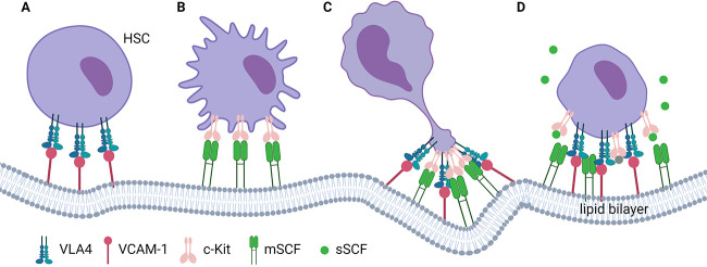
In this issue, Hao et al. addressed this question by using a supported lipid bilayer (SLB) system to model the interaction between HSCs and membrane-bound cytokines, including SCF (4). SLBs present an advantage over conventional immobilization methods; they allow the lateral mobility of membrane-bound proteins and clustering of receptors and signaling complexes, thus resembling the lipid bilayer of plasma membrane in vivo. Focusing on HSC cytokines that may be presented as membrane-bound forms in the bone marrow niche, the authors performed an imaging screen in vitro using SLBs and found that mSCF but not soluble SCF (sSCF) induced mSCF/cKIT clustering and the formation of membrane protrusions on HSCs. While mSCF alone was sufficient to promote cell protrusions, HSCs required both mSCF and VCAM-1 for large, polarized protrusions. They followed HSCs at different time points after exposure to VCAM-1 and mSCF by scanning electron microscopy and observed that HSCs first formed diffuse mSCF clusters and multifocal thin protrusions and then proceeded to a polarized, clustered morphology with larger and thicker protrusions. Using a controlled sheer stress device, Hao et al. showed that these polarized protrusions had a functional consequence on the adhesion strength of HSCs. mSCF and VCAM-1 dramatically increased the adhesion of HSCs to SLB compared with VCAM-1 or mSCF alone. Interestingly, the effect was more prominent in HSCs compared with their immediate downstream progenies, multipotent progenitors. This phenotype was also specific to ligands presented on SLB because the effect was canceled when the cytokines were directly immobilized onto the glass surface. Then, they had a close look into the cytoskeletal organization of HSCs in the presence of both mSCF and VCAM-1 on SLB. They found that F-actin and myosin IIa concentrated at the protrusion, which led them to speculate that the cytoskeleton remodeling mediates the formation of the polarized morphology. Indeed, chemical inhibitors blocking myosin contraction, actin polymerization, or Rho-associated protein kinase disrupted the formation of the large and polarized protrusion. The authors noted that phosphatidylinositol 3-kinase (PI3K) also localized with mSCF/cKIT clusters, so they further assessed the contribution of the PI3K/Akt pathway to the polarized morphology of HSCs by using total internal reflection fluorescence microscopy and PI3K and Akt chemical inhibitors. PI3K/Akt activation contributed downstream of the mSCF–VCAM-1 synergy to regulating HSC cell adhesion and polarized mSCF/cKIT distribution. In addition, PI3K signaling enhanced the nuclear retention of FOXO3a, a crucial factor for HSC self-renewal; this enhancement was induced by mSCF but lessened by sSCF. Intriguingly, sSCF also competed with mSCF and abrogated the effect of the mSCF–VCAM-1 synergy on polarized protrusion formation. However, whether and how PI3K transmits the mSCF–VCAM-1 synergy into proliferation or quiescence cues in HSCs requires further investigation. Taken together, these data suggest that mSCF and VCAM-1 synergize to induce polarized protrusions on HSCs, which regulates their adhesion to the niche (Fig. 1). These protrusions share many features with the immunological synapse (5), which points toward the existence of a similar model for stem cells, “stem cell synapse,” where HSCs interact with and receive a variety of signals from their niche cells.
Figure 1.
VCAM-1 and mSCF synergistically promote the formation of polarized protrusions (stem cell synapse) on HSCs. (A and B) VCAM-1 or mSCF alone does not induce apparent polarized morphology on HSCs. The signaling and adhesion of HSCs to the niche is not at its full potential. (C) VCAM-1 and mSCF together induce robust receptor clustering on HSCs, optimal signaling, and strong adhesion. (D) sSCF can competitively disrupt the polarized protrusions on HSCs. The figure was created with BioRender.com.
While the study by Hao et al. sheds light on how niche signals, particularly mSCF, regulate HSCs, several outstanding questions remain. First, even though many hematopoietic cells express cKIT (some of them even express higher levels than HSCs), HSCs respond to mSCF + VCAM-1 the strongest by recruiting the most mSCF to clusters. What is the specific mechanism in HSCs underlying this specificity? Second, SCF is produced both as mSCF and sSCF in vivo, through alternative splicing and proteolytic cleavage; if mSCF is mainly responsible for anchoring HSCs in the niche, what is the function of sSCF in vivo? Does sSCF modulate the available pool of mSCF? Third, robust maintenance of HSCs in culture has been challenging. HSCs can be maintained in a system composed of sSCF, thromopoietin (TPO), fibronectin, and polyvinyl alcohol (6). Tethering cytokines to SLB elicits more physiological response from HSCs compared with soluble cytokines or direct immobilization. Does SLB improve maintenance of HSCs in in vitro culture? Fourth, some cytokines, such as TPO, act on HSCs in a long-range manner (7). How do these systemic cytokines induce robust signaling in HSCs? Do they participate in the stem cell synapse even if they are not the initiators? Finally, do stem cells and their niche interact by forming similar synapses in other stem cell systems? Answering these questions will deepen our understanding of the stem cell niche and help integrate the niche component into potential, more successful applications in regenerative medicine.
Acknowledgments
This work was supported by the National Heart, Lung and Blood Institute (R01HL153487 and R01HL155868). L. Ding was supported by the Rita Allen Foundation, the Irma T. Hirschl Trust Research Award, the Schaefer Research Scholar Program, and the Leukemia and Lymphoma Society Scholar Award.
The authors declare no competing financial interests.
References
- 1.Ding, L., et al. 2012. Nature. 10.1038/nature10783 [DOI] [Google Scholar]
- 2.Morrison, S.J., and Scadden D.T.. 2014. Nature. 10.1038/nature12984 [DOI] [PMC free article] [PubMed] [Google Scholar]
- 3.Barker, J.E. 1994. Exp. Hematol. [Google Scholar]
- 4.Hao, J., et al. 2021. J. Cell Biol. 10.1083/jcb.202010118 [DOI] [Google Scholar]
- 5.Huppa, J.B., and Davis M.M.. 2003. Nat. Rev. Immunol. 10.1038/nri1245 [DOI] [PubMed] [Google Scholar]
- 6.Wilkinson, A.C., et al. 2020. Nat. Protoc. 10.1038/s41596-019-0263-2 [DOI] [PMC free article] [PubMed] [Google Scholar]
- 7.Decker, M., et al. 2018. Science. 10.1126/science.aap8861 [DOI] [Google Scholar]


