The thermography of the plantar skin has been used to assess the feet complications of the patient with Type 2 Diabetes Mellitus, T2DM,1-3 particularly, for the follow up of the treatments of the Peripheral Artery Diseases, PAD.4 Furthermore, the photoplethysmogram (PPG), may be used to observe the blood flow on the toes.5 Then, as a complement to these results, in this communication, the thermograms and the PPG of a female patient with T2DM, at the before and after revascularization surgery were compared with those of a female with no signs neither symptoms of PAD.
The thermograms, acquired with a Flir® E6 thermal camera, were used to quantify the average temperature of the angiosomes6 of the foot (Tang). While, the PPG were photographed from the screen of an Hergon® pulse oximeter, located on the second toe of the assessed foot. Since the thermography is totally not-invasive, and, the pulse oximeter is almost cero invasive, at the time of the data acquisition (ie, thermograms and PPG photograph), the consent was obtained verbally.
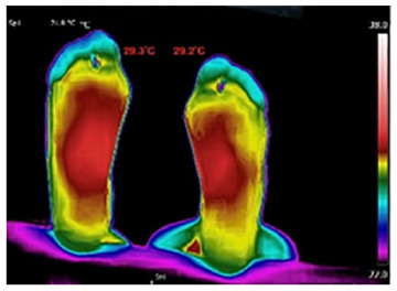
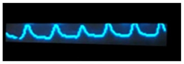
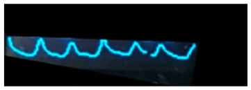
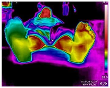
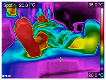
The results are resumed in the Table 1, firstly, the row (a) shows the thermogram of the plantar skin and the PPG of the healthy individual. The thermogram showed the gradual decrement of the temperature, from the middle arc to the toes and the heel. The Tang were 29.2°C and 29.3°C for the right and left foot, respectively. The PPG, for the second toe of each foot showed the rhythmic slopes (abrupt up and down smooth), and the same number of peaks. In the row (b), the thermograms and the PPGs are from the female patient with T2DM at the day before a revascularization surgery, the Tang difference, between the feet, is just 1.3°C, the PPG is almost flat in the affected foot and normal in the other one. Finally, after the revascularization surgery, the row (c) shows that, the Tang difference, between the feet, is 5.4°C and the PPGs are normal for both toes.
Table 1.
The Thermograms and the PPG Signals of (a) Healthy Female Individual, 55 years old, No Signs of Symptoms of PAD, and a Female Patient, 66 years old, with T2DM Established 18 years ago, (b) The Day Before, and (c) After the Revascularization Surgery.
| Thermogram | T(ang) (°C) right foot | T(ang) (°C) left foot | PPG right | PPG left | |
|---|---|---|---|---|---|
| a) |
|
29.3 | 29.2 |
|
|
| b) |
 Before surgery |
24.7 | 26.0 |

|

|
| c) |
 After surgery |
30.7 | 25.3 |

|

|
The findings presented here, even when they come from very specific PAD events, show objective and relevant information for the health professional, that is, as the protocol to get a thermogram requires that the patient lay down, for 10 to 15 minutes, and a little of thermal stress the feet, hence, this technique provides information about the central nervous and the peripheral vascular system.2 Furthermore, the PPG provides, specifically, evidence about the blood passing through the toes, very useful in the PAD. Thus, the evolution of the PAD can be assessed and followed up by these subclinical parameters (temperature distribution, the Tang and the form of the PPG). The main advantages of these techniques are, (1) qualitative, by knowing the temperature distribution and the form of the PPG, (2) quantitative, by the computation of the Tang, (3) no human-dependent.
Acknowledgments
The authors would like to thank to Dr. Hugo Moreno and Dr. Francisco Regalado from the Hospital General de Mexico, for their kindly comments on several aspects of this work.
Footnotes
Abbreviations: PAD, peripheral artery disease; PPG, photoplethysmogram; T2DM: type 2 diabetes mellitus.
Declaration of Conflicting Interests: The author(s) declared no potential conflicts of interest with respect to the research, authorship, and/or publication of this article.
Funding: The author(s) received no financial support for the research, authorship, and/or publication of this article.
ORCID iD: Francisco-J Renero-C  https://orcid.org/0000-0003-1273-4725
References
- 1.Ilo A, Romsi P, Mäkelä J.Infrared thermography and vascular disorders in diabetic feet. J Diabetes Sci Technol. 2020;14(1):28-36. doi: 10.1177/19322968198712 [DOI] [PMC free article] [PubMed] [Google Scholar]
- 2.Renero-C FJ.The thermoregulation of healthy individuals, overweight–obese, and diabetic from the plantar skin thermogram: a clue to predict the diabetic foot. Diabet Foot Ankle. 2017;8(1):1361298. doi: 10.1080/2000625X.2017.1361298 [DOI] [PMC free article] [PubMed] [Google Scholar]
- 3.Renero-C FJ.The abrupt temperature changes in the plantar skin thermogram of the diabetic patient: looking in to prevent the insidious ulcers. Diabet Foot Ankle. 2018;9(1):1430950. doi: 10.1080/2000625X.2018.1430950 [DOI] [PMC free article] [PubMed] [Google Scholar]
- 4.Ilo A, Romsi P, Pokela M, Mäkelä J.Infrared thermography follow-up after lower limb revascularization. J Diabet Sci Technol. Published online March 20, 2020. doi: 10.1177/19322968209123 [DOI] [PMC free article] [PubMed] [Google Scholar]
- 5.Kagaya Y, Ohura N, Suga H, Eto H, Takushima A, Harii K.‘Real angiosome’ assessment from peripheral tissue perfusion using tissue oxygen saturation foot-mapping in patients with critical limb ischemia. Eur J Vasc Endovasc Surg. 2014;47(4):433-441. doi: 10.1016/j.ejvs.2013.11.011 [DOI] [PubMed] [Google Scholar]
- 6.Attinger CE, Evans KK, Bulan E, Blume P, Cooper P.Angiosomes of the foot and ankle and clinical implications for limb salvage: reconstruction, incisions, and revascularization. Plast Reconstr Surg. 2006;117:261-276. [DOI] [PubMed] [Google Scholar]


