



Abstract
More than a decade ago, the ENCODE and NIH Epigenomics Roadmap consortia organized large multilaboratory efforts to profile the epigenomes of >110 different mammalian somatic cell types. This generated valuable publicly accessible datasets that are being mined to reveal genome-wide patterns of a variety of different epigenetic parameters. This consortia approach facilitated the powerful and comprehensive multiparametric integrative analysis of the epigenomes in each cell type. However, no germ cell types were included among the cell types characterized by either of these consortia. Thus, comprehensive epigenetic profiling data are not generally available for the most evolutionarily important cells, male and female germ cells. We discuss the need for reproductive biologists to generate similar multiparametric epigenomic profiling datasets for both male and female germ cells at different developmental stages and summarize our recent effort to derive such data for mammalian spermatogonial stem cells and progenitor spermatogonia.
Keywords: epigenetic profiling, chromatin, cell fate, spermatogonial stem cells, progenitor spermatogonia
The ENCODE and NIH Epigenome Roadmap consortia have generated valuable datasets profiling epigenomes of >110 different mammalian somatic cell types, but none of these have included profiles of even a single germ cell type.
In multicellular eukaryotic organisms such as mammals, differential cell fate and function are manifest on the basis of differential gene expression, which, in turn, is regulated by cell-type specific transcription factor networks functioning within differential epigenetic landscapes. The past decade has witnessed phenomenal progress in our ability to characterize genome-wide epigenetic programming and begin to understand how different epigenetic parameters and/or genomic elements work in tandem to initiate and sustain differential cell fates. At the most proximal level, gene expression is regulated by promoter regions with directly or indirectly bound protein factors and non-coding RNAs (ncRNAs) that influence the recruitment of RNA polymerase to initiate transcription [1]. In addition, most, if not all, promoter complexes are themselves regulated by interactions with enhancers that also bind factors and ncRNAs necessary to establish promoter-enhancer interactomes [2]. The epigenetic landscape consists of two-dimensional (2D) modifications to DNA or chromatin that influence interactions between sequence elements and bound factors/ncRNAs, along with three-dimensional (3D) interactions among these 2D features. Most of these interactions require favorable, decondensed/open/accessible chromatin states that provide DNA-binding factors access to their cognate binding sequences, ultimately facilitating formation of the RNA polymerase/preinitiation complex at the transcription start site. Chromatin accessibility is regulated by histone modifications and associated chromatin remodeling complexes as well as by DNA methylation patterns [3, 4]. Thus, epigenetic regulation of gene expression involves a collection of individual epigenomic parameters that together comprise a complex code that programs cell-type-specific gene expression patterns that dictate cell fate.
The ENCODE and NIH Roadmap Epigenomics and related projects have found that while there are only ~20,000 regulated protein-encoding genes in the mammalian genome, there are >1 million candidate enhancers in distal intergenic regions [5, 6]. This results in a “one to many” functional relationship between promoters and enhancers, with multiple enhancers interacting with each active promoter, and with distinct sets of enhancers driving the expression of distinct sets of genes in distinct cell types [7]. Importantly, enhancers often do not interact with promoters of immediately neighboring genes but rather skip over intermediary genes to interact with more distal target promoters [8]—hence, the need to directly assay promoter-enhancer interactions in each cell type. Once mapped, factors that either direct the formation, or interpret the programming of these interactions can be predicted on the basis of motif enrichment or de novo motif prediction methods [9, 10] and then directly validated by gene knockout), knockdown, or over-expression assays [6]. Single-cell methods have added high resolution to these assays [11], such that they are now being used by the International Human Epigenome Consortium (IHEC) to assess >1000 epigenomes in human somatic cells in a manner that will also describe the extent of heterogeneity within these processes (http://ihec-epigenomes.org/research/reference-epigenome-standards/).
These concepts have emerged from decades of research describing epigenetic mechanisms that impact gene transcription [12]. Initially, studies of epigenetic programming associated with regulation of gene expression focused on individual epigenetic parameters including DNA methylation, histone modifications, chromatin accessibility, and ncRNAs, among others. These studies were followed by large multi-institutional cooperative studies including the ENCODE [13] and NIH Epigenomics Roadmap [6] consortia, which characterized epigenomes of well over 100 different somatic cell types, as well as the ongoing IHEC Consortium (http://ihec-epigenomes.org) effort to describe >1000 human somatic epigenomes. These large-scale studies facilitated the parallel analysis of multiple epigenetic parameters simultaneously, which clearly revealed that in nature these parameters do not function in isolation. Rather they contribute to a complex combinatorial epigenetic code that directs the formation of at least 15 different chromatin states, which, in turn, regulate complex gene expression patterns dictating cell fate and function [5]. These chromatin states vary with respect to accessibility to the transcriptional machinery in ways that may distinguish highly active or terminally repressed transcription, or may differ in much more subtle ways to predispose the initiation of transcription or control different levels of ongoing transcription.
These findings have led to the emerging field of “multi-omics” and the multiparametric integrative analyses necessary to derive comprehensive descriptions of epigenetic landscapes associated with cell-type specific transcriptomes [11]. Analyses conducted as parallel assessments of individual parameters in a given cell type can now be performed simultaneously to assess epigenetic and gene expression parameters for individual cell types [11]. Interestingly, recent studies have suggested that epigenetic profiles associated with cell-type-specific gene expression patterns can become “primed” prior to the actual initiation of expression [14]. This may be manifest as the establishment of promoter-enhancer loops, or of partial epigenetic landscapes that, by themselves, are not sufficient to activate transcription, but are components of the final programming that is necessary to activate transcription. Such epigenetic priming is presumed to commit cells to specific developmental fates [14].
Strikingly, despite these extensive analyses of epigenetic programming in numerous different somatic cell types, neither the ENCODE nor NIH Roadmap Epigenomics consortia included analysis of even a single mammalian germ cell type. While it may seem reasonable to assume epigenetic mechanisms regulating differentiation-pathway specific gene expression patterns will be similar in germ and somatic cell types, there are unique features of epigenetic programming in germ cells. Thus, despite significant differentiation during gametogenesis, germ cells must retain the epigenetic potential to regulate transcriptomes needed to initiate the development of all cell types following fertilization. This raises questions about the extent to which epigenetic programming in germ cells may be similar to that in somatic cells. Of more direct clinical relevance, understanding how unique epigenetic programming directs specific germ cell fates and functions will contribute to the elucidation of mechanisms that may malfunction to cause infertility, or that may be manipulated to treat infertility or inhibit reproduction.
With respect to epigenetic programming, germ cells are the most dynamic cell type in the body. Their complex development transforms them into vehicles that transmit parent-specific epigenetic programs to the next generation. This involves waves of genome-wide reprogramming to globally reset inherited epigenetic programming [15], plus locus-specific reprogramming to direct sexually dimorphic differentiation patterns [16] and the unique requirements of meiosis and genetic recombination [17]. Finally, gametes are the mediators of inter- or transgenerational epigenetic inheritance. Thus, germ cell epigenomes represent the route by which environmental exposures or lifestyle choices in one generation can predispose adverse phenotypic effects such as susceptibility to disease in subsequent generations [18].
Bulk- and single-cell (sc)-RNA-sequencing (RNA-seq) studies of spermatogenic cells have included an emphasis on spermatogonial stem cells (SSCs) [19–28]. As demonstrated by Ralph Brinster’s seminal demonstration of spermatogonial transplantation [29–31], SSCs are pivotal to spermatogenesis. Initial specification of SSC fate establishes the renewable seminiferous epithelium. Conversely, the loss of SSC fate as SSCs give rise to progenitor spermatogonia marks commitment to spermatogenic differentiation. Recent advances in the ability to identify and selectively recover [32, 33] and culture [34] SSCs have facilitated expanded studies of their regenerative capacity [32]. At the same time, remarkably powerful methods to assess genome-wide patterns of gene expression [35, 36], and a variety of epigenetic parameters (“multi-omics”) at either the aggregate or single-cell level [11] have accelerated the ability to comprehensively characterize cell-type specific transcriptomes and epigenomes as exemplified by the ENCODE and Epigenomic Roadmap studies of somatic cell types noted above [5, 6]. Recently, we merged these advances to perform one of the first comprehensive multi-omics assessments of any mammalian germ cell type similar to those conducted in multiple somatic cell types by the previous consortia studies. Specifically, we exploited the ability to recover highly enriched populations of SSCs and progenitors along with the potential to conduct multi-omic epigenomic profiling to assess differential gene expression and associated epigenetic programming that distinguish these two spermatogonial subtypes. This exemplifies the benefit of applying ENCODE/Epigenomics Roadmap-like analysis to germ cells.
SSCs descend from non-self-renewing prospermatogonia and either self-renew to sustain the regenerative SSC population or give rise to non-self-renewing, non-regenerative progenitor spermatogonia that are primed to enter the spermatogenic differentiation pathway. The initial specification, subsequent maintenance during self-renewal, or loss coincident with entry into spermatogenic differentiation, of SSC cell fate forms the basis of spermatogenesis. These represent substantive transitions in spermatogonial subtype fate that are associated with differential gene expression plus accompanying differential epigenetic programming (Figure 1). Published RNA-seq databases chronicle differential gene expression associated with different spermatogonial subtype cell fates (Table 1). We complemented this spermatogonial-subtype-specific gene expression data with multi-omic analysis of DNA methylation, chromatin accessibility, and histone modification patterns to relate differential epigenetic programming to differential gene expression in SSCs and progenitor spermatogonia.
Figure 1.
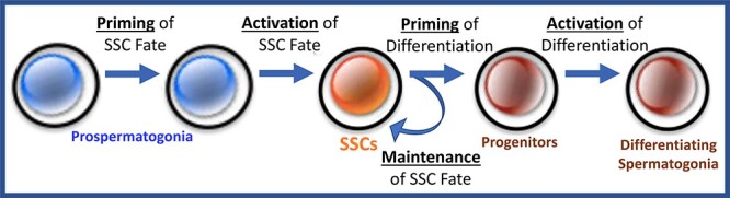
Specification, maintenance and loss of SSC fate. Suggested transitions in epigenetic programming associated with SSC fate and function.
Table 1.
Spermatogonial subtype-specific genes
| Spermatogonial subtype | # subtype-specific genes | Timing of activation |
|---|---|---|
| Foundational SSCs | 320 | P3-P6 |
| First-wave differentiating | 619 | P6-P8 |
| Steady-state SSCs | 257 | P30-P60 |
| Steady-state differentiating | 497 | P30-P60 |
Specifically, we conducted a parallel assessment of nine different parameters in aliquots of each cell sample, including gene expression by bulk and scRNA-seq, six different histone modifications by chromatin immunoprecipitation-sequencing (ChIP-seq), chromatin accessibility by the assay for transposase-accessible chromatin-sequencing (ATAC-seq), and DNA methylation by methylated DNA immunoprecipitation-sequencing (MeDIP-seq). Despite the very close lineage relationship of these two spermatogonial subtypes, we could detect 1131 statistically significant differentially expressed genes (DEGs) distinguishing SSCs and progenitors. For several of the epigenetic parameters we assessed in association with DEGs, we did not observe differences between the two spermatogonial subtypes. However, we did detect differences in the abundance of two histone modifications—histone H3, lysine 27, acetylation or trimethylation (= H3K27ac or H3K27me3) at promoters and enhancers associated with the DEGs. Specifically, DEGs consistently showed elevated H3K27ac and decreased H3K27me3 in association with genes upregulated in either spermatogonial subtype.
Interestingly, we found similar patterns of hypomethylated DNA, enhanced chromatin accessibility, and enrichment of H3K4me1,2,3 at promoters and enhancers of DEGs in both subtypes, regardless of whether these genes were up- or downregulated in each. We interpreted this to indicate how subtle changes in epigenetic programming can promote differences in gene expression leading to distinct cell fates. Thus, it appears that in two such closely related cellular subtypes, genes expressed in one subtype are not likely to be found in a terminally repressed state in the other subtype. Rather, these loci appear to be transcriptionally potentiated in both cell types as evidenced by hypomethylation of DNA, open chromatin, and H3K4me1-3 at promoters and enhancers of these genes in both subtypes but are more finely tuned to be up- or downregulated by differences in just a couple different histone modifications (H3K27ac vs me3) as well as potential differences in associated binding of certain transcription factors. Thus, in this case, we view hypomethylation of DNA, enhanced chromatin accessibility, and enrichment of H3K4me1-3 at promoters and enhancers as indicators of a permissive or “potentiated” chromatin state. At these same sites, enrichment of either H3K27ac or H3K27me3 appear to function as instructive marks that precisely regulate differential expression of fate/function-determining genes in each spermatogonial subtype in a positive or negative manner, respectively. We suspect that subsequent changes in enrichment of other histone modifications, DNA methylation, and/or chromatin accessibility may then become established to stabilize these differences in cell fate as progenitors give rise to later spermatogenic cell types.
We also were able to confirm the “one to many” functional relationship between promoters and enhancers of DEGs [7], as well as the potential for enhancers to be differentially programmed in different cellular subtypes [38]. Thus, in association with the 1131 DEGs distinguishing SSCs and progenitors, we found a total of 11,549 active, 15,392 poised, and 22,490 primed enhancers of which 6003, 6337, and 13,612, respectively, were differentially programmed between the two subtypes [37]. This further highlights the complexity of epigenetic programming associated with different cellular fates or functions, and the need to simultaneously assess multiple epigenetic parameters in a manner that can then be interpreted as an integrated multiparametric code.
Changes in cell fate or function are associated with differential gene expression, as evidenced by the 1131 DEGs we found distinguishing SSCs and progenitors, and those are often regulated in a coordinated manner, such that a relatively small number of key regulatory factors can orchestrate the differential expression of a much larger number of genes [39]. Promoters or enhancers are the sites at which these factors typically function by either dictating (writing or erasing) or interpreting (reading) differential epigenetic programming. Thus, candidates for master regulatory factors that coordinate specific transcriptomes associated with each cell type or subtype can be identified by examining differentially programmed promoters or enhancers for over-representation of specific factor binding motifs. In association with DEGs that distinguish SSCs and progenitors, we found 67, 49, and 79 over-represented factor-binding sites within active, poised, or primed enhancers, respectively. We further investigated a small subset of these candidate regulatory factors using immunohistochemical staining to detect differential overall expression of specific factors, and locus-specific ChIP to detect differential factor binding at enhancers of DEGs within the two spermatogonial subtypes. While still preliminary, this exemplifies additional information that can be gleaned from ENCODE-like, multiparametric epigenomic profiling of different germ cell types or subtypes.
Our recent study complements many other published genome-wide assessments of one or more epigenetic parameters in one or more male germ cell types in mammals (Table 2). However, our recent study of epigenomic programming associated with differential gene expression distinguishing SSCs and progenitors illustrates the extensive insight that can be gathered using the multiparametric epigenomic profiling methodologies developed by the ENCODE and NIH Roadmap Epigenomics consortium projects that have been applied to more than 100 different somatic cell types. Hopefully, this approach will now be applied to many of the different cell types recognized within the male and female germ cell lineages, given the several specific advantages that could accrue from in-depth, multiparametric, integrative profiling of germline cells. Little is currently known about how germline epigenetic programming may impact certain phenomena that are common to gametogenesis in both sexes, such as meiosis and genetic recombination. Interestingly, recent studies of the 3D epigenomic interactome in spermatocytes have revealed that many of the interactions that are found in essentially all somatic cell types as well as premeiotic spermatogenic cell types disappear during male meiosis [40]. A question under current discussion in the SSC field is whether the subset of developing prospermatogonia that gives rise to foundational SSCs is predetermined on the basis of unique epigenetic programming that potentiates activation of the SSC-specific transcriptome [41], or whether all prospermatogonia are similarly programmed but those which give rise to foundational SSCs somehow respond differentially to unique developmental cues, possibly based on intratesticular positioning [42]. Finally, the phenomenon of transgenerational epigenetic inheritance is clearly mediated via the germ line, and studies of environmentally induced disruptions of epigenetic programming (epimutations) have shown that these defects are often more robustly transmitted via the male germ line [18]. Thus, understanding the normal dynamics of epigenetic programming during male germ line development and spermatogenesis will provide a critically important and relevant context in which to study how environmentally induced epimutations enter the germ line, avoid being erased by genome-wide germline epigenetic reprogramming, and are transmitted to subsequent generations via the sperm. Indeed, how epimutations induced in the male germ line by environmental effects or lifestyle choices are able to escape the dynamic, genome-wide reprogramming that occurs in the germ line of an exposed male’s descendants is a mystery that has remained unanswered since the discovery of transgenerational epigenetic inheritance [18].
Table 2.
Published studies of male germ cell epigenetic programming*
| Study | Cell types examined# | Epigenetic parameters examined^ |
|---|---|---|
| Cheng et al., 2020 [37] | mSSCs, mProg | DNA Me, Chrom ACC, H3K4me1/2/3, H3K9me3, H3K27me3, H3K27ac |
| Chen et al., 2020 [43] | mSgonia, mScytes | H3K4me1/3, H3K27ac, H3K36me3 |
| Huang et al., 2020 [44] | mScytes, mStids | H3K4me3, H3K36me3 |
| Lismer et al., 2020 [45] | mScytes, mSperm | H3K4me3, H3K27me3 |
| Tatehana et al., 2020 [46] | mPcytes, mStids, | H3K4me2/3, H3K27ac, H3K27me3, H3K79me2/3 |
| Ernst et al., 2019 [19] | mPcytes, mStids, mSperm | H3K4me3, H3K27ac, H3K9me3 |
| Liu et al., 2019 [47] | mSgonia, mSytes, mStids, mSperm | DNA Me, H3K9me3 |
| Skinner et al., 2019 [48] | rPGCs, rPgonia, rSgonia, rScytes, | DNA Me rStids, rSperm |
| Ben Maamar et al., 2018 [49] | rSperm | DNA Me, H3 |
| Maezawa et al., 2018 [50] | mPgonia, mSgonia, mCytes, mStids | H3K4me2/3, H3K27me3 |
| Tomizawa et al., 2018 [51] | mSSCs | H3K4me3, H3K27me3 |
| Guo et al., 2017 [21] | hSSCs | DNA Me, Chrom Acc |
| Shnorhavorian et al., 2017 [52] | hSperm | DNA Me |
| Hammoud et al., 2015 [53] | mPGCs, mSSCs, mSgonia, mStids | DNA Me, H3K27me3 |
| Kubo et al., 2015 [54] | mPgonia, mSgonia | DNA Me |
| Lambrot et al., 2015 [55] | mSgonia, mSperm | H3.3, H3K4me2/3, H3K27me3 |
| Siklenka et al., 2015 [56] | mSperm | H3.3, H3K4me2/3, H3K27me3 |
| Zamudio et al., 2015 [57] | mSgonia, mScytes, mStids | H3K4me3, H3K9me2 |
| Hammoud et al., 2014 [53] | mPGCs, mSgonia, mStids | DNA Me, H3K4me1/3, H3K9ac, H3K27me3, H3K27ac |
| Radford et al., 2014 [58] | mSperm | DNA Me |
*An exemplary list not meant to be exhaustive.
#Species prefixes: h = human; m = mouse; r = rat. Cell types: PGCs = primordial germ cells; Pgonia = prospermatogonia; SSCs = spermatogonial stem cells; Prog = progenitor spermatogonia; Sgonia = spermatogonia; Scytes = spermatocytes; Stids = spermatids; Sperm = spermatozoa.
^DNA Me = DNA methylation; Chrom Acc = chromatin accessibility; H3K4me1/2/3, H3K27me3; H3K9me3, H3K27me3, H3K27ac, H3K36me3, H3K79me2/3 = histone modifications; H3 = histone H3.
Data availability
All data referred to in this manuscript has been deposited in publicly available archives as described in the original publications describing these datasets. In particular, data related to epigenomic profiling of SSCs and progenitor spermatogonia can be found in Cheng et al., 2020 [37].
Adherence to Ethical Guidelines for Publication of Research Results in Biology of Reproduction
Adherence to proper guidelines was previously confirmed in the original publications of any data described in this manuscript.
Conflict of Interest: The authors have declared that no conflict of interest exists.
Footnotes
†Grant Support: This work was supported by NIH grants R01 HD78679 and P50 HD98593, and the Robert J. Kleberg, Jr. and Helen C. Kleberg Foundation, and the Nancy Smith Hurd Foundation.
Contributor Information
John R McCarrey, Department of Biology, University of Texas at San Antonio, San Antonio, TX USA.
Keren Cheng, Department of Biology, University of Texas at San Antonio, San Antonio, TX USA.
References
- 1.Stewart TA, Bellvé AR, Leder P. Transcription and promoter usage of the Myc gene in normal somatic and spermatogenic cells. Science 1984; 226:707–710. [DOI] [PubMed] [Google Scholar]
- 2.Spurrell CH, Dickel DE, Visel A. The ties that bind: mapping the dynamic enhancer-promoter interactome. Cell 2016; 167:1163–1166. [DOI] [PMC free article] [PubMed] [Google Scholar]
- 3.Guo F, Lin L, Jingyun L, Wu X, Hu B, Zhu P, Wen L, Tang F. Single-cell multi-omics sequencing of mouse early embryos and embryonic stem cells. Cell Res 2017; 27:165–183. [DOI] [PMC free article] [PubMed] [Google Scholar]
- 4.Bannister AJ, Kouzarides T. Regulation of chromatin by histone modifications. Cell Res 2011; 21:381–395. [DOI] [PMC free article] [PubMed] [Google Scholar]
- 5.Landt SG, Marinov K, Kundaje A, Kheradpour P, Pauli F, Batzoglou S, Bernstein BE, Bickel P, Brown JB, Cayting P, Chen Y, DeSalvo G et al. ChIP-seq guidelines and practices of the ENCODE and modENCODE consortia. Genome Res 2012; 22:1813–1831. [DOI] [PMC free article] [PubMed] [Google Scholar]
- 6.Roadmap Epigenomics Consortium et al. Integrative analysis of 111 reference human epigenomes. Nature 2015; 518:31–330. [DOI] [PMC free article] [PubMed] [Google Scholar]
- 7.Javierre BM, Burren OS, Wilder SP, Kreuzhuber R, Hill SM, Sewitz S, Cairns J, Wingett SW, Várnai C, Thiecke MJ, Burden F, Farrow S et al. Lineage-specific genome architecture links enhancers and non-coding disease variants to target gene promoters. Cell 2016; 167:1369–1384. [DOI] [PMC free article] [PubMed] [Google Scholar]
- 8.Misfud B, Tavares-Cadete F, Young AN, Sugar R, Schoenfelder S, Ferreira L, Wingett SW, Andrews S, Grey W, Ewels PA, Herman B et al. Mapping long-range promoter contacts in human cells with high-resolution capture Hi-C. Nat Genet 2015; 47:598–606. [DOI] [PubMed] [Google Scholar]
- 9.Bailey TL, Boden M, Buske FA, Frith M, Grant CE, Clementi L, Ren J, Li WW, Noble WS. MEME suite: tools for motif discovery and searching. Nucleic Acids Res 2009; 37:W202–W208. [DOI] [PMC free article] [PubMed] [Google Scholar]
- 10.Heinz S, Benner C, Spann N, Bertolino E, Lin YC, Laslo P, Cheng JX, Murre C, Singh H, Glass CK. Simple combinations of lineage-determining transcription factors prime cis-regulatory elements required for macrophage and B cell identities. Mol Cell 2010; 38:576–589. [DOI] [PMC free article] [PubMed] [Google Scholar]
- 11.Argelaguet R, Clark SJ, Mohammed H, Stapel LC, Krueger C, Kapourani CA, Imaz-Rosshandler I, Lohoff T, Xiang Y, Hanna CW, Smallwood S, Ibarra-Soria X et al. Multi-omics profiling of mouse gastrulation at single-cell resolution. Nature 2019; 576:487–491. [DOI] [PMC free article] [PubMed] [Google Scholar]
- 12.Gibney ER, Nolan CM. Epigenetics and gene expression. Heredity 2010; 105:4–13. [DOI] [PubMed] [Google Scholar]
- 13.ENCODE Project Consortium . An integrated encyclopedia of DNA elements in the human genome. Nature 2012; 489:57–74. [DOI] [PMC free article] [PubMed] [Google Scholar]
- 14.Gokbuget D, Blelloch R. Epigenetic control of transcriptional regulation in pluripotency and early differentiation. Development 2019; 146:dev164772. [DOI] [PMC free article] [PubMed] [Google Scholar]
- 15.Lee HJ, Hore TA, Reik W. Reprogramming the methylome: erasing memory and creating diversity. Cell Stem Cell 2014; 14:710–719. [DOI] [PMC free article] [PubMed] [Google Scholar]
- 16.Slizynski BM. Sexual dimorphism in mouse gametogenesis. Gen Res 1960; 1:477–486. [Google Scholar]
- 17.Bowles J, Koopman P. Retinoic acid, meiosis, and germ cell fate in mammals. Development 2007; 134:3401–3411. [DOI] [PubMed] [Google Scholar]
- 18.Anway MD, Cupp AS, Uzumcu M, Skinner MK. Epigenetic transgenerational actions of endocrine disruptors and male fertility. Science 2005; 308:1466–1469. [DOI] [PMC free article] [PubMed] [Google Scholar]
- 19.Ernst C, Eling N, Martinez-Jimenez CP, Marioni JC, Odom DT. Staged developmental mapping and X chromosome transcriptional dynamics during mouse spermatogenesis. Nat Commun 2019; 10:1251. [DOI] [PMC free article] [PubMed] [Google Scholar]
- 20.Green CD, Ma Q, Manske GL, Shami AN, Zheng X, Marini S, Moritz L, Sultan C, Gurczynski SJ, Moore BB, Tallquist MD, Li JZ et al. A comprehensive roadmap of murine spermatogenesis defined by single-cell RNA-seq. Dev Cell 2018; 46:651–667.e10. [DOI] [PMC free article] [PubMed] [Google Scholar]
- 21.Guo J, Grow EJ, Yi C, Mlcochova H, Maher GJ, Lindskog C, Murphy PJ, Wike CL, Carrell DT, Goriely A, Hotaling JM, Cairns BR. Chromatin and single-cell RNA-seq profiling reveal dynamic signaling and metabolic transitions during human spermatogonial stem cell development. Cell Stem Cell 2017; 21:533–546.e6. [DOI] [PMC free article] [PubMed] [Google Scholar]
- 22.Hermann BP, Cheng K, Singh A, Roa-de la Cruz L, Mutoji KN, Chen I-C, Gildersleeve H, Lehle JD, Mayo M, Westernströer B, Law NC, Oatley MJ et al. The mammalian spermatogenesis single-cell transcriptome, from spermatogonial stem cells to spermatids. Cell Rep 2018; 25:1650–1667.e8. [DOI] [PMC free article] [PubMed] [Google Scholar]
- 23.Jung M, Wells D, Rusch J, Ahmad S, Marchini J, Myers SR, Conrad DF. Unified single-cell analysis of testis gene regulation and pathology in five mouse strains. ELife 2019; 8:e43966. [DOI] [PMC free article] [PubMed] [Google Scholar]
- 24.Law NC, Oatley MJ, Oatley JM. Developmental kinetics and transcriptome dynamics of stem cell specification in the spermatogenic lineage. Nat Commun 2019; 10:2787. [DOI] [PMC free article] [PubMed] [Google Scholar]
- 25.Liao JY, Ng SH, Luk AC, Suen HC, Qian Y, Lee AWT, Tu JJ, Fung JCL, Tang NLS, Feng B, Chan WY, Fouchet P et al. Revealing cellular and molecular transitions in neonatal germ cell differentiation using single cell RNA sequencing. Development 2019; 146:dev174953. [DOI] [PubMed] [Google Scholar]
- 26.Sohni A, Tan K, Song HW, Burow D, de Rooij DG, Laurent L, Hsieh TC, Rabah R, Hammoud SS, Vicini E, Wilkinson MF. The neonatal and adult human testis defined at the single-cell level. Cell Rep 2019; 26:1501–1517.e4. [DOI] [PMC free article] [PubMed] [Google Scholar]
- 27.Tan K, Song HW, Wilkinson MF. Single-cell RNAseq analysis of testicular germ and somatic cell development during the perinatal period. Development 2020; 147:dev183251. [DOI] [PMC free article] [PubMed] [Google Scholar]
- 28.Velte EK, Niedenberger BA, Serra ND, Singh A, Roa-DeLaCruz L, Hermann BP, Geyer CB. Differential RA responsiveness directs formation of functionally-distinct spermatogonial populations at the initiation of spermatogenesis in the mouse. Development 2019; 146:dev173088. [DOI] [PMC free article] [PubMed] [Google Scholar]
- 29.Brinster RL, Avarbock MR. Germline transmission of donor haplotype following spermatogonial transplantation. Proc Natl Acad Sci (USA) 1994; 91:11303–11307. [DOI] [PMC free article] [PubMed] [Google Scholar]
- 30.Brinster RL, Zimmermann JW. Spermatogenesis following male germ-cell transplantation. Proc Natl Acad Sci (USA) 1994; 91:11298–11302. [DOI] [PMC free article] [PubMed] [Google Scholar]
- 31.Kubota H, Brinster RL. Spermatogonial stem cells. Biol Reprod 2018; 99:52–74. [DOI] [PMC free article] [PubMed] [Google Scholar]
- 32.Chan F, Oatley MJ, Kaucher AV, Yang QE, Bieberich CJ, Shashikant CS, Oatley JM. Functional and molecular features of the Id4+ germline stem cell population in mouse testes. Genes Dev 2014; 28:1351–1362. [DOI] [PMC free article] [PubMed] [Google Scholar]
- 33.Helsel AR, Yang Q-E, Oatley MJ, Lord T, Sablitzky F, Oatley JM. ID4 levels dictate the stem cell state in mouse spermatogonia. Development 2017; 144:146928–146634. [DOI] [PMC free article] [PubMed] [Google Scholar]
- 34.Kubota H, Brinster RL. Culture of rodent spermatogonial stem cells, male germline stem cells of the postnatal animal. Methods Cell Biol 2008; 86:59–84. [DOI] [PMC free article] [PubMed] [Google Scholar]
- 35.Lister R, O’Malley RC, Tonti-Filippini J, Gregory BD, Berry CC, Harvey Millar A, Ecker JR. Highly integrated single-base resolution maps of the epigenome in Arabidopsis. Cell 2008; 133:523–536. [DOI] [PMC free article] [PubMed] [Google Scholar]
- 36.Mortazavi A, Williams BA, McCue K, Schaeffer L, Wold B. Mapping and quantifying mammalian transcriptomes by RNA-Seq. Nat Methods 2008; 5:621–628. [DOI] [PMC free article] [PubMed] [Google Scholar]
- 37.Cheng K, Chen I-C, Cheng C-H E, Mutoji K, Hale BJ, Hermann BP, Geyer CB, Oatley JM, JR MC. Unique epigenetic programming distinguishes regenerative spermatogonial stem cells in the developing mouse testis. iScience 2020; 23:101596. [DOI] [PMC free article] [PubMed] [Google Scholar]
- 38.Wang D, Garcia-Bassets I, Benner C, Li W, Su X et al. Reprogramming transcription by distinct classes of enhancers functionally defined by eRNA. Nature 2011; 474:390–394. [DOI] [PMC free article] [PubMed] [Google Scholar]
- 39.Remenyi A, Scholer HR, Wilmanns M. Combinatorial control of gene expression. Nat Struct Mol Biol 2004; 11:812–815. [DOI] [PubMed] [Google Scholar]
- 40.Vara C, Paytuví-Gallart A, Cuartero Y, le Dily F, Garcia F, Salvà-Castro J, Gómez-H L, Julià E, Moutinho C, Aiese Cigliano R, Sanseverino W, Fornas O et al. Three-dimensional genomic structure and cohesin occupancy correlate with transcriptional activity during spermatogenesis. Cell Rep 2019; 28:352–367. [DOI] [PMC free article] [PubMed] [Google Scholar]
- 41.McCarrey JR. Transition of prenatal prospermatogonia to postnatal spermatogonia. In: The Biology of Mammalian Spermatogonia. JM Oatley & MD Griswold, eds. Springer. New York. 2017; 23–38. [Google Scholar]
- 42.Yoshida S. From cyst to tubule: Innovations in vertebrate spermatogenesis. Wiley Interdiscip Rev Dev Biol 2016; 5:119–131. [DOI] [PMC free article] [PubMed] [Google Scholar]
- 43.Chen Y, Lyu R, Rong B, Zheng Y, Lin Z et al. Refined spatial temporal epigenomic profiling reveals intrinsic connection between PRDM9-mediated H3K4me3 and the fate of double-stranded breaks. Cell Res 2020; 30:256–268. [DOI] [PMC free article] [PubMed] [Google Scholar]
- 44.Huang T, Yuan S, Gao L, Li M, Yu X, Zhan J, Yin Y, Liu C, Zhang C, Lu G, Li W, Liu J et al. The histone modification reader ZCWPW1 links histone methylation to PRDM9-induced double-strand break repair. Eife 2020; 9:e53459. [DOI] [PMC free article] [PubMed] [Google Scholar]
- 45.Lismer A, Siklenka K, Lafleur C, Dumeaux V, Kimmins S. Sperm histone H3 lysine 4 trimethylation is altered in a genetic mouse model of transgenerational epigenetic inheritance. Nucleic Acids Res 2020; 48:11380–11393. [DOI] [PMC free article] [PubMed] [Google Scholar]
- 46.Misako Tatehana M, Kimura R, Mochizuki K, Inada H, Osumi N. Comprehensive histochemical profiles of histone modification in male germline cells during meiosis and spermiogenesis: comparison of young and aged testes in mice. PLoS One 2020; 15:e0230930. [DOI] [PMC free article] [PubMed] [Google Scholar]
- 47.Liu Y, Zhang Y, Yin J, Gao Y, Li Y et al. Distinct H3K9me3 and DNA methylation modifications during mouse spermatogenesis. J Biol Chem 2019; 294:18714–18725. [DOI] [PMC free article] [PubMed] [Google Scholar]
- 48.Skinner MK, Nilsson E, Sadler-Riggleman I, Beck D, Ben Maamar M, McCarrey JR. Transgenerational sperm DNA methylation epimutation developmental origins following ancestral vinclozolin exposure. Epigenetics 2019; 14:721–739. [DOI] [PMC free article] [PubMed] [Google Scholar]
- 49.Ben Maamar M, Sadler-Riggleman I, Beck D, McBirney M, Nilsson E, Klukovich R, Xie Y, Tang C, Yan W, Skinner MK. Alterations in sperm DNA methylation, non-coding RNA expression, and histone retention mediate vinclozolin-induced epigenetic transgenerational inheritance of disease. Environ Epigenet 2018; 4:dvy010. [DOI] [PMC free article] [PubMed] [Google Scholar]
- 50.Maezawa S, Hasegawa K, Yukawa M, Kubo N, Sakashita A et al. Polycomb protein SCML2 facilitates H3K27me3 to establish bivalent domains in the male germline. Proc Natl Acad Sci USA 2018; 115:4957–4962. [DOI] [PMC free article] [PubMed] [Google Scholar]
- 51.Tomizawa SI, Kobayashi Y, Shirakawa T, Watanabe K, Mizoguchi K, Hoshi I, Nakajima K, Nakabayashi J, Singh S, Dahl A, Alexopoulou D, Seki M et al. Kmt2b conveys monovalent and bivalent H3K4me3 in mouse spermatogonial stem cells at germline and embryonic promoters. Development 2018; 145:dev169102. [DOI] [PubMed] [Google Scholar]
- 52.Shnorhavorian M, Schwartz SM, Stansfeld B, Sadler-Riggleman I, Beck D, Skinner MK. Differential DNA methylation regions in adult human sperm following adolescent chemotherapy: potential for epigenetic inheritance. PLoS One 2017; 12:e0170085. [DOI] [PMC free article] [PubMed] [Google Scholar]
- 53.Hammoud SS, Low DHP, Yi C, Carrell DT, Guccione E, Cairns BR. Chromatin and transcription transitions of mammalian adult germline stem cells and spermatogenesis. Cell Stem Cell 2014; 15:239–253. [DOI] [PubMed] [Google Scholar]
- 54.Kubo N, Toh H, Shirane K, Shirakawa T, Kobayashi H, Sato T, Sone H, Sato Y, Tomizawa SI, Tsurusaki Y, Shibata H, Saitsu H et al. DNA methylation and gene expression dynamics during spermatogonial stem cell differentiation in the early postnatal mouse testis. BMC Genomics 2015; 16:624. [DOI] [PMC free article] [PubMed] [Google Scholar]
- 55.Lambrot R, Lafleur C, Kimmins S. The histone demethylase KDM1A is essential for the maintenance and differentiation of spermatogonial stem cells and progenitors. FASEB J 2015; 29:4402–4416. [DOI] [PubMed] [Google Scholar]
- 56.Siklenka K, Erkek S, Godmann M, Lambrot R, McGraw S, Lafleur C, Cohen T, Xia J, Suderman M, Hallett M, Trasler J, Peters AH et al. Disruption of histone methylation in developing sperm impairs offspring health transgenerationally. Science 2015; 350:aab2006. [DOI] [PubMed] [Google Scholar]
- 57.Zamudio N, Barau J, Teissandier A, Walter M, Borsos M, Servant N, Bourc'his D. DNA methylation restrains transposons from adopting a chromatin signature permissive for meiotic recombination. Genes Dev 2015; 29:1256–1270. [DOI] [PMC free article] [PubMed] [Google Scholar]
- 58.Radford EJ, Ito M, Shi H, Corish JA, Yamazawa K, Isganaitis E, Seisenberger S, Hore TA, Reik W, Erkek S, Peters AHFM, Patti ME et al. In utero undernourishment perturbs the adult sperm methylome and is linked to metabolic disease transmission. Science 2014; 345:1255903. [DOI] [PMC free article] [PubMed] [Google Scholar]
Associated Data
This section collects any data citations, data availability statements, or supplementary materials included in this article.
Data Availability Statement
All data referred to in this manuscript has been deposited in publicly available archives as described in the original publications describing these datasets. In particular, data related to epigenomic profiling of SSCs and progenitor spermatogonia can be found in Cheng et al., 2020 [37].