CONFLICTS OF INTEREST
None declared.
To The Editor:
INTRODUCTION
It was with great interest that we read the letter written by Martelli Júnior et al (2021). 1 To our knowledge, it is the first study to evaluate the frequency of alterations in salivary glands by COVID‐19 in Brazil. The COVID‐19 is a defiant infectious disease due to systemic complications and potential sequelae. 2 The invasion of oral epithelial cells by SARS‐CoV‐2 may result in damage to glandular parenchyma. 3 However, few studies have focussed on long follow‐up of oral disorders in patients post‐COVID‐19. With this in mind, we reported the case of persistent hyposalivation 1 year after the initial infection by SARS‐CoV‐2.
CLINICAL CASE
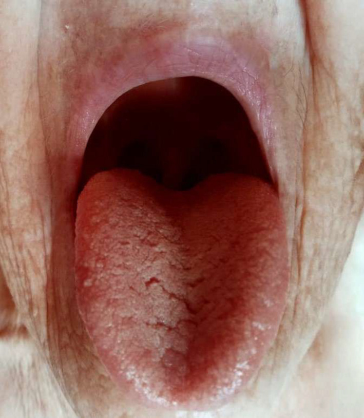
An 85‐year‐old female presented on June 15, 2020, with fever, cough and shortness of breath. The physical examination revealed 38℃ body temperature and 91% oxygen saturation in room air. The patient was hospitalized for 10 days with daily oxygen supplementation and administration of corticosteroids and antibiotics. Posteriorly, the RT‐PCR result confirmed the diagnosis of COVID‐19. After the gradual restoration of saturation levels, the patient was released. In subsequent days, the patient's complaints of hyposalivation, dry mouth and progressive worsening of symptoms, including difficulty in swallowing caught our attention (Figure 1). For this reason, she was conducted to our oral medicine clinic and is under specialist care with the use of sialagogues, artificial saliva and psychological assistance. There was no previous history of hyposalivation or any salivary gland disorder.
FIGURE 1.

Aspect of dorsal surface of the tongue with marked presence of grooves and fissures. Note to the pronounced fungiform papillae and redness as well as flaking in upper lips
DISCUSSION
SARS‐CoV‐2, similarly to other viruses, may induce an inflammatory process in salivary glands with related cases of parotitis. 4 In his groundbreaking work, Soares et al (2021) 3 found immunohistochemically high expression of ACE2 in vessels and ductal structures, demonstrated that intraoral SGs may be involved in COVID‐19 pathogenesis. Consequently, the damage to acinar cells is repaired by fibroblast proliferation and subsequent fibrous repair and hyperplasia are accompanied by stenosis of the ducts of salivary glands, resulting in hyposecretion. 5 The linkage between vascular events in patients with COVID‐19 and alterations in salivary glands is still unclear, however, some paths can be taken to elucidate this potential correlation.
Haemostatic abnormalities have been related to COVID‐19 in the form of venous thromboembolism, amongst them, the antiphospholipid syndrome—an autoimmune disorder, 6 that may reflect even in salivary glands physiology. 7 Moreover, coagulation favours fibrosis, especially by activation of mesenchymal cells that differentiated in myofibroblasts and contribute to the high deposition of extracellular matrix components and eventual loss of tissue function. 8
Another topic is the occurrence of the ‘post‐COVID syndrome’ that includes persistent symptoms (4–12 weeks after initial infection). 2 The duration of oral sequelae of COVID‐19 is unpredictable and the risk of permanent lesions is not ruled out. Besides, the medical literature has reported significant damage in patients’ brains and lungs, 9 which reveals a potential lifetime alteration.
Finally, our report reinforces the need for a multidisciplinary team monitoring patients affected by SARS‐CoV‐2, to achieve optimal treatment of symptoms and improve their well‐being and quality of life.
da Mota Santana LA, Sousa‐e‐Silva N, Gonçalo RIC, de Oliveira EM, de Oliveira Corrêa R, Moreno A, et al. Persistent hyposalivation in patients after COVID‐19 infection: Temporary or lasting alteration? Oral Surg. 2021;00:1–2. 10.1111/ors.12660
REFERENCES
- 1. Martelli Júnior H, Gueiros LA, de Lucena EG, Coletta RD. Increase in the number of Sjögren's syndrome cases in Brazil in the COVID‐19 Era. Oral Dis. 2021; 10.1111/odi.13925 [DOI] [PMC free article] [PubMed] [Google Scholar]
- 2. Nalbandian A, Sehgal K, Gupta A, Madhavan MV, McGroder C, Stevens JS, et al. Post‐acute COVID‐19 syndrome. Nat Med. 2021;27(4):601–15. 10.1038/s41591-021-01283-z [DOI] [PMC free article] [PubMed] [Google Scholar]
- 3. Soares CD, Mosqueda‐Taylor A, Hernandez‐Guerrero JC, de Carvalho MGF, de Almeida OP. Immunohistochemical expression of angiotensin‐converting enzyme 2 in minor salivary glands during SARS‐CoV‐2 infection. J Med Virol. 2021;93(4):1905–6. 10.1002/jmv.26723 [DOI] [PubMed] [Google Scholar]
- 4. Riad A, Kassem I, Badrah M, Klugar M. Acute parotitis as a presentation of COVID‐19? Oral Dis. 2020; 10.1111/odi.13571 [DOI] [PubMed] [Google Scholar]
- 5. Tsuchiya H. Oral symptoms associated with COVID‐19 and their pathogenic mechanisms: a literature review. Dent J (Basel). 2021;9(3):32– 10.3390/dj9030032 [DOI] [PMC free article] [PubMed] [Google Scholar]
- 6. Amezcua‐Guerra LM, Rojas‐Velasco G, Brianza‐Padilla M, Vázquez‐Rangel A, Márquez‐Velasco R, Baranda‐Tovar F, et al. Presence of antiphospholipid antibodies in COVID‐19: case series study. Ann Rheum Dis. 2020; 10.1136/annrheumdis-2020-218100 [DOI] [PubMed] [Google Scholar]
- 7. Basu R, De Hazra O, Mondal S, Ghosh S, Ghosh S. Parotid swelling due to external carotid artery thrombus‐a rare manifestation in antiphospholipid syndrome. Oxf Med Case Rep. 2019;2019(6): 10.1093/omcr/omz052 [DOI] [PMC free article] [PubMed] [Google Scholar]
- 8. Mercer PF, Chambers RC. Coagulation and coagulation signalling in fibrosis. Biochim Biophys Acta. 2013;1832(7):1018–27. 10.1016/j.bbadis.2012.12.013 [DOI] [PubMed] [Google Scholar]
- 9. Wang F, Kream RM, Stefano GB. Long‐term respiratory and neurological sequelae of COVID‐19. Med Sci Monit. 2020;26:e928996. 10.12659/MSM.928996 [DOI] [PMC free article] [PubMed] [Google Scholar]


