

Conflicts of interest
Dr. Tosti acts as a consultant for DS Laboratories, Monat Global, Almirall, Tirthy Madison, Eli Lilly, Leo Pharmaceuticals, Bristol Myers Squibb and P&G. Dr. Morrison and Edward Hadeler have nothing to disclose.
Funding sources
None.
According to recent data, up to 20% of patients with COVID‐19 have cutaneous manifestations. Nails can also develop abnormalities during and after infection. In this article, we review the nail findings observed in patients with COVID‐19.
We reviewed the PubMed and Embase databases to identify all articles up to May 2021 that have described nail findings in association with COVID‐19.
A total of 70 studies were reviewed including 61 studies on chilblain‐like lesions, which are one of the most widely identified cutaneous findings associated with the COVID‐19 pandemic.
Nine studies described specific nail findings (Table 1). Three of these findings [Beau lines, 1 transverse leukonychia, 1 , 2 and onychomadesis (Figure 1a)] are commonly seen with other systemic disease, including viral infection, and are likely the consequence of high fever and/or severe illness. One finding, paronychia, was seen in association with chilblain‐like lesions, 3 and three nail findings (the red half‐moon sign 4 , 5 (Figure 1d), the transverse orange discoloration 6 and the diffuse red‐white nail bed discoloration 7 ) are novel and potentially related to the microvascular injury due to COVID‐19. Of note, an orange‐brownish discoloration of the nail in a transverse pattern, the most similar finding to date, has been described in patients with Kawasaki disease, which shares a similar inflammatory response component to COVID‐19.
Table 1.
Nail findings described during the COVID‐19 pandemic, including studies documenting nail findings associated with COVID‐19 infection and studies describing nail involvement in patients with chilblain‐like lesions
|
Nail finding Study title |
Author, month, year, country | Patient characteristics | COVID‐19 disease course/associated symptoms and treatment | Onset of nail symptoms and resolution | Detailed description of cutaneous and nail findings | Time to nail symptom resolution | Additional Comments |
|---|---|---|---|---|---|---|---|
| Studies documenting nail findings associated with COVID‐19 | |||||||
|
Beau lines and leukonychia Beau’s Lines and Leukonychia in a COVID‐19 Patient Case report |
Ide, November, 2020, Japan | 68 years old, male | 18‐day hospital stay, received hydroxychloroquine 400 mg/day for 7 days, methylprednisolone 0.5 mg/kg/day for 5 days. | 1.5 months after diagnosis of COVID‐19 | White horizonal nail striae and sunken nails clinically defined as leukonychia and Beau lines | Unknown | |
|
Beau lines Beau lines associated with COVID‐19 Case report |
Alobaida, September, 2020, Canada* | 45 years old, male | Presented with diarrhea, fever, shortness of breath. Symptoms lasted 10 days and no hospital admission was required. | 3.5 months after diagnosis of COVID‐19 | Horizontal grooves over fingernails and toenails, most noticeably over his great toenails bilaterally, with a horizontal groove 5 mm from the proximal nailfold, clinically defined as Beau lines | Unknown | Toenail growth (approximately 1.62 mm per month) used to link distance of Beau lines from proximal nailfold to time of COVID‐19 infection |
|
Transverse leukonychia Transverse leukonychia (Mees’ lines) nail alterations in a COVID‐19 patient Case report |
Fernandez‐Nieto, November, 2020, Spain | 47 years old, male | Admitted to hospital with mild COVID‐19 bilateral pneumonia, treated with lopinavir/ritonavir 100mg/400mg BID for 10 days with good response and no need for oxygen. Labs notable for mild lymphopenia (830 cells/μL, range 1000–4500 cells/μL) and slight elevation of D‐dimer (1330 ng/mL, range 0–500 ng/mL). | 5 days after diagnosis of COVID‐19 | Transverse, non‐blanchable white lines on all fingernails, which progressively migrated with the growth of the nail and persisted at time of visit, clinically defined as Mees’ lines, or transverse leukonychia. | Unknown | |
|
Onychomadesis Onychomadesis following COVID‐19 infection: Is there a relationship? Case report |
Senturk, November, 2020, Turkey* | 47 years old, female | Patient was hospitalized and received hydroxychloroquine, azithromycin, oseltamivir, and ceftriaxone. | 3 months after hospitalization for COVID‐19 | Finger and toenails were detached, and new healthy nails were growing from the proximal matrix, clinically defined as onychomadesis | Unknown | Patient had pre‐existing hypertension and diabetes mellitus, continued these medications during hospital course |
|
Orange discoloration in transverse pattern Transverse orange nail lesions following SARS‐CoV‐2 infection Case report |
Tammaro, December, 2020, Italy | 89 years old, female | Patient presented with cough and asthenia. A nasal PCR was negative for COVID‐19. 16 weeks later the patient presented with orange nail discolorations. A blood test was positive for IgG against SARS‐CoV‐2 and ferropenic anemia. She also developed sarcopenia at this time | 16 weeks after initial symptoms | Orange discolorations at the end of nail beds, following the shape of the lunula | Unchanged one month following the discovery of the nail discolorations | |
|
Convex red half‐moon The red half‐moon nail sign: a novel manifestation of coronavirus infection Case report |
Neri, November, 2020, Italy | 60 years old, female | Patient presented with history of fever (>38 degrees Celsius) and cough. 7 days after these symptoms the patient had dyspnea associated with anosmia and ageusia. The patient had a normal chest x‐ray, but chest CT showed bilateral ground‐glass opacities, leading to a diagnosis of bilateral interstitial pneumonia. Diagnosis was confirmed by nasal PCR swab. Patient was hospitalized, therapy included hydroxychloroquine, lopinavir/ritonavir, ceftriaxone, heparin, and oxygen. Patient experienced complete remission of respiratory symptoms 10 days after treatment. | 2 weeks after initial onset of symptoms of COVID‐19 | Distally convex half‐moon shaped red band surrounding the distal margin of the lunula appeared on all nails, denied associated symptoms and no other skin manifestations. One month follow up, bands still present and wider. | Ongoing at follow up one month after initial presentation | |
|
Convex red half‐moon COVID‐19 and nail manifestation: be on the lookout for the red half‐moon nail sign Case report |
Méndez‐Flores, August, 2020, Mexico | 37 years old, female | Patient presented with anosmia, dry cough, persistent fever, relatively normal O2 saturation (>92%), positive nasal swab PCR confirmed SARS‐CoV‐2 infection. Managed at home, no oxygen therapy required. | 2 days after initial onset of symptoms of COVID‐19 | Red‐violet bands in the nail bed, above the nail lunula | 1 week after initial presentation | |
|
Red‐white nailbed discoloration Heterogenous red‐white discoloration of the nail bed and distal onycholysis in a patient with COVID‐19 Case report |
Demir, May, 2021, Turkey | 23 years old, male | Patient presented with history of fever, sore throat and joint pain, four months prior to onset of nail discoloration. | 4 months after initial onset of symptoms of COVID‐19 | Heterogenous red‐white discoloration in all nails; two round onycholytic areas surrounded by erythema in the distal part of the second nail on the left hand | Unknown | |
|
Nailfold video capillaroscopy (NVC) findings in patients with coronavirus disease 2019 Nailfold capillaroscopy findings in patients with coronavirus 2019: Broadening the spectrum of COVID‐19 microvascular involvement Prospective observational study |
Natalello, January, 2021, Italy | 82 patients (mean age 58.8 ± 13.2 years, 68.3% male) |
Patients were affected by COVID‐19 pneumonia, diagnosed by laboratory test (nasopharyngeal PCR) and suggestive chest imaging. (n, %): (11, 13.4%) smoked, (25, 30.5%) had hypertension, (9, 11%) had diabetes, (4, 4.9%) had rheumatic disease, (50, 61%) had a BMI > 25kg/m2, (8, 9.8%) had acral symptoms, (47, 57.3%) required oxygen therapy, (5, 6.1%) were admitted to the ICU, (21, 25.6%) received Anti‐IL6R therapy, (39, 47.5%) received enoxaparin therapy, (8, 9.8%) had PTE or DVT. 28 patients enrolled during hospitalization, 54 enrolled after discharge. |
Duration from onset of symptoms was 37.3 ± 23.1 days |
Abnormalities classifiable as non‐specific patterns in 53 patients (64.6%). Findings: Precapillary edema (80.5%), enlarged capillaries (61%), sludge flow (53.7%), meandering capillaries and reduced capillary density (50%). Acute COVID‐19 patients, compared to recovered patients, showed higher prevalence of hemosiderin deposits as a result of micro‐hemorrhages (p = .027), micro‐thrombosis (p < 0.016), sludge flow (p = 0.001) and precapillary edema (p < 0.001). Recovered patients showed higher prevalence of enlarged capillaries (p < 0.001), loss of capillaries (p = 0.002), meandering capillaries (p < 0.001), and empty dermal papillae. |
Unknown | |
| Studies describing nail involvement with chilblains‐like manifestations of COVID‐19 | |||||||
|
Subungual erythema Two cases of COVID‐19 presenting with a clinical picture resembling chilblains: first report from the Middle East Case series |
Alarmthan, May, 2020, Kuwait* | 27‐year‐old female and 35‐year‐old female |
PCR positive in both patients, patients had reported recent travel to UK. No additional information provided on disease course or treatment |
Unknown |
Red‐purple papules on the dorsal aspect of the fingers on both hands; patient 2 had diffuse erythema in the subungual area of her right thumb |
Unknown | |
|
Periungual erythema Chilblains in children in the setting of COVID‐19 pandemic Retrospective case series |
Andina, May, 2020, Spain* | 22 patients (13 male, 9 female); median age: 12 (range: 6–17) |
Respiratory symptoms (cough or rhinorrhea) (9, 41%), GI symptoms (abdominal pain or diarrhea) (2, 9%), shortness of breath 0, fever 0. Household contact with probable case of COVID‐19 12 (55%), confirmed case of COVID‐19 1 (4%). PCR positive in 1, negative in 18. |
Duration of lesions before consultation ranged from 1 to 28 days (median 7 days). |
Feet affected in all 22 cases: acrally located, erythematous‐violaceous or purpuric macules on the toes and lateral aspects of the feet and heels. The tips and periungual or distal subungual areas of the toes were commonly involved. 3 patients showed similar lesions on fingers, located predominantly on periungual areas. Dermoscopy recorded in 10 patients: signs observed included erythema, dilated capillaries, ischemic areas, purpuric dots, and hyperpigmentation. Pruritus (9, 41%), and mild pain (7, 32%) present in some cases. Skin biopsy obtained in 6 patients, all showed similar results: superficial and deep angiocentric and eccrinotropic lymphocytic infiltrate, papillary dermal edema, vacuolar degeneration of the basal layer and lymphocytic exocytosis to the epidermis and acrosyringia. Features of lymphocytic vasculopathy seen in all cases. |
Lesions showed marked improvement or almost complete resolution 3–5 weeks after onset. | |
|
Erythematous macules around the distal nailfolds Clustered cases of acral perniosis: Clinical features, histopathology, and relationship to COVID‐19 Case series |
Cordoro, May, 2020, United States* | 6 patients (age range: 12–17 years; 5 male, 1 female) |
2 siblings from one family reported rhinorrhea, congestion, sore throat, and subjective fevers 1 week prior to onset of skin lesions; none of the patients had cough, shortness of breath, or changes in smell or taste. All 6 patients had contact with adults who had mild, transient upper respiratory infection symptoms 1–2 weeks prior to the onset of skin lesions. None had known contact with confirmed COVID‐19 cases. |
1 week after presentation of other COVID‐19 systemic symptoms and or contact with adults who had mild upper respiratory infection symptoms |
Nearly all described lesions as itchy and few reported tenderness in context of swelling. Red violaceous macules and dusky, purpuric plaques scattered on the mid and distal aspects of toes. More severely affected digits were edematous with overlying superficial bullae and focal hemorrhagic crust. None of the digits appeared ischemic or necrotic. Several patients had scattered petechial and purpuric macules on the heels, soles, and distal aspect of the dorsal feet and a predominant distribution along the lateral foot, a few had subtle erythematous macules around the distal nailfolds. Half had livedo reticularis involving the flexor surfaces of the forearms, dorsal hands, and dorsal feet. 2 biopsies: superficial and deep lymphocytic infiltrate that also abuts junctional zone, where vacuolar change and purpura noted. Hemorrhagic parakeratosis found in stratum corneum. Dermal infiltrate was tightly perivascular and also perieccrine and intramural lymphocytes ("lymphocytic vasculitis") present in thin muscular walls of small vessels. No evidence of thrombosis in vessels. Immunofluorescence negative for immunoreactant deposition in all cases |
Unknown | All PCR negative. COVID‐19 IgM‐ and IgG negative. |
|
Paronychia Are chilblain‐like acral lesions really indicative of COVID‐19? A prospective study and literature review Prospective study |
Docampo‐Simon, September, 2020, Spain |
58 patients (median age: 14, range 3 months–85 years), male 29 (50%), female 29 (50%). |
Experienced COVID symptoms: Yes (11, 21.2%), No 41 (78.8%). Exposure or contact with confirmed case 12 (21.8%), suspected case 7 (12.1%), none 36 (65.5%). PCR positive in 1 (1.7%) |
Time from development of lesions to PCR test: median 12 days, range: 1–28 days; time from COVID‐19 symptoms to development of lesions (n = 11), median 7 days (0‐42 days) |
Hands (9, 15.5%), feet (36, 62.1%), hands and feet (13, 22.4%). Symptoms: pain (17, 32.1%), pruritus (20, 37.7%), pain and pruritus (5, 8.6%), asymptomatic (11, 20.8%). Morphology: chilblain‐like (42, 72.4%), purpuric (3, 5.2%), maculopapular (3, 5.2%), vesiculobullous (3, 5.2%), eczematous (3, 5.3%), paronychia (2, 3.4%), ulcer (1, 1.7%), desquamation (1, 1.7%). |
Unknown | |
|
Periungual erythema Histological findings in chilblain lupus‐like COVID lesions: in search of an answer to understand their aetiology Case report |
Rodriguez‐Villa Lario, October 2020, Spain* | 17‐year‐old male | Caregiver to patient convalescing from COVID pneumonia | 2 days of evolution |
Periungual erythema in second and third finger toe; 2 days of evolution Punch biopsy showed marked hydropic degeneration of the basal layer, isolates of necrotic keratinocyte. In papillary and reticular dermis, a moderate lymphocyte infiltration around the vessels as sleeves. The endothelium was conspicuously predominant without visualizing fibrinoid necrosis. Dense perieccrine infiltration. Positive CD123 around vessels and sweat glands. |
Unknown | Blood analysis revealed elevation of IgA. PCR negative. Serologies showed positive IgG, negative IgM. |
|
Periungual erythema and onychomadesis Are SARS‐CoV‐2 IgA antibodies in paediatric patients with chilblain‐like lesions indicative of COVID‐19 asymptomatic or paucisymptomatic infection? Prospective study |
Diociaiuti, January, 2021, Italy* | 30 patients (all adolescents) | 17 patients (group A), belonged to previous published series (2 lost to follow up), underwent second serology testing for SARS‐CoV‐2. Group B consisted of 13 new patients who underwent PCR and serology. | Fever, headache, sore throat, 1 month before (1 patient); fever, 2 months before (1 patient); sore throat, fever, diarrhea, 1.5 months before (1 patient); fever cough, 2 months before (1 patient); flu‐like symptoms, 1 month before (2 patients); asthenia, headache, 1 month before (1 patient); asymptomatic with positive PCR, 1 month before (1 patient); negative (20 patients) |
Group B: 3 patients reported flu‐like symptoms 3–4 weeks before skin lesion, 1 patient developed chilblain after proving positive to SARS‐CoV‐2; other patients presented cutaneous manifestations 2–8 weeks before screening visit. All patients presented with swelling, erythematous‐violaceous‐purpuric macules, pustules and crusts on the toes, in some cases the heels, lateral foot aspect and soles Group A: 4 patients had residual periungual toe erythema, 4 presented with toenail onychomadesis at follow‐up visit (5–7 weeks after first consultation) Serology specific for S1‐specific IgA and IgG in 30 patients showed 16 positive (53.3%), IgG detectable in 5 (16.6%). |
Unknown | |
|
Peeling around the nails What are COVID toes? A case study Case report |
Beuscher, December, 2020, United States* | 45‐year‐old female | March 12 2020: Patient presented with diarrhea, dry cough, sore throat, eye irritation, swollen lymph nodes, abdominal pain, intermittent hypoxia as low as 84, chest pain during deep inhalation, altered sense of smell | 7 days after altered sensations (neuropathic‐type symptoms) in her feet | April 19: Presented with hot and itchy and tingling toes and peeling around the nails. | Symptoms ongoing after 21 days | COVID test negative 21 days after symptom onset |
|
Nail fold telangiectasia COVID‐19 associated chilblain‐like lesions in an asymptomatic doctor Case report |
Hadjieconomou, July 2020, United Kingdom* | Woman, no age provided | No other symptoms described. | Cutaneous symptoms started 2 days before COVID‐19 diagnosed in her partner. | 2‐week history of burning, itching of her fingers and toes, with erythematous and purple papules. Erosion present on her fingers, and nail fold telangiectasia was seen. | Unknown | |
|
Dermoscopy features of nails in patients with chilblains Dermoscopy features of COVID‐19 related chilblains in children and adolescents Prospective study |
Navarro, December, 2020, Spain | 12 patients (children and adolescents) | No other symptoms described. | Unknown | Background area present in all cases; predominant color was red in 18 pictures, brown in 11, purple in 10, grey in 2; most pictures (31) contained areas of other colors within the areas whereas in 10 (24.4%) there was only one homogenous color present; globules seen in 38 (92.7%) and prominent in 32, mild in 6; reticule observed in 12 images (29.3%); other features found were splinter hemorrhages in nails (3 image), dilated capillaries in nail folds with loss of polarity (2 images) and subcorneal hemorrhagic dots (1 image). | Unknown |
41 dermoscopy pictures obtained from 12 patients. Three main dermascopic features described: a background area, globules, and reticule. Background area is the predominant background color in the lesion (ranging from red, purple, brown to grey); globules are round oval structures of red to purple color; the network reticule is a mesh of grey‐brown interconnected lines usually located peripherally within the background macule. |
Not included in the references as only 10 references are allowed as per the letter format.
Figure 1.
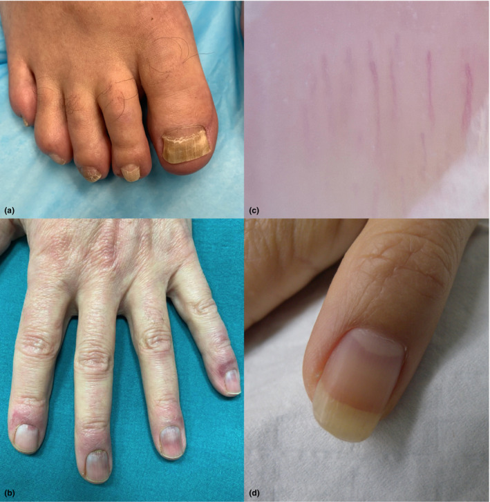
(a) Onychomadesis involving all toenails. This picture was taken 4 months after the patient was hospitalized for 30 days, including 10 days in ICU, because of severe COVID‐19 infection. (b) Chilblain‐like lesions, located distally on the fingers around the nail folds (Courtesy of Dr. Maria Pia De Padova, Bologna, Italy). (c) Nail plate dermoscopy showing leukonychia and dilated and tortuous capillaries. This picture was taken 3 months after patient had a mild COVID‐19 infection. FotoFinder R 50X. (d) Red discoloration of the nail arranged in a convex half‐moon shape, located distally on the lunula. In this patient, this was associated with orange discoloration of the distal nail plate. All fingernails were affected. This picture was taken 2 months after the patient had COVID‐19 infection not requiring hospitalization.
COVID‐19’s effects on the nail blood vessels were documented by Navarro et al. in 12 pediatric patients with COVID‐19–related chilblains, described in Table 1. 8 At dermoscopy of the nail fold and hyponychium, they found a red background with globules, indicative of vascular damage.
We also documented dilated and tortuous capillaries at dermoscopy in a patient with transverse leukonychia after COVID‐19 infection (Figure 1c).
The presence of microvascular abnormalities was confirmed by a capillaroscopy study of the nails of 82 patients, enrolled during hospitalization for COVID‐19 (28), or shortly after discharge. 9 Using nail video capillaroscopy (NVC), the authors observed microvascular abnormalities in all patients, which are described in detail in Table 1 . Findings varied between acutely ill and discharged patients, providing visual evidence of a vascular pathogenic component to COVID‐19 infection.
Chilblain‐like lesions are a commonly reported manifestation involving the digits. They are also known as ‘COVID toes,’ even though they can also affect fingers, and they present as erythematous, purpuric, papules and macules on the dorsal phalanges, nail folds and digital pulps (Figure 1b). Chilblain‐like lesions are predominantly found in children and adolescents. Nail findings have been reported in association with chilblain‐like lesions. In a prospective study conducted by Docampo‐Simon et al., two patients with chilblain‐like lesions also presented with paronychia. 3 A causative link between COVID‐19 and chilblains has not been firmly established despite an increase in the prevalence of these lesions during the pandemic.
Studies suggest that this well‐known manifestation of COVID‐19 infection may be the consequence of an exacerbated INF‐α response. Very few patients presenting with chilblain‐like lesions have other symptoms of COVID‐19 infection, and only a few test positive for the infection when presenting with the lesions. It has been suggested that the overproduction of INF‐α, which is produced at declining rates with age, may lead to rapid control of viral infection, thereby protecting younger patients from more severe disease and resulting in lower rates of positive nasopharyngeal swabs. The study by Hubiche et al. 10 also demonstrates that an increase in INF‐α in patients with chilblain‐like lesions could help young patients clear the virus rapidly. Other nail findings described in patients with chilblains include periungual erythema, peeling around the nail, nail fold telangiectasia and erythematous macules around the distal nail folds.
Our review of the literature did not reveal an association of nail disease with poor outcome for patients.
Acknowledgements
The patients in this manuscript have given written informed consent to the publication of their case details.
This paper has not been previously published or posted and is not under consideration elsewhere.
References
- 1. Ide S, Morioka S, Inada M, Ohmagari N. Beau’s lines and leukonychia in a COVID‐19 patient. Intern Med 2020; 59(24): 3259. 10.2169/internalmedicine.6112-20. [DOI] [PMC free article] [PubMed] [Google Scholar]
- 2. Fernandez‐Nieto D, Jimenez‐Cauhe J, Ortega‐Quijano D, Diaz‐Guimaraens B, Dominguez‐Santas M, Martinez‐Rubio J. Transverse leukonychia (Mees’ lines) nail alterations in a COVID‐19 patient. Dermatol Ther 2020; 33(6): e13863. 10.1111/dth.13863. [DOI] [PMC free article] [PubMed] [Google Scholar]
- 3. Docampo‐Simón A, Sánchez‐Pujol MJ, Juan‐Carpena G et al. Are chilblain‐like acral skin lesions really indicative of COVID‐19? A prospective study and literature review. J Eur Acad Dermatol Venereol 2020; 34(9): e445–e447. 10.1111/jdv.16665. [DOI] [PMC free article] [PubMed] [Google Scholar]
- 4. Neri I, Guglielmo A, Virdi A, Gaspari V, Starace M, Piraccini BM. The red half‐moon nail sign: a novel manifestation of coronavirus infection. J Eur Acad Dermatol Venereol 2020; 34(11): e663–e665. 10.1111/jdv.16747. [DOI] [PMC free article] [PubMed] [Google Scholar]
- 5. Méndez‐Flores S, Zaladonis A, Valdes‐Rodriguez R. COVID‐19 and nail manifestation: be on the lookout for the red half‐moon nail sign. Int J Dermatol. 2020; 59(11): 1414. 10.1111/ijd.15167. [DOI] [PubMed] [Google Scholar]
- 6. Tammaro A, Adebanjo GAR, Erasmus H‐P et al. Transverse orange nail lesions following SARS‐CoV‐2 infection. Dermatol Ther 2020; 34(1): e14688. 10.1111/dth.14688 [DOI] [PMC free article] [PubMed] [Google Scholar]
- 7. Demir B, Yuksel EI, Cicek D, Turkoglu S. Heterogeneous red‐white discoloration of the nail bed and distal onycholysis in a patient with COVID‐19. J Eur Acad Dermatol Venereol 2021; 35: e551–e553. 10.1111/jdv.17347. [DOI] [PMC free article] [PubMed] [Google Scholar]
- 8. Navarro L, Andina D, Noguera‐Morel L, Hernández‐Martín A, Colmenero I, Torrelo A. Dermoscopy features of COVID‐19‐related chilblains in children and adolescents. J Eur Acad Dermatol Venereol. 2020; 34(12): e762–e764. 10.1111/jdv.16800. [DOI] [PMC free article] [PubMed] [Google Scholar]
- 9. Natalello G, De Luca G, Gigante L et al. Nailfold capillaroscopy findings in patients with coronavirus disease 2019: Broadening the spectrum of COVID‐19 microvascular involvement. Microvasc Res 2021; 133: 104071. 10.1016/j.mvr.2020.104071. [DOI] [PMC free article] [PubMed] [Google Scholar]
- 10. Hubiche T, Cardot‐Leccia N, Le Duff F et al. Clinical, laboratory, and interferon‐alpha response characteristics of patients with chilblain‐like lesions during the COVID‐19 pandemic. JAMA Dermatol 2021; 157(2): 202. 10.1001/jamadermatol.2020.4324. [DOI] [PMC free article] [PubMed] [Google Scholar]