PLOS ONE logoLink to PLOS ONE
. 2021 Sep 17;16(9):e0257563. doi: 10.1371/journal.pone.0257563

Isothermal amplification and fluorescent detection of SARS-CoV-2 and SARS-CoV-2 variant virus in nasopharyngeal swabs

Les Jones 1,2, Abhijeet Bakre 1,2, Hemant Naikare 1,3, Ravindra Kolhe 2,4, Susan Sanchez 1,2, Yung-Yi C Mosley 1,3, Ralph A Tripp 1,2,*
Editor: Etsuro Ito5
PMCID: PMC8448339  PMID: 34534259

Abstract

The COVID-19 pandemic caused by the SARS-CoV-2 is a serious health threat causing worldwide morbidity and mortality. Real-time reverse transcription PCR (RT-qPCR) is currently the standard for SARS-CoV-2 detection. Although various nucleic acid-based assays have been developed to aid the detection of SARS-CoV-2 from COVID-19 patient samples, the objective of this study was to develop a diagnostic test that can be completed in 30 minutes without having to isolate RNA from the samples. Here, we present an RNA amplification detection method performed using reverse transcription loop-mediated isothermal amplification (RT-LAMP) reactions to achieve specific, rapid (30 min), and sensitive (<100 copies) fluorescent detection in real-time of SARS-CoV-2 directly from patient nasopharyngeal swab (NP) samples. When compared to RT-qPCR, positive NP swab samples assayed by fluorescent RT-LAMP had 98% (n = 41/42) concordance and negative NP swab samples assayed by fluorescent RT-LAMP had 87% (n = 59/68) concordance for the same samples. Importantly, the fluorescent RT-LAMP results were obtained without purification of RNA from the NP swab samples in contrast to RT-qPCR. We also show that the fluorescent RT-LAMP assay can specifically detect live virus directly from cultures of both SARS-CoV-2 wild type (WA1/2020), and a SARS-CoV-2 B.1.1.7 (alpha) variant strain with equal sensitivity to RT-qPCR. RT-LAMP has several advantages over RT-qPCR including isothermal amplification, speed (<30 min), reduced costs, and similar sensitivity and specificity.

Introduction

COVID-19 is a pandemic disease caused by SARS-CoV-2 [1, 2]. The animal reservoir for SARS-CoV-2 is unknown, but it is believed to be of zoonotic origin as SARS-CoV-2 isolates from bats and pangolins are genetically related to human isolates [3, 4]. SARS-CoV-2 has initiated a pandemic, and according to the WHO Coronavirus (COVID-19) Detailed Surveillance Data Dashboard, currently there are more than 170 million confirmed COVID-19 cases and more than 3.8 million deaths from COVID-19 worldwide. SARS-CoV-2 is an RNA virus with a genome that encodes four structural proteins, i.e. the spike (S) protein, the envelope (E) protein, the membrane (M) protein and the nucleocapsid (N) protein, and the remainder polyproteins that are cleaved into various non-structural proteins important for replication [5, 6]. SARS-CoV-2 infects a variety of cell types by binding to its receptor, i.e. angiotensin-converting enzyme-2 (ACE2) by the receptor-binding domain (RBD) on the spike (S) protein that is proteolytically-activated by human serine proteases [7, 8]. Cell entry depends on binding of the surface unit, S1, of the S protein to ACE2 to facilitate viral attachment to the target cells, and entry requires S protein cleavage at the S1/S2 and the S2’ site and allows fusion of viral and cellular membranes, a process driven by the S2 subunit [9]. The virus is transmitted from person to person through aerosol [10, 11]. The pathological symptoms of infection can vary but include influenza-like respiratory illness, dry cough, fever, and pulmonary complications [12], however many SARS-CoV-2 infected individuals are asymptomatic and capable of increasing the spread of the disease [13, 14]. Researchers are currently testing 100 vaccines in clinical trials on humans, and several have reached the final stages of testing. Three COVID-19 vaccines to date have been authorized and recommended for use in the United States, i.e. the Pfizer-BioNTech mRNA vaccine, the Moderna mRNA vaccine, and the Janssen-Johnson & Johnson viral vector vaccine [15]. Therefore, precautions to control disease and infection prevention practices are critical and need to be supported by diagnostics, which is a crucial component when caring for a patient with suspected or confirmed SARS-CoV-2 infection.

In addition to the Centers for Disease Control and Prevention (CDC) RT-PCR SARS-CoV-2 test [16], many other diagnostic tests have recently become available. RT-qPCR is the gold standard in diagnostic testing but its use is expensive, time-consuming, and labor-intensive. An advantage of RT-qPCR is the ability to tune the reaction using multiple temperatures allowing for multiplex target discrimination, optimized primer annealing, denaturation, and extension. Further, these properties can be adjusted at each cycle, and RT-qPCR can monitor reaction progress by non-specific dyes (e.g., SYBR Green) or TaqMan probes [17]. However, isothermal detection platforms offer key advantages over RT-qPCR platforms in terms of reduced cost, speed, and reduced labor yet have the same sensitivity. Specifically, isothermal detection platforms allow amplification at constant temperatures and avoid the need for thermal cycling required for RT-qPCR. Here, we describe a one-step isothermal amplification/detection method that is amenable to lab-based or POC/field deployment. Loop-mediated isothermal amplification (LAMP) reactions use a strand-displacing DNA polymerase (and reverse transcriptase for RNA targets) with four to six primers resulting in exponential amplification [18, 19]. LAMP is a one-step reaction allowing all the primers and enzymes to be incubated isothermally in a single step. Reverse transcription LAMP (RT-LAMP) is useful for detecting RNA-based viruses as it combines conventional LAMP with a reverse transcriptase enzyme, allowing for simultaneous reverse transcription and amplification. The endpoint for LAMP can be real-time detection using fluorescence or absorbance [20]. The RT-LAMP assay has been shown to detect SARS-CoV-2 in ≤30 minutes using patient samples such as saliva and nasopharyngeal swabs [21, 22]. However, to achieve these results, the sample preparation includes a viral RNA extraction step that increases the cost and time required to perform the assay. The LAMP detection step is performed using direct visual assessment of color change using an indicator to show a positive reaction, e.g. a shift from dark blue to lighter blue using hydroxynaphthol blue [23, 24], or pink to yellow using creosol red [25]. However, the assessment of color change has issues including unexpected signals derived from primer-dimer and/or non-primer reactions. Fluorescent dyes can be used in the LAMP reaction for fluorescence monitoring which improves detection and solves the problem of non-primer-derived signals [26, 27]. Thus, RT-LAMP has several advantages over RT-qPCR, and when fluorescence is used as an endpoint for RT-LAMP there is also improved detection arguing fluorescent RT-LAMP superiority [28]. Further, fluorescent RT-LAMP can be deployed as a point-of-care (POC) test filling a role that is vital for disease management and control.

Materials and methods

Fluorescent RT-LAMP reactions

All RT-LAMP reactions were performed using a Warm Start LAMP 2X master mix (New England Biolabs, Ipswitch, MA, USA) and an AriaMX Real-Time PCR System machine (Agilent Technologies, Santa Clara, CA) for 30 min and monitored once per minute for fluorescence in the SYBR/FAM channel. Reactions contained warm Start LAMP Kit 2X Master Mix (12.5 μl NEB), LAMP primer mix (10X, 2.5 μl, IDT), assay target (RNA, or live virus, or clinical specimen, 2 μl), SYBR-like fluorescent dye diluted 8-fold in deionized water (50X, 0.5 μl, NEB) and deionized water (7.5 μl) for a total of 25 μl. The 10X LAMP primer mix contained FIP/ BIP at 10μM, LF/ LB at 2 μM and 4 μM, respectively, prepared in deionized RNAse DNAse free water. Primers were designed from multiple alignments of SARS-CoV-2 N gene (nts 251–468) with (USA-WA1/2020, GenBank accession number MN985325.1) as a reference genome using PrimerExplorer V software. Primers were commercially synthesized by Integrated DNA technologies (IDT, Coralville, IA) (S1 Table). Final concentrations of RT-LAMP primers LAMP primers (S1 Table) were used for the fluorescent reaction: FIP/BIP 1.0 μM, F3/B3, and LF/LB primers were used at 0.2 μM and 0.4 μM, respectively.

Preparation of RT-LAMP templates

NP swab samples / cell culture derived virus

For NP swabs, a 20 μl aliquot was mixed 1:1 (v:v) with 2X LAMP Lysis Buffer (LLB): 2% Triton X-100 (Sigma, St. Louis, MO) adjusted to pH = 8.0 with 2M Tris-HCL (Sigma, St. Louis, MO) and 80 U/ml Proteinase K (NEB) followed by incubation at room temperature (RT) for 15 min, then incubation at 95°C for 10 min to inactivate the Proteinase K. The patient sample lysate (2 μl) was used as target directly in the fluorescent RT-LAMP reactions without further processing. Fluorescent RT-LAMP reactions using virus-infected cell culture supernatant samples were used to detect live viruses. SARS-CoV-2 (USA-WA1/2020, GenBank: MN985325.1), and alpha variant B1.1.7 (20I/501Y.V1, GenBank: MW422255.1), human coronavirus strains, OC43 (GenBank: AY585228.1), 229E (GenBank: AF304460.1), and NL63 (GenBank: AY567487.2) (BEI Resources, USA) were used to infect Vero cells and cultured to 107 PFU/ml. A 20 μl aliquot of the infected cell culture supernatants was mixed with 20 μl of 2X LLB and incubated at RT for 15 min followed by 10 min 95°C incubation to inactivate Proteinase K. Fluorescent RT-LAMP was performed using 2 μl of the sample lysate.

Fluorescent RT-LAMP reaction using virus-infected cell culture supernatant samples

An identical process was used to detect live viruses. SARS-CoV-2 (USA-WA1/2020, GenBank: MN985325.1), and alpha variant B.1.1.7 (20I/501Y.V1, GenBank: MW422255.1) were inoculated on Vero cells and cultured to 107 pfu/ml. A 20 μl aliquot of the infected cell culture supernatants was mixed with 20 μl of 2X LLB and incubated at RT for 15 min followed by 10 min 95°C incubation to inactivate Proteinase K. Fluorescent RT-LAMP was performed using 2 μl of the sample lysate. We also examined human coronavirus strains, OC43 (GenBank: AY585228.1), 229E (GenBank: AF304460.1), and NL63 (GenBank: AY567487.2), all of which were obtained from BEI Resources and cultured using Vero cells for detection in fluorescent RT-LAMP as specificity controls.

Optimizing the limit of detection/fluorescent end-point cutoff

The fluorescent LAMP reaction contained an intercalating fluorescent SYBR-like dye with excitation (λExci = 497 nm) and emission (λEmis = 520 nm) when complexed with the double-stranded DNA LAMP reaction product. This allowed for easy monitoring of the accumulation of double-stranded DNA in the RT-LAMP reaction with a real-time thermocycler using a standard SYBR/FAM channel for fluorescence. Using a log dilution series (ranging from 108 to 10 copies of the N gene) of purified RNA templates described below (N gene IVT RNA and viral genomic RNA), we measured the signal from the fluorescent RT-LAMP reaction at 1 min intervals analogous to a standard dye-based RT-qPCR reaction utilizing a 30 min total time cut-off. The results were compared to the cycle threshold (Ct) values obtained from our RT-qPCR assay (described below) for the same purified RNA templates. In the fluorescent RT-LAMP reaction, the purified RNA templates representing ~100 copies or more of the N gene generated strong fluorescence by 30 min and were readily detectable by RT-qPCR. RNA templates diluted beyond 10 copies of the N gene did not generate fluorescence signal within 30 min in fluorescent RT-LAMP and were negative in RT-qPCR with a cut-off of 40 cycles. Based on this observation, and consistent with other related RT-LAMP publications [29, 30], we used fluorescence at 30 min to determine whether a sample was positive or negative.

RT-qPCR

Serial log dilutions of a 2019-nCoV_N_positive control plasmid (IDT) containing the SARS-CoV-2 (Wuhan-Hu-1 strain, GenBank: MN908947) N gene from 105 copies/μl to 1 copy/μl, or SARS-CoV-2 genomic viral RNA (vRNA), or SARS-CoV-2 N gene in vitro transcribed RNA (IVT RNA) from 1ng/μl to 0.1fg/μl corresponding to 107 copies/μl to 1 copies/μl, or 108 copies/μl to 10 copies/μl, respectively, were prepared in nuclease-free water. Nuclease-free water was used as a no-template control for all reactions. The SARS-CoV-2 RNA templates were added to LunaScript RT SuperMix (NEB) at 2 μl in a 20 μl reaction and reverse transcribed for 10 min at 55°C. The cDNA from this reaction was used directly at 1 μl in a 20 μl reaction with Luna Universal Probe qPCR Master Mix (NEB) and 1 μl of the SARS-CoV-2 CDC EUA N1 probe assay (IDT) and amplified for 40 cycles in a two-step reaction (15s/95°C, and 30s/60°C) after an initial 1 min pre-amplification hold at 95°C on an AriaMx Real-Time PCR System (Agilent). SARS-CoV-2 plasmid DNA was RT-qPCR amplified by adding 1 μl to a 20 μl reaction with the same qPCR master mix and probe assay using the same two-step amplification protocol after an initial 3 min pre-amplification hold at 95°C. Results from the N gene control plasmid were used to construct a standard curve relating the N gene copy number to RT-qPCR Ct values (S3 Fig). The N gene copy numbers of the SARS-CoV-2 IVT RNA or SARS-CoV-2 genomic vRNA per unit mass were extrapolated from this curve.

For analysis of clinical samples, RNA was extracted from 200 μl of patient nasopharyngeal swab liquid in saline using the RNAdvance Viral Reagent Kit (Beckman Coulter, Indianapolis, IN) and eluted in a final volume of 40 μl of nuclease-free water. RT-qPCR analysis of purified RNA from patient samples as described above was performed using the CDC EUA N1 probe assay and the CDC EUA human RNAseP internal control probe assay to validate sample RNA extraction in separate reactions.

SARS-CoV-2 RNA

Genomic RNA from SARS-CoV-2 (USA-WA1/2020 GenBank: MN985325.1) and the SARS-CoV-2 variant B.1.1.7 (SARS-CoV-2/human/USA/SEARCH-5574-SAN/2020 GenBank: MW422255.1) was purified from 200 μl of virus-infected Vero cell culture supernatant using an RNAeasy kit (Qiagen) according to the manufacturer’s instructions and eluted in 40 μl of nuclease-free deionized water and stored at -80°C.

In vitro transcribed (IVT) RNA

The 1260 bp complete coding sequence of SARS-CoV-2 Wuhan-Hu-1 strain N gene (GenBank: MN908947) was amplified from 2019-nCoV_N_Positive Control plasmid (IDT) using primers (IDT) SARS2N_F and SARS2N_R (S1 Table) in standard end-point PCR with Q5 DNA polymerase (NEB). The PCR band representing the N gene coding sequence was subcloned into plasmid pMiniT2.0 (NEB) with the 5’ end of the coding sequence proximal to the vector-provided T7 polymerase promoter and confirmed by Sanger sequencing. HiScribe T7 RNA Synthesis Kit (NEB) was used to produce the SARS-CoV-2 N gene single-stranded positive-sense RNA via in-vitro transcription from the pMiniT2.0 construct. The IVT RNA was digested with DNase1 (NEB) and purified using RNAClean XP (Beckman Coulter). Purified IVT RNA was eluted in nuclease-free water, adjusted to a final concentration of 1 μg/μl in nuclease-free water, and stored at -80°C.

Analysis of NP swab samples

Deidentified samples were collected from consenting adult volunteers by the Georgia National Guard or the University of Georgia (UGA) COVID-19 Task Force following written approval and were tested from frozen -80°C samples collected by the Georgia Taskforce for COVID-19, the Georgia National Guard, or the University of Georgia Diagnostic Laboratories. NP samples were collected using a sterile swab applicator that was placed in 1 mL of saline. The University of Georgia (UGA) Diagnostic Laboratories determined the SARS-CoV-2 status of the NP samples using an Applied Biosystems TaqPath COVID-19 EUA assay (ThermoFisher) to detect SARS-CoV-2 RNA in a multiplex RT-qPCR format. The assay is specific for three SARS-CoV-2 genome regions, i.e. spike (S) gene, nucleocapsid (N) gene, and orf-1ab. We analyzed the same NP samples using the CDC EUA N1 RT-qPCR assay for the detection of SARS-CoV-2 RNA. The CDC EUA N1 RT-qPCR assay is singleplex and specific for the SARS-CoV-2 N gene. Fifty SARS-CoV-2 positive samples and 84 SARS-CoV-2 negative samples were tested in our laboratory using the CDC EUA N1 RT-qPCR assay to detect the N gene sequence. All NP samples were subsequently tested in a fluorescent RT-LAMP assay for comparative SARS-CoV-2 diagnostic analysis with CDC EUA N1 RT-qPCR assay.

Results

Analytical sensitivity of fluorescent RT-LAMP using IVT RNA and genomic viral RNA

We examined the sensitivity of the fluorescent RT-LAMP assay, by comparison, to the CDC EUA N1 RT-qPCR assay. The limit of detection (LOD) in the RT-qPCR assay using SARS-CoV-2 (Wuhan-Hu-1) IVT RNA was 0.1 fg of RNA corresponding to 10 copies of the N gene sequence (Fig 1). The LOD by RT-qPCR was 100 copies of purified viral genomic RNA from SARS-CoV-2 USA-WA1/2020 (S1A Fig). The fluorescent RT-LAMP assay rapidly and sensitively detected SARS-CoV-2 IVT RNA with a LOD of approximately 10–100 N gene copies with efficient isothermal amplification across seven logs of target concentration (Fig 2). The fluorescent RT-LAMP assay can detect 100 copies of SARS-CoV-2 (USA-WA1/2020 GenBank: MN985325.1) viral genomic RNA equaling the sensitivity of RT-qPCR (S1B Fig).

Fig 1. SARS-CoV-2 RT-qPCR CDC EUA N1 probe assay.
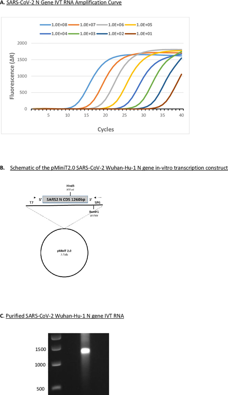
Fig 1

A. RT-qPCR (CDC EUA N1 probe assay) targeting SARS-CoV-2 Wuhan-Hu-1 N gene in vitro transcribed (IVT) RNA. Log dilution series of N gene IVT RNA ranging from 108 to 10 copies of the N gene underwent reverse transcription. Fluorescence data were collected and processed using a 40 cycle cut-off on an AriaMx Real-Time PCR System. B. Schematic of the pMiniT2.0 SARS-CoV-2 Wuhan-Hu-1 N gene in vitro transcription construct with T7 RNA polymerase promoter driving full-length (1260 nt) N gene RNA production. C. DNase-treated and purified SARS-CoV-2 Wuhan-Hu-1 N gene IVT RNA on a non-denaturing 1% agarose gel stained with ethidium bromide.

Fig 2. SARS-CoV-2 fluorescent RT-LAMP assay.
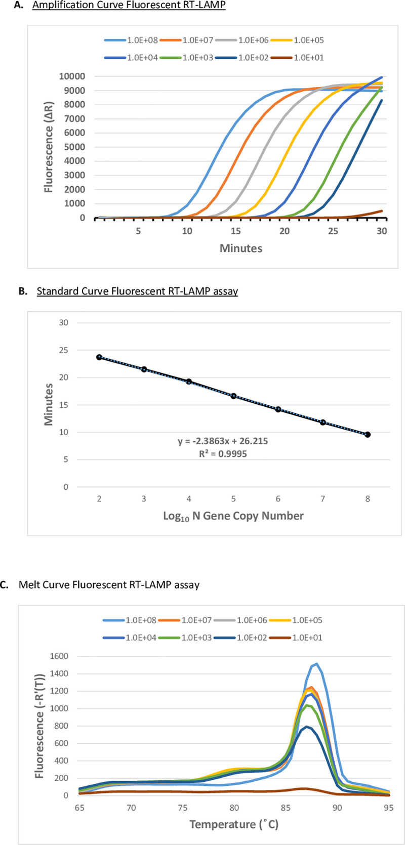
Fig 2

A. Fluorescent RT-LAMP assay targeting SARS-CoV-2 Wuhan-Hu-1 N gene IVT (GenBank: MN908947) RNA. Log-dilution series of N gene IVT RNA ranging from 108 to 10 copies of the N gene was added to fluorescent RT-LAMP reactions containing six LAMP primers (FIP/BIP, F3/B3, and LF/LB) and an SYBR-like fluorescent dye. Reactions were performed on an AriaMx Real-Time PCR System at 65°C for 30 min with fluorescence monitored in the FAM/SYBR channel. Fluorescence values were plotted against the elapsed reaction time (min) to generate the amplification curve. B. Standard Curve of fluorescent RT-LAMP assay C. Melt curve of the fluorescent RT-LAMP products produced from the reactions in Fig 2A.

Fluorescent RT-LAMP detects the SARS-CoV-2 B.1.1.7 (alpha) variant

SARS-CoV-2 (WA1/2020) or SARS-CoV-2 B1.1.7 (alpha) variant (20I/501Y.V1) infected (MOI = 0.1) Vero cell culture supernatants were examined by fluorescent RT-LAMP. We demonstrated that the fluorescent RT-LAMP assay successfully detected the SARS-CoV-2 B1.1.7 (alpha) variant with equal sensitivity to that of SARS-CoV-2 at a LOD of approximately 100 pfu/ml (Fig 3A and 3B). To confirm RT-LAMP specificity, we tested human coronavirus strains, OC43 (GenBank: AY585228.1), 229E (GenBank: AF304460.1), and NL63 (GenBank: AY567487.2) infected (MOI = 1.0) Vero cells. All human coronavirus strains tested did not produce a detectable fluorescent signal in the fluorescent RT-LAMP assay (Fig 3C). The sensitivity of fluorescent RT-LAMP detection of both the variant and the wild-type SARS-CoV-2 strains compared favorably with the RT-qPCR results for RNA purified from an equivalent amount of SARS-CoV-2 infected Vero cell culture supernatant (Fig 4).

Fig 3. SARS-CoV-2 live virus fluorescent RT-LAMP assay.
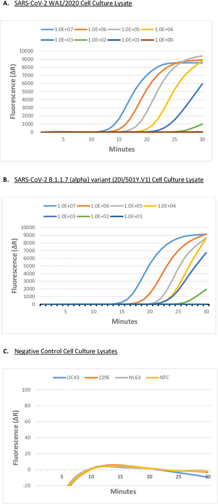
Fig 3

A. Fluorescent RT-LAMP assay targeting SARS-CoV-2 (USA-WA1/2020 GenBank: MN985325.1), and B. Fluorescent RT-LAMP assay targeting SARS-CoV-2 variant B1.1.7 variant (20I/501Y.V1) in infected Vero cell culture. Culture supernatants were log-serially diluted from 107 pfu/ml to 10 pfu/ml. Fluorescent RT-LAMP was performed using six LAMP primers (FIP/BIP, F3/B3, and LF/LB), and SYBR-like fluorescent dye. Reactions were performed on an AriaMx Real-Time PCR System at 65°C for 30 min with fluorescence monitored in the FAM/SYBR channel. Fluorescence values were plotted against the elapsed reaction time (min) to generate the amplification curves. Virus-infected Vero cell culture supernatants below 102 pfu/ml were not detected by 30 min. C. No Template Control (NTC), and heterologous human coronavirus strains at 106 pfu/ml, OC43 (GenBank: AY585228.1), 229E (GenBank: AF304460.1), and NL63 (GenBank: AY567487.2) in infected Vero cells failed to produce fluorescence signal over the background in 30 min in the fluorescence RT-LAMP assay.

Fig 4. SARS-CoV-2 live virus CDC EUA N1 RT-qPCR assay.
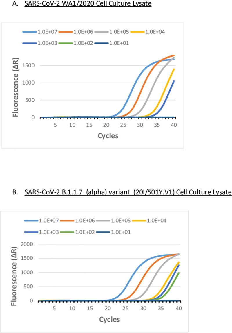
Fig 4

A. RT-qPCR using CDC EUA N1 probe assay targeting SARS-CoV-2 (WA1/2020) and, B. targeting SARS-CoV-2 B.1.1.7 (Alpha) (20I/501Y.V1) in infected Vero cells. Culture supernatants were log-serially ranging from 107 pfu/ml to 10 pfu/ml. RNA was purified from cell culture supernatants and reverse transcribed and cDNA was used in an RT-qPCR reaction in the CDC EUA N1 Probe Assay. Fluorescence data were collected and processed using a 40 cycle cut-off on an AriaMx Real-Time PCR System.

Fluorescent RT-LAMP detects SARS-CoV-2 in nasopharyngeal swab samples

We determined the presence of SARS-CoV-2 directly in clinical nasopharyngeal swab samples collected from patients using fluorescent RT-LAMP. Nasal swabs and NP swabs from suspected COVID-19 patients were collected by personnel from the Georgia COVID-19 Task Force and the University of Georgia Diagnostic Laboratories and confirmed positive or negative for SARS-CoV-2 using an Applied Biosystems TaqPath COVID-19 EUA kit to detect SARS-CoV-2 RNA in a multiplex RT-qPCR format. In comparing fluorescent RT-LAMP to RT-qPCR, we chose to evaluate the singleplex N gene-specific SARS-CoV-2 CDC EUA N1 RT-qPCR assay since it shares close target specificity with our fluorescent RT-LAMP assay (S1 Table). Using the same NP samples, we used the CDC EUA N1 RT-qPCR assay on both SARS-CoV-2 positive (n = 50) and SARS-CoV-2 negative (n = 84) NP samples in triplicate. Positive samples in the CDC EUA N1 RT-qPCR probe assay with Ct values <40 for each of the three repeat samples were considered N gene positives in our comparative analysis. We determined that 84% (n = 42/50) of the previously established SARS-CoV-2 positive NP samples were N gene-positive (Ct<40) by the CDC EUA N1 RT-qPCR assay for each of three repeat measurements (Table 1). The mean Ct of the SARS-CoV-2 N gene-positive NP samples (n = 42) ranged from 16 to 34 cycles with an overall mean Ct of 25.7, 95% CI [2427] using the SARS-CoV-2 CDC EUA N1 RT-qPCR assay. Negative samples with no Ct value in the CDC EUA N1 RT-qPCR assay for any of three repeat measurements were considered true N gene negatives. For the previously confirmed SARS-CoV-2 negative NP samples, we determined that 81% (n = 68/84) gave matching N gene negative results, i.e. no Ct value in any of the three repeat measurements using the SARS-CoV-2 CDC EUA N1 RT-qPCR assay (Table 2).

Table 1. Fluorescent RT- LAMP on SARS-CoV-2 positive NP swab samples.

SARS-CoV-2 CDC EUA N1 RT-qPCR Mean Ct [95%CI] Number of Samples RT-LAMP Concordant Results (%) RT-LAMP Discordant Results (%) Mean RT-LAMP (min) [95% CI]
Below assay cut-off (Ct<40) M = 25.7, 95% CI [24.0–27.3] 42 41 (98) 1 (2) mean = 23
95% CI [2224]
No Ct 8 1 (12) 7 (88) na
Positive samples 50 42 (84) 8 (16) na

Patient samples were previously confirmed as SARS-CoV-2 positive or negative by the UGA Diagnostic Laboratories using the ThermoFisher Applied Biosystems TaqPath COVID-19 EUA Combo Kit to detect SARS-CoV-2 RNA in a multiplex RT-qPCR format specific for three SARS-CoV-2 genome regions: S gene, N gene, and orf-1ab. We further analyzed the same NP samples by RT-qPCR using the singleplex CDC EUA N1 probe assay with a cut-off of 40 cycles, and the N gene-specific fluorescent RT-LAMP assay with a cut-off at 30 min. FIP/BIP, F3/B3, and LF/LB primers and an SYBR-like fluorescent dye were used in the RT-LAMP assay. The SARS-CoV-2 positive sample fluorescent RT-LAMP results (n = 50) were grouped according to their CDC EUA N1 RT-qPCR assay result: samples below the assay cut-off (Ct<40) n = 42/50, or samples above the assay cut-off (no Ct) n = 8/50. The number of positive samples having a fluorescent RT-LAMP assay value less than the 30 min assay cut-off (n = 41/42) has concordant results to RT-qPCR for these samples. One specimen (n = 1/42) had discordant results in the fluorescent RT-LAMP assay as it failed to produce a signal above the background in the 30 min assay time. The average fluorescent RT-LAMP assay value (23 minutes) is calculated for the group of positive samples (n = 41) at the 95%CI, 23 [2224]. Samples having (no Ct) result in RT-qPCR (8/50) had discordant fluorescent RT-LAMP results signified by a positive signal in the RT-LAMP assay (n = 7/8). Fluorescent LAMP is measured by the time in minutes required to generate fluorescence signal (ΔR) over baseline in the assay.

Table 2. Fluorescent RT- LAMP of SARS-CoV-2 negative NP swab samples.

SARS-CoV-2 CDC EUA N1 RT-qPCR Result Number of Samples RT-LAMP Concordant Results (%) RT-LAMP Discordant Results (%)
No Ct 68 59 (87) 9 (13)
Below assay cut-off (Ct<40) 16 7 (44) 9 (56)
All Negative samples 84 66 (79) 18 (21)

NP samples were previously confirmed as SARS-CoV-2 positive or negative by the UGA Diagnostic Laboratories using the ThermoFisher Applied Biosystems TaqPath COVID-19 EUA Kit to detect SARS-CoV-2 RNA in a multiplex RT-qPCR format specific for three SARS-CoV-2 genome regions: S gene, N gene, and orf-1ab. We further analyzed the same NP samples by RT-qPCR using the singleplex CDC EUA N1 probe assay with a cut-off of 40 cycles, and the N gene-specific fluorescent RT-LAMP assay with a cut-off at 30 min. FIP/BIP, F3/B3, and LF/LB primers and an SYBR-like fluorescent dye were used in the RT-LAMP assay. The SARS-CoV-2 negative sample fluorescent RT-LAMP results (n = 84) were grouped according to their CDC EUA N1 RT-qPCR assay result: samples showing No Ct (n = 68/84), or samples having Ct below the assay cut-off Ct<40 (n = 16/84). N = 68 of the negative NP samples failed to signal in our RT-qPCR assay, as expected. Of this group, n = 59/68 gave concordant fluorescent RT-LAMP assay results failing to signal above baseline in the 30 min assay time, and n = 9/68 gave discordant fluorescent RT-LAMP assay results signified by a positive fluorescent RT-LAMP signal in the 30 min assay time. Some of the negative NP samples gave RT-qPCR Ct values below the assay cut-off (n = 16/84) suggesting a positive result. This is unexpected from negative samples but likely is related to the differential specificities between our singleplex CDC EUA N1 RT-qPCR assay and the multiplex RT-qPCR assay used by the UGA Diagnostic Laboratories. Of this group of samples, n = 7/16 also gave a positive fluorescent RT-LAMP signal, while n = 9/16 failed to signal above baseline in the 30 min assay time. Fluorescent RT-LAMP is measured by the time in minutes required to generate fluorescence signal (ΔR) over baseline in the assay.

The fluorescent RT-LAMP assays were performed in triplicate using the same NP samples that were previously determined to be positive for SARS-CoV-2 (n = 50) by the UGA Diagnostic Laboratories. The fluorescent RT-LAMP assay values for the SARS-CoV-2 N gene-positive NP samples ranged from 18 to 28 minutes with an overall mean of 23 minutes and 95% CI [2224]. The results for N gene-positive NP samples showed 98% (n = 41/42) concordance with RT-qPCR for SARS-CoV-2 positivity (Table 1). N gene NP samples with no Ct result in the CDC EUA N1 RT-qPCR assay (8/50) had discordant fluorescent RT-LAMP results as indicated by a positive signal in the RT-LAMP assay (n = 7/8).

A fluorescent RT-LAMP assay was performed on the same NP samples that were previously shown to be SARS-CoV-2 negative (n = 84) by the UGA Diagnostic Laboratories. Results for the fluorescent RT-LAMP assay on the N gene negative NP samples showed 87% (n = 59/68) concordance with RT-qPCR for SARS-CoV-2 (Table 2), while 13% (n = 9/68) of N gene negative samples gave discordant fluorescent RT-LAMP assay results as determined by a positive result in the 30 minute assay time. When compared to RT-qPCR, N gene-positive samples assayed by fluorescent RT-LAMP had 98% (n = 41/42) concordance, while fluorescent RT-LAMP values were 87% (n = 59/68) concordant with RT-qPCR. Importantly, the fluorescent RT-LAMP assay results were obtained without purification of RNA from the NP samples in contrast to the RT-qPCR assay protocol.

Discussion

The benchmark method for the detection of SARS-CoV-2 from patient samples is the RT-qPCR assay requiring RNA extraction, highly trained laboratory personnel, and expensive equipment and reagents. Currently, the majority of clinical diagnosis for SARS-CoV-2 are performed by central testing laboratories that typically takes >24h for return of results. These clinical RT-qPCR assays are used typically with symptomatic patients, are not required to be low-cost, but rely on their analytical sensitivity to produce a definitive diagnosis even if only a single testing opportunity is available. Diagnostic assays used in surveillance are designed to detect the prevalence of SARS-CoV-2. These tests should be cost-effective, rapid, and easy to perform allowing for frequent testing. We sought to improve upon SARS-CoV-2 diagnostic testing by reducing the time for confirmation of a result, lowering costs to acquire the results, and developing a reliable, sensitive, and specific rapid assay. Many emerging studies have shown that RT-LAMP can meet these requirements for screening and testing for SARS-CoV-2 and potentially be a complementary tool to RT-qPCR [18, 31, 32]. Several RT-LAMP studies have focused on developing point-of-care (POC) diagnostic assays by detection of SARS-CoV-2 RNA in clinical samples [33, 34]. Other POC RT-LAMP studies have developed assays to allow multiplexing of more than one genomic target, while others have focused on optimizing RT-LAMP conditions to improve detection of SARS-CoV-2 genomes [35], but direct detection of the virus has been limited.

In this study, we show that fluorescent RT-LAMP has similar performance to RT-qPCR in detecting SARS-CoV-2 from the same clinical samples, and importantly, that fluorescent RT-LAMP can sensitively and reliably detect virus from only minimally pre-processed nasal swabs. Despite differences in SARS-CoV-2 detection in NP samples between the multiplex assay and the RT-LAMP assay, we believe that comparative sensitivity and specificity is important for accelerating diagnostic applications. Likely, the broad target specificity associated with the multiplex RT-qPCR assay used by the UGA Diagnostic Laboratories may in part account for the difference in sensitivity we obtained on the same NP samples using the singleplex CDC EUA N1 RT-qPCR assay. It is also possible that RNA degradation of the NP samples may have occurred by the time we obtained them as they were stored in saline and subsequently underwent several freeze-thaw cycles which may have contributed to differences in sensitivity associated with our RT-qPCR assay.

Of the group of SARS-CoV-2 positive NP samples from the UGA Diagnostic Laboratories, we found 84% (n = 42/50) to be N gene-positive as determined by our RT-qPCR assay. Importantly, fluorescent RT-LAMP assay results were 98% (n = 41/42) concordant as N gene-positive samples. Eight SARS-CoV-2 positive NP samples determined by the UGA Diagnostic Laboratories failed to amplify the N gene target in our RT-qPCR assay. However, the internal control amplicon (human RNase P) amplified in all NP samples tested s that the extracted RNA was intact. We found n = 7/8 positive NP samples by the fluorescent RT-LAMP assay for the failed RT-qPCR NP samples which was discordant with our RT-qPCR result for these NP samples but in agreement with NP samples showing SARS-CoV-2 positive standing by the UGA Diagnostic Laboratories. This may suggest that sample preparation differences (e.g. RNA purification for RT-qPCR or the minimal pre-processing for RT-LAMP) may contribute to the different results. We show that the fluorescent RT-LAMP assay sensitivity is more than sufficient to reliably detect SARS-CoV-2 in NP samples to virus load, as SARS-CoV-2 patients have been reported to have a range from 641 copies/mL to 1011 copies/mL with a median of 104–105 [36]. Of the SARS-CoV-2 negative NP samples from the UGA Diagnostic Laboratories, 81% (n = 68/84) were N gene negatives as determined by our RT-qPCR assay. Fluorescent RT-LAMP assay results were 87% (n = 59/68) concordant confirming N gene negative samples, and 16 NP negative samples were positive in our RT-qPCR assay. These results likely represent RT-qPCR false-positives that were SARS-COV-2 negative as determined by the UGA Diagnostic Laboratories with differences likely owing to the broad-specificity of the RT-qPCR assay. The N gene negative NP specimens by RT-LAMP were 13% (n = 9/68) with positive results discordant with RT-qPCR results.

We show that the fluorescent RT-LAMP assay sensitively and specifically detects wild-type SARS-CoV-2 and the B.1.1.7 (Alpha) variant in virus-infected Vero cell culture supernatants after the minimal pre-processing steps. The fluorescent RT-LAMP assay primers were designed based on the nucleotide sequence of the N gene reported for the SARS-CoV-2 (WA1/2020) strain. A majority of the sequence divergence associated with variant strains is located in the S gene, while the N gene nucleotide sequence is highly conserved. The N gene RT-LAMP primer sequences are 100% conserved among the wild type and variant SARS-CoV-2 strains (S2 Fig). This conservation allows our RT-LAMP assay to detect various SARS-CoV-2 strains. Also, the specificity of our fluorescent RT-LAMP assay is shown by the failure of three closely related human coronavirus strains, i.e. OC43, 229E, and NL63, to give any positive signal over the baseline in this 30 minute assay (S2 Fig).

Overall, the fluorescent RT-LAMP assay was shown to be positive for >98% of NP samples (n = 41/42) that were N gene-positive samples by RT-qPCR, and negative for 87% (n = 59/68) of NP samples that were N gene negative samples by RT-qPCR. Discordant results in RT-qPCR and fluorescent RT-LAMP could represent false positives from spurious amplification from primer-dimer sometimes associated with RT-LAMP assays, sample contamination, variability among different RT-qPCR tests detecting different or multiple SARS-CoV-2 amplicons, or increased sensitivity of RT-LAMP compared to RT-qPCR. RT-LAMP employing six primers has high-specificity for selected amplification targets when compared to standard PCR utilizing only two primers, or RT-qPCR employing an additionally labeled probe oligo. Careful design of the six LAMP primers, particularly the inner primers FIP and BIP to avoid self-complementarity, and optimized formulation can help to mitigate these concerns. Our data shows that the fluorescent RT-LAMP assay is comparable to RT-qPCR and useful for detecting SARS-CoV-2 infected NP samples having a moderate viral load. Importantly, the viral load does not indicate either the course of infection or COVID-19 disease severity because the viral load is not representative of the overall viral burden of an infected individual, and the level of SARS-CoV-2 is not the only attribute affecting disease in an individual [3739].

Supporting information

S1 Fig. SARS-CoV-2 WA-1 USA-WA1/2020 detection.

A. RT-qPCR using a CDC EUA N1 probe assay targeting purified genomic RNA from SARS-CoV-2 USA-WA1/2020. Log dilution series of purified genomic viral RNA underwent reverse transcription and cDNA was used in a CDC EUA N1 Probe Assay. Fluorescence data were collected and processed using a 40 cycle cut-off on an AriaMx Real-Time PCR System. B. Fluorescent RT-LAMP assay targeting purified SARS-CoV-2 USA-WA1/2020 RNA. Log dilution series of purified genomic RNA ranging from 108 to 10 copies of the N gene was added to fluorescent RT-LAMP reactions containing six LAMP primers (FIP/BIP, F3/B3, and LF/LB) and an SYBR-like fluorescent dye. Reactions were performed on an AriaMx Real-Time PCR System and fluorescence values were plotted against the elapsed reaction time (min) to generate the amplification curve.

(DOCX)

S2 Fig. SARS-CoV-2 and human coronavirus N gene sequences.

Nucleocapsid (N) gene multiple sequence alignment (T-Coffee) of SARS-CoV-2 strains: SARS2_WA1 (USA-WA1/2020 GenBank: MN985325.1), SARS2_Wuhan1 (Wuhan-Hu-1 GenBank: MN908947), SARS2_VAR_B117 (B.1.1.7 Alpha SARS-CoV-2/human/USA/SEARCH-5574-SAN/2020 GenBank: MW422255.1); and closely-related human coronavirus strains: HuCoV_OC43(GenBank: AY585228.1), HuCoV_NL63 (GenBank: AY567487.2), and HuCoV_229E (GenBank: AF304460.1) used in the study. Position of the six fluorescent RT-LAMP assay primers and their conservation (100%) among the wild-type and variant SARS-CoV-2 strains accounts for assay specificity for the variant strain, SARS2 B.1.1.7. (alpha). Divergence of the heterologous human coronavirus strain sequences results in no signal in the fluorescent RT-LAMP assay for these targets.

(DOCX)

S3 Fig. SARS-CoV-2 N gene positive control plasmid RT-qPCR.

RT-qPCR using the CDC EUA N1 probe assay targeting SARS-COV-2 N gene positive control plasmid (IDT). Log dilution series of plasmid DNA ranging from 105 to 10 copies of the SARS-CoV-2 N gene used in the CDC EUA N1 Probe Assay. Fluorescence data were collected and processed using a 40 cycle cut-off on an AriaMx Real-Time PCR System and Ct values were plotted against the Log10 of the starting N gene copy numbers to generate the standard curve.

(DOCX)

S1 Table. SARS-CoV-2 fluorescent RT-LAMP primers.

N gene cloning primers, fluorescent RT-LAMP assay primers created using free Primer Explorer V software, and SARS-CoV-2 CDC EUA N1 RT-qPCR assay primers and probe. Numbering according to SARS-CoV-2 Wuhan-Hu-1 (GenBank: MN908947.3).

(DOCX)

S1 Raw images

(PDF)

Data Availability

All relevant data are within the manuscript and its Supporting Information files.

Funding Statement

Funding was through the Georgia COVID-19 Task Force and the Georgia Research Alliance to RT. The funders had no role in study design, data collection and analysis, decision to publish, or preparation of the manuscript.

References

  • 1.Krammer F. SARS-CoV-2 vaccines in development. Nature. 2020;586(7830):516–27. Epub 2020/09/24. doi: 10.1038/s41586-020-2798-3 . [DOI] [PubMed] [Google Scholar]
  • 2.Poland GA, Ovsyannikova IG, Kennedy RB. SARS-CoV-2 immunity: review and applications to phase 3 vaccine candidates. Lancet. 2020;396(10262):1595–606. Epub 2020/10/17. doi: 10.1016/S0140-6736(20)32137-1 ; PubMed Central PMCID: PMC7553736. [DOI] [PMC free article] [PubMed] [Google Scholar]
  • 3.Mahdy MAA, Younis W, Ewaida Z. An Overview of SARS-CoV-2 and Animal Infection. Front Vet Sci. 2020;7:596391. Epub 2020/12/29. doi: 10.3389/fvets.2020.596391; PubMed Central PMCID: PMC7759518. [DOI] [PMC free article] [PubMed] [Google Scholar]
  • 4.Irving AT, Ahn M, Goh G, Anderson DE, Wang LF. Lessons from the host defences of bats, a unique viral reservoir. Nature. 2021;589(7842):363–70. Epub 2021/01/22. doi: 10.1038/s41586-020-03128-0 . [DOI] [PubMed] [Google Scholar]
  • 5.Whittaker GR, Daniel S, Millet JK. Coronavirus entry: how we arrived at SARS-CoV-2. Curr Opin Virol. 2021;47:113–20. Epub 2021/03/22. doi: 10.1016/j.coviro.2021.02.006 ; PubMed Central PMCID: PMC7942143. [DOI] [PMC free article] [PubMed] [Google Scholar]
  • 6.Das A, Ahmed R, Akhtar S, Begum K, Banu S. An overview of basic molecular biology of SARS-CoV-2 and current COVID-19 prevention strategies. Gene Rep. 2021:101122. Epub 2021/04/07. doi: 10.1016/j.genrep.2021.101122; PubMed Central PMCID: PMC8012276. [DOI] [PMC free article] [PubMed] [Google Scholar]
  • 7.Alturki SO, Alturki SO, Connors J, Cusimano G, Kutzler MA, Izmirly AM, et al. The 2020 Pandemic: Current SARS-CoV-2 Vaccine Development. Front Immunol. 2020;11:1880. Epub 2020/09/26. doi: 10.3389/fimmu.2020.01880; PubMed Central PMCID: PMC7466534. [DOI] [PMC free article] [PubMed] [Google Scholar]
  • 8.Costa LB, Perez LG, Palmeira VA, Macedo ECT, Ribeiro VT, Lanza K, et al. Insights on SARS-CoV-2 Molecular Interactions With the Renin-Angiotensin System. Front Cell Dev Biol. 2020;8:559841. Epub 2020/10/13. doi: 10.3389/fcell.2020.559841; PubMed Central PMCID: PMC7525006. [DOI] [PMC free article] [PubMed] [Google Scholar]
  • 9.Hoffmann M, Kleine-Weber H, Schroeder S, Kruger N, Herrler T, Erichsen S, et al. SARS-CoV-2 Cell Entry Depends on ACE2 and TMPRSS2 and Is Blocked by a Clinically Proven Protease Inhibitor. Cell. 2020;181(2):271–80 e8. Epub 2020/03/07. doi: 10.1016/j.cell.2020.02.052 ; PubMed Central PMCID: PMC7102627. [DOI] [PMC free article] [PubMed] [Google Scholar]
  • 10.Sosnowski TR. Inhaled aerosols: their role in COVID-19 transmission including biophysical interactions in the lungs. Curr Opin Colloid Interface Sci. 2021:101451. Epub 2021/03/31. doi: 10.1016/j.cocis.2021.101451; PubMed Central PMCID: PMC7989069 personal relationships that could have appeared to influence the work reported in this paper. [DOI] [PMC free article] [PubMed] [Google Scholar]
  • 11.Allam M, Cai S, Ganesh S, Venkatesan M, Doodhwala S, Song Z, et al. COVID-19 Diagnostics, Tools, and Prevention. Diagnostics (Basel). 2020;10(6). Epub 2020/06/21. doi: 10.3390/diagnostics10060409; PubMed Central PMCID: PMC7344926. [DOI] [PMC free article] [PubMed] [Google Scholar]
  • 12.To KK, Sridhar S, Chiu KH, Hung DL, Li X, Hung IF, et al. Lessons learned 1 year after SARS-CoV-2 emergence leading to COVID-19 pandemic. Emerg Microbes Infect. 2021;10(1):507–35. Epub 2021/03/06. doi: 10.1080/22221751.2021.1898291 ; PubMed Central PMCID: PMC8006950. [DOI] [PMC free article] [PubMed] [Google Scholar]
  • 13.Syangtan G, Bista S, Dawadi P, Rayamajhee B, Shrestha LB, Tuladhar R, et al. Asymptomatic SARS-CoV-2 Carriers: A Systematic Review and Meta-Analysis. Front Public Health. 2020;8:587374. Epub 2021/02/09. doi: 10.3389/fpubh.2020.587374; PubMed Central PMCID: PMC7855302. [DOI] [PMC free article] [PubMed] [Google Scholar]
  • 14.Wiersinga WJ, Rhodes A, Cheng AC, Peacock SJ, Prescott HC. Pathophysiology, Transmission, Diagnosis, and Treatment of Coronavirus Disease 2019 (COVID-19): A Review. JAMA. 2020;324(8):782–93. Epub 2020/07/11. doi: 10.1001/jama.2020.12839 . [DOI] [PubMed] [Google Scholar]
  • 15.Canedo-Marroquin G, Saavedra F, Andrade CA, Berrios RV, Rodriguez-Guilarte L, Opazo MC, et al. SARS-CoV-2: Immune Response Elicited by Infection and Development of Vaccines and Treatments. Front Immunol. 2020;11:569760. Epub 2020/12/29. doi: 10.3389/fimmu.2020.569760; PubMed Central PMCID: PMC7759609. [DOI] [PMC free article] [PubMed] [Google Scholar]
  • 16.Moore NM, Li H, Schejbal D, Lindsley J, Hayden MK. Comparison of Two Commercial Molecular Tests and a Laboratory-Developed Modification of the CDC 2019-nCoV Reverse Transcriptase PCR Assay for the Detection of SARS-CoV-2. J Clin Microbiol. 2020;58(8). Epub 2020/05/29. doi: 10.1128/JCM.00938-20; PubMed Central PMCID: PMC7383545. [DOI] [PMC free article] [PubMed] [Google Scholar]
  • 17.Nygaard V, Hovig E. Methods for quantitation of gene expression. Front Biosci (Landmark Ed). 2009;14:552–69. Epub 2009/03/11. doi: 10.2741/3262 . [DOI] [PubMed] [Google Scholar]
  • 18.Moehling TJ, Choi G, Dugan LC, Salit M, Meagher RJ. LAMP Diagnostics at the Point-of-Care: Emerging Trends and Perspectives for the Developer Community. Expert Rev Mol Diagn. 2021;21(1):43–61. Epub 2021/01/22. doi: 10.1080/14737159.2021.1873769 . [DOI] [PubMed] [Google Scholar]
  • 19.Augustine R, Hasan A, Das S, Ahmed R, Mori Y, Notomi T, et al. Loop-Mediated Isothermal Amplification (LAMP): A Rapid, Sensitive, Specific, and Cost-Effective Point-of-Care Test for Coronaviruses in the Context of COVID-19 Pandemic. Biology (Basel). 2020;9(8). Epub 2020/07/28. doi: 10.3390/biology9080182; PubMed Central PMCID: PMC7464797. [DOI] [PMC free article] [PubMed] [Google Scholar]
  • 20.Zhang X, Lowe SB, Gooding JJ. Brief review of monitoring methods for loop-mediated isothermal amplification (LAMP). Biosens Bioelectron. 2014;61:491–9. Epub 2014/06/21. doi: 10.1016/j.bios.2014.05.039 . [DOI] [PubMed] [Google Scholar]
  • 21.Chaouch M. Loop-mediated isothermal amplification (LAMP): An effective molecular point-of-care technique for the rapid diagnosis of coronavirus SARS-CoV-2. Rev Med Virol. 2021:e2215. Epub 2021/01/22. doi: 10.1002/rmv.2215; PubMed Central PMCID: PMC7995099. [DOI] [PMC free article] [PubMed] [Google Scholar]
  • 22.Sagredo-Olivares K, Morales-Gomez C, Aitken-Saavedra J. Evaluation of saliva as a complementary technique to the diagnosis of COVID-19: a systematic review. Med Oral Patol Oral Cir Bucal. 2021. Epub 2021/02/21. doi: 10.4317/medoral.24424. [DOI] [PMC free article] [PubMed] [Google Scholar]
  • 23.Park JY, Park S, Park YR, Kang DY, Kim EM, Jeon HS, et al. Reverse-transcription loop-mediated isothermal amplification (RT-LAMP) assay for the visual detection of European and North American porcine reproductive and respiratory syndrome viruses. J Virol Methods. 2016;237:10–3. Epub 2016/08/21. doi: 10.1016/j.jviromet.2016.08.008 . [DOI] [PubMed] [Google Scholar]
  • 24.Bakre AA, Jones LP, Bennett HK, Bobbitt DE, Tripp RA. Detection of swine influenza virus in nasal specimens by reverse transcription-loop-mediated isothermal amplification (RT-LAMP). J Virol Methods. 2021;288:114015. Epub 2020/12/04. doi: 10.1016/j.jviromet.2020.114015; PubMed Central PMCID: PMC7799534. [DOI] [PMC free article] [PubMed] [Google Scholar]
  • 25.Ji J, Xu X, Wu Q, Wang X, Li W, Yao L, et al. Simple and visible detection of duck hepatitis B virus in ducks and geese using loop-mediated isothermal amplification. Poult Sci. 2020;99(2):791–6. Epub 2020/02/08. doi: 10.1016/j.psj.2019.12.024 ; PubMed Central PMCID: PMC7587725. [DOI] [PMC free article] [PubMed] [Google Scholar]
  • 26.Garneret P, Coz E, Martin E, Manuguerra JC, Brient-Litzler E, Enouf V, et al. Performing point-of-care molecular testing for SARS-CoV-2 with RNA extraction and isothermal amplification. PLoS One. 2021;16(1):e0243712. Epub 2021/01/12. doi: 10.1371/journal.pone.0243712; PubMed Central PMCID: PMC7799764. [DOI] [PMC free article] [PubMed] [Google Scholar]
  • 27.Gonzalez-Gonzalez E, Lara-Mayorga IM, Rodriguez-Sanchez IP, Zhang YS, Martinez-Chapa SO, Santiago GT, et al. Colorimetric loop-mediated isothermal amplification (LAMP) for cost-effective and quantitative detection of SARS-CoV-2: the change in color in LAMP-based assays quantitatively correlates with viral copy number. Anal Methods. 2021;13(2):169–78. Epub 2021/01/06. doi: 10.1039/d0ay01658f . [DOI] [PubMed] [Google Scholar]
  • 28.Naji E, Fadajan Z, Afshar D, Fazeli M. Comparison of Reverse Transcription Loop-Mediated Isothermal Amplification Method with SYBR Green Real-Time RT-PCR and Direct Fluorescent Antibody Test for Diagnosis of Rabies. Jpn J Infect Dis. 2020;73(1):19–25. Epub 2019/09/03. doi: 10.7883/yoken.JJID.2019.009 . [DOI] [PubMed] [Google Scholar]
  • 29.Le Thi N, Ikuyo T, Nguyen Gia B, Truong Thai P, Vu Thi TV, Bui Minh V, et al. A clinic-based direct real-time fluorescent reverse transcription loop-mediated isothermal amplification assay for influenza virus. J Virol Methods. 2020;277:113801. Epub 2019/12/16. doi: 10.1016/j.jviromet.2019.113801. [DOI] [PubMed] [Google Scholar]
  • 30.Tomita N, Mori Y, Kanda H, Notomi T. Loop-mediated isothermal amplification (LAMP) of gene sequences and simple visual detection of products. Nat Protoc. 2008;3(5):877–82. Epub 2008/05/03. doi: 10.1038/nprot.2008.57 . [DOI] [PubMed] [Google Scholar]
  • 31.Wei S, Suryawanshi H, Djandji A, Kohl E, Morgan S, Hod EA, et al. Field-deployable, rapid diagnostic testing of saliva for SARS-CoV-2. Sci Rep. 2021;11(1):5448. Epub 2021/03/23. doi: 10.1038/s41598-021-84792-8; PubMed Central PMCID: PMC7943555. [DOI] [PMC free article] [PubMed] [Google Scholar]
  • 32.Shabani E, Dowlatshahi S, Abdekhodaie MJ. Laboratory detection methods for the human coronaviruses. Eur J Clin Microbiol Infect Dis. 2021;40(2):225–46. Epub 2020/09/29. doi: 10.1007/s10096-020-04001-8 ; PubMed Central PMCID: PMC7520381. [DOI] [PMC free article] [PubMed] [Google Scholar]
  • 33.Rai P, Kumar BK, Deekshit VK, Karunasagar I, Karunasagar I. Detection technologies and recent developments in the diagnosis of COVID-19 infection. Appl Microbiol Biotechnol. 2021;105(2):441–55. Epub 2021/01/05. doi: 10.1007/s00253-020-11061-5 ; PubMed Central PMCID: PMC7780074. [DOI] [PMC free article] [PubMed] [Google Scholar]
  • 34.Ganguli A, Mostafa A, Berger J, Aydin MY, Sun F, Ramirez SAS, et al. Rapid isothermal amplification and portable detection system for SARS-CoV-2. Proc Natl Acad Sci U S A. 2020;117(37):22727–35. Epub 2020/09/02. doi: 10.1073/pnas.2014739117 ; PubMed Central PMCID: PMC7502724. [DOI] [PMC free article] [PubMed] [Google Scholar]
  • 35.Schermer B, Fabretti F, Damagnez M, Di Cristanziano V, Heger E, Arjune S, et al. Rapid SARS-CoV-2 testing in primary material based on a novel multiplex RT-LAMP assay. PLoS One. 2020;15(11):e0238612. Epub 2020/11/03. doi: 10.1371/journal.pone.0238612; PubMed Central PMCID: PMC7605681 to SHERLOCK technology filed by the Broad Institute, with the specific aim of ensuring this technology can be made freely, widely, and rapidly available for research and deployment (US provisional application no. 62/975,743, "CRISPR Effector System Based Coronavirus Diagnostics"). This does not alter our adherence to PLOS ONE`s policies on sharing data and materials. O.O.A., J.S.G., and F.Z. are co-founders, scientific advisors, and hold equity interests in Sherlock Biosciences, Inc., and Pine Trees Health, Inc. F.Z. is also a co-founder of Editas Medicine, Beam Therapeutics, Pairwise Plants, and Arbor Biotechnologies. NAT is employed by New England Biolabs, Inc, manufacturer of the LAMP reagents and some additional enzymes described in this manuscript. All of the aforementioned does not alter our adherence to PLOS ONE`s policies on sharing data and materials. The remaining authors declare no competing financial interests. [DOI] [PMC free article] [PubMed] [Google Scholar]
  • 36.Pan Y, Zhang D, Yang P, Poon LLM, Wang Q. Viral load of SARS-CoV-2 in clinical samples. The Lancet Infectious Diseases. 2020;20(4):411–2. doi: 10.1016/S1473-3099(20)30113-4 [DOI] [PMC free article] [PubMed] [Google Scholar]
  • 37.Ge H, Wang X, Yuan X, Xiao G, Wang C, Deng T, et al. The epidemiology and clinical information about COVID-19. Eur J Clin Microbiol Infect Dis. 2020;39(6):1011–9. Epub 2020/04/16. doi: 10.1007/s10096-020-03874-z ; PubMed Central PMCID: PMC7154215. [DOI] [PMC free article] [PubMed] [Google Scholar]
  • 38.Ouassou H, Kharchoufa L, Bouhrim M, Daoudi NE, Imtara H, Bencheikh N, et al. The Pathogenesis of Coronavirus Disease 2019 (COVID-19): Evaluation and Prevention. J Immunol Res. 2020;2020:1357983. Epub 2020/07/17. doi: 10.1155/2020/1357983; PubMed Central PMCID: PMC7352127 publication of this article. [DOI] [PMC free article] [PubMed] [Google Scholar]
  • 39.Attaway AH, Scheraga RG, Bhimraj A, Biehl M, Hatipoglu U. Severe covid-19 pneumonia: pathogenesis and clinical management. BMJ. 2021;372:n436. Epub 2021/03/12. doi: 10.1136/bmj.n436. [DOI] [PubMed] [Google Scholar]

Decision Letter 0

Etsuro Ito

31 May 2021

PONE-D-21-13279

Isothermal Amplification and Fluorescent Detection of SARS-CoV-2 and SARS-CoV-2 Variant Virus in Nasal Swabs

PLOS ONE

Dear Dr. Tripp,

Thank you for submitting your manuscript to PLOS ONE. After careful consideration, we feel that it has merit but does not fully meet PLOS ONE’s publication criteria as it currently stands. Therefore, we invite you to submit a revised version of the manuscript that addresses the points raised during the review process.

Please pay attention to the methods. We have to notice this point in PLOS ONE very much.

Please submit your revised manuscript by Jul 15 2021 11:59PM. If you will need more time than this to complete your revisions, please reply to this message or contact the journal office at plosone@plos.org. When you're ready to submit your revision, log on to https://www.editorialmanager.com/pone/ and select the 'Submissions Needing Revision' folder to locate your manuscript file.

Please include the following items when submitting your revised manuscript:

  • A rebuttal letter that responds to each point raised by the academic editor and reviewer(s). You should upload this letter as a separate file labeled 'Response to Reviewers'.

  • A marked-up copy of your manuscript that highlights changes made to the original version. You should upload this as a separate file labeled 'Revised Manuscript with Track Changes'.

  • An unmarked version of your revised paper without tracked changes. You should upload this as a separate file labeled 'Manuscript'.

If you would like to make changes to your financial disclosure, please include your updated statement in your cover letter. Guidelines for resubmitting your figure files are available below the reviewer comments at the end of this letter.

If applicable, we recommend that you deposit your laboratory protocols in protocols.io to enhance the reproducibility of your results. Protocols.io assigns your protocol its own identifier (DOI) so that it can be cited independently in the future. For instructions see: http://journals.plos.org/plosone/s/submission-guidelines#loc-laboratory-protocols. Additionally, PLOS ONE offers an option for publishing peer-reviewed Lab Protocol articles, which describe protocols hosted on protocols.io. Read more information on sharing protocols at https://plos.org/protocols?utm_medium=editorial-email&utm_source=authorletters&utm_campaign=protocols.

We look forward to receiving your revised manuscript.

Kind regards,

Etsuro Ito

Academic Editor

PLOS ONE

Journal Requirements:

When submitting your revision, we need you to address these additional requirements.

1. Please ensure that your manuscript meets PLOS ONE's style requirements, including those for file naming. The PLOS ONE style templates can be found at

https://journals.plos.org/plosone/s/file?id=wjVg/PLOSOne_formatting_sample_main_body.pdf and

https://journals.plos.org/plosone/s/file?id=ba62/PLOSOne_formatting_sample_title_authors_affiliations.pdf

2. Thank you for including your ethics statement:  "Samples from consenting adult volunteers undergoing COVID-19 testing and collected anonymously by the Georgia Taskforce for COVID-19, the Georgia National Guard, or from the University of Georgia Diagnostic Laboratory. ".   

Please amend your current ethics statement to confirm that your named institutional review board or ethics committee specifically approved this study.

Once you have amended this/these statement(s) in the Methods section of the manuscript, please add the same text to the “Ethics Statement” field of the submission form (via “Edit Submission”).

For additional information about PLOS ONE ethical requirements for human subjects research, please refer to http://journals.plos.org/plosone/s/submission-guidelines#loc-human-subjects-research.

3. Please provide additional details regarding participant consent. In the ethics statement in the Methods and online submission information, please ensure that you have specified what type you obtained (for instance, written or verbal, and if verbal, how it was documented and witnessed). If your study included minors, state whether you obtained consent from parents or guardians. If the need for consent was waived by the ethics committee, please include this information.

4. PLOS ONE now requires that authors provide the original uncropped and unadjusted images underlying all blot or gel results reported in a submission’s figures or Supporting Information files. This policy and the journal’s other requirements for blot/gel reporting and figure preparation are described in detail at https://journals.plos.org/plosone/s/figures#loc-blot-and-gel-reporting-requirements and https://journals.plos.org/plosone/s/figures#loc-preparing-figures-from-image-files. When you submit your revised manuscript, please ensure that your figures adhere fully to these guidelines and provide the original underlying images for all blot or gel data reported in your submission. See the following link for instructions on providing the original image data: https://journals.plos.org/plosone/s/figures#loc-original-images-for-blots-and-gels.

In your cover letter, please note whether your blot/gel image data are in Supporting Information or posted at a public data repository, provide the repository URL if relevant, and provide specific details as to which raw blot/gel images, if any, are not available. Email us at plosone@plos.org if you have any questions.

5. We note that you have included the phrase “data not shown” in your manuscript. Unfortunately, this does not meet our data sharing requirements. PLOS does not permit references to inaccessible data. We require that authors provide all relevant data within the paper, Supporting Information files, or in an acceptable, public repository. Please add a citation to support this phrase or upload the data that corresponds with these findings to a stable repository (such as Figshare or Dryad) and provide and URLs, DOIs, or accession numbers that may be used to access these data. Or, if the data are not a core part of the research being presented in your study, we ask that you remove the phrase that refers to these data.

6. We note you have included a table to which you do not refer in the text of your manuscript. Please ensure that you refer to Tables 2-4 in your text; if accepted, production will need this reference to link the reader to the Table.

Reviewers' comments:

Reviewer's Responses to Questions

Comments to the Author

1. Is the manuscript technically sound, and do the data support the conclusions?

The manuscript must describe a technically sound piece of scientific research with data that supports the conclusions. Experiments must have been conducted rigorously, with appropriate controls, replication, and sample sizes. The conclusions must be drawn appropriately based on the data presented.

Reviewer #1: Yes

Reviewer #2: Partly

Reviewer #3: Yes

Reviewer #4: Yes

**********

2. Has the statistical analysis been performed appropriately and rigorously?

Reviewer #1: N/A

Reviewer #2: No

Reviewer #3: Yes

Reviewer #4: I Don't Know

**********

3. Have the authors made all data underlying the findings in their manuscript fully available?

The PLOS Data policy requires authors to make all data underlying the findings described in their manuscript fully available without restriction, with rare exception (please refer to the Data Availability Statement in the manuscript PDF file). The data should be provided as part of the manuscript or its supporting information, or deposited to a public repository. For example, in addition to summary statistics, the data points behind means, medians and variance measures should be available. If there are restrictions on publicly sharing data—e.g. participant privacy or use of data from a third party—those must be specified.

Reviewer #1: Yes

Reviewer #2: No

Reviewer #3: Yes

Reviewer #4: Yes

**********

4. Is the manuscript presented in an intelligible fashion and written in standard English?

PLOS ONE does not copyedit accepted manuscripts, so the language in submitted articles must be clear, correct, and unambiguous. Any typographical or grammatical errors should be corrected at revision, so please note any specific errors here.

Reviewer #1: Yes

Reviewer #2: No

Reviewer #3: Yes

Reviewer #4: Yes

**********

5. Review Comments to the Author

Please use the space provided to explain your answers to the questions above. You may also include additional comments for the author, including concerns about dual publication, research ethics, or publication ethics. (Please upload your review as an attachment if it exceeds 20,000 characters)

Reviewer #1: The authors have documented the development of a Fluorescent LAMP detection method that can be completed in 30 minutes from minimally pre-processed nasal swabs. It shows to be faster and sensitive enough, when compared to RT-PCR, the globally accepted standard technique for detecting SARS-CoV-2.

Repeated information in Introduction second paragraph: “reduced cost, speed…sensitivity”. It can be consolidated.

Materials and Methods, RT-LAMP reactions, second paragraph:

Please specify which kind of sample in: “For patient sample fluorescent…”

The micro units must be spelled correctly as symbol, not “u”.

Same paragraph as above, word specificity is misspelled.

Fourth paragraph: …”QPCR machine”…, use thermal cycler or thermocycler.

Tables 2 through 4 are not cited in the main text. This needs to be corrected.

One important issue addressed in this paper is that the proposed method has the capacity to detect different SARS-CoV-2 variants. It would help the paper to (briefly) discuss why that is technically possible.

Reviewer #2: This work is part of a series of studies that aim to develop precise and fast methods to detect the SARS-CoV2 virus in patient samples. The objective of the study is to develop a diagnostic test that can be completed in 30 min without isolating RNA from the samples using Fluorescence as a read-out.

The novel finding in this study is that their method has the potential to work with more than one virus variant. RT-LAMP methods that have no prior RNA isolation have been published as well as RT-LAMP using fluorescence as read-out. For further references review Thompson et al., 2020 https://doi.org/10.1016/j.snr.2020.100017. Compared to other published methods the results shown in this article show low sensitivity and specificity.

The data presented should not be separated according to the RT-qPCR Ct’s. When testing patients we cannot know which are going to have a higher or lower viral load, therefore sensitivity and specificity of the (whole) method should be given.

The article needs to improve the language quality and has almost no mention of other current RT-LAMP SARS-CoV2 detection methods. Comparing the proposed RT-LAMP fluorescence diagnostic test against RT-qPCR makes sense since it is the standard diagnostic test, but adding a full comparison seems appropriate as well as some emphasis on why do we need more options. The discussion should address better why is this method better or different.

Major revisions.

1. The abstract lacks the main findings of the paper and a small conclusion. A clear explanation on why do we need a better or different method should be added.

2. To date, there have been over 163 million cases and over 3 million deaths worldwide. What is “CONSIDERABLE morbidity and mortality” and how does it “affect 12 million people” This data has no reference so I couldn’t find what the affected 12 million people were referring to. All data should have a reference. An update in the number of cases can be found here: https://coronavirus.jhu.edu/map.html

3. It is highly recommended to have an English editor. The document has numerous grammar mistakes and lacks the general flow of the ideas from general to particular.

4. The introduction needs to be focused on the article’s topic. The first paragraph starts with the origin of the coronavirus, then statistics, pathogenic mechanisms, viral transmission, symptoms and vaccines. A lot of subjects are mentioned in a very superficial way. The description of the interaction between the S1 Unit of the S protein of the virus with the ACE2 of the cell is very confusing and adds no value to the paper. It would be better to have a well explained current testing methods focused introduction that really introduces the paper and has one clear goal: why is developing new testing methods necessary?

Many articles on RT-LAMP SARS-CoV2 detection methods with no prior RNA isolation have been published and there is mention of any of these in the introduction. RT-LAMP methods that use fluorescence as readout have also been published before. Since none of this is novel, they should be mentioned.

If there are any issues in the current LAMP detection methods, how common are these? How does it affect sensitivity? Why would we want to change them? All references used here are with other virus, there are articles that test SARS-CoV2 with RT-LAMP.

5. Reorganizing and adding more subtitles in the method section could make it easier to understand. Start with the SARS-CoV-2 RNA, IVT RNA and how were patient samples obtained headings. Then explain the treatments.

Separate the protocols for Vero cells infections, RT-qPCR and LAMP in Vero cells and then Patient samples treatment: RT-qPCR with RNA isolation and LAMP. Try not to add results in the method section.

6. Add a statement on if the samples where obtained with IRB permission and maintained in a deidentified manner, etc

7. The LOD does not determine the sensitivity of the test.

8. Why are some assays and controls not shown? They can be added to the Supplementary files.

9. Results are very hard to understand. Clustering sensitivity and specificity of each sample type might help: Address first the differences between the RT-qPCR from the official labs and the RT-qPCR done in your lab. It would also be helpful to add different names or an extra letter to differentiate this. Then address the sensitivity and specificity of the RT-LAMP.

The text does not match the Tables: For table 1, where are the negative samples? Or how did we get the 81% indicated in the text? Tables 2 and 3 are not referenced in the text.

10. Why are the tables divided depending on the Ct values? And why is this not in the text? Separating the data according to Ct values makes the RT-LAMP look good, I understand it could be interesting to mention that in the discussion, but the sensitivity and specificity of the method should include all samples.

11. According to the method section, there are 50 positive samples and 84 negative ones. Table 1 and 2 add up for the 50 positive samples. Why are there only 16 negative ones in table 3 and why do they have a Ct if they are negative?

12. Discussion should be focused on comparing this method with the other available methods.

Minor comments.

1. Real time reverse transcription PCR is abbreviated RT-qPCR not RT-PCR or qRT-PCR. Make this consistent in all the text.

2. “SARS-CoV-2 is an RNA virus with a genome that encodes four (MAIN?) proteins...” I would add the main because in the same sentence it says it encodes other proteins.

3. Do not number the reagents in a PCR/LAMP reaction; write the methods in a scientific way. Also, number 4 is repeated in the RT-LAMP setup reaction.

4. Change the u to μ.

5. What liquid sample are we talking about in the second paragraph of the Materials & Methods?

6. In methods: change -“soak” to inactivate Proteinase K- to –incubate-. Check for spelling minor mistakes (specicity).

7. Change Cq values to Ct

8. The sentence: “The viral load in SARS-CoV-2 patients is reported to range from 641 copies/mL to 1011 copies/mL with a median of 104 – 105” doesn’t make sense.

9. Forgot some () when referring to Figures in the text, and check how to cite the Figures in the text according to the Plos publishing guidelines.

10. Add confidence intervals to statistics.

11. Change the Ct value to ranges. Ct values < 40 include form 0 to 40. A range from 31-40 would be appropriate.

13. On the figures: Check that numbers and letters match. Each figure should have a title in the figure legend.

• Table: RT-qPCR

• On table 3. There is a Ct value of 16 in sample 12. Is this correct?

Reviewer #3: This article describes an RNA amplification detection method using loop-mediated isothermal amplification (LAMP) to achieve specific, rapid (30 min) and sensitive. There are few major comments that the authors should provide more evidence and justification.

1. There are two qRT-PCR methods used in this study, the authors need to clarify the differences and name it differently to avoid confusion.

2. Analytical sensitivity of 100 copies is not high as compared to RT-LAMP of previous studies, this has to be justified in ‘Discussion’.

3. SARS-CoV-2 positive samples with qRTPCR Ct values >30 were only 65% (n = 13/20) detected by fluorescent RT-LAMP assay. Better explanation must be provided to justify the low sensitivity and possible ways of improvement.

3. Similarly for false positive results, proper reasons should be provided and ways to avoid contamination.

5. The authors sought to improve upon SARS-CoV-2 diagnostic testing by reducing the time and costs to acquire the results. However, in this assay, a AriaMX Real-Time PCR System machine us still needed which is costly.

Reviewer #4: Comments to the authors.

In the presented manuscript the authors developed a novel assay employing a LAMP method to detect SARS-CoV-2 and SARS-CoV-2 variant virus. In this COVID-19 pandemic, LAMP method can be alternative to qPCR. Therefore, this study is very valuable. However, this article possesses some technical shortcomings and requires considerable revision as listed below.

Major comments are as follows:

1. Authors must obtain IRB approval and show IRB approval number in the manuscript.

2. Abstract should include actual data in this study. (e.g. Sensitivity and specificity of LAMP method in clinical samples.)

3. Authors should confirm whether LAMP products match with the expected sequences by direct sequencing.

4. Authors should discuss the accuracy of the LAMP method and qPCR using sensitivity and specificity referring to the CDC 2019-nCov qPCR assay. A 2×2 table may help readers to understand the accuracy of each method. (e.g. Table 1 in J Med Microbiol. 2015 Nov;64(11):1335-1340.)

5. Authors should show the easily understandable interpretation of Table 1-4.

If they discuss sensitivity, specificity, and false positives, a 2×2 table is easier to understand. If they discuss the viral load, Table 1-4 is indispensable.

6. Authors should avoid including the discussions in the result.

・The viral load in SARS-CoV-2 patients is reported to range from 641 copies/mL to 1011 copies/mL with a median of 104 - 105 [31].

・Form ‘Importantly’ to ‘when the Ct value was <30 and >40 for negative fluorescent RT-LAMP result’.

Minor comments are as follows:

7. The title of Figure 2 is the same as that of figure 3. Authors should set appropriate titles.

8. In the legend of figure 3. A, B between ‘and’ and ‘Fluorescent RT-LAMP assay’ is a typo?

9. Authors should confirm the titles of Figure 4.

The title of A is SARS CoV-2 WA-1 Cell culture lysate?

The title of B is SARS CoV-2 Variant B. 1. 1. 7 variant cell culture lysate?

**********

6. PLOS authors have the option to publish the peer review history of their article (what does this mean?). If published, this will include your full peer review and any attached files.

If you choose “no”, your identity will remain anonymous but your review may still be made public.

Do you want your identity to be public for this peer review? For information about this choice, including consent withdrawal, please see our Privacy Policy.

Reviewer #1: Yes: Jorge A. Huete-Pérez

Reviewer #2: No

Reviewer #3: No

Reviewer #4: No

While revising your submission, please upload your figure files to the Preflight Analysis and Conversion Engine (PACE) digital diagnostic tool, https://pacev2.apexcovantage.com/. PACE helps ensure that figures meet PLOS requirements. To use PACE, you must first register as a user. Registration is free. Then, login and navigate to the UPLOAD tab, where you will find detailed instructions on how to use the tool. If you encounter any issues or have any questions when using PACE, please email PLOS at figures@plos.org. Please note that Supporting Information files do not need this step.

Attachment

Submitted filename: Reviewer comments Isothermal Amplification and Fluorescent Detection of SARS-CoV-2.docx

PLoS One. 2021 Sep 17;16(9):e0257563. doi: 10.1371/journal.pone.0257563.r002

Author response to Decision Letter 0


30 Jun 2021

PONE-D-21-13279: Isothermal Amplification and Fluorescent Detection of SARS-CoV-2 and SARS-CoV-2 Variant Virus in Nasal Swabs

Reviewer #1: The authors have documented the development of a Fluorescent LAMP detection method that can be completed in 30 minutes from minimally pre-processed nasal swabs. It shows to be faster and sensitive enough, when compared to RT-PCR, the globally accepted standard technique for detecting SARS-CoV-2.

Repeated information in Introduction second paragraph: “reduced cost, speed…sensitivity”. It can be consolidated.

• We have consolidated the Introduction as suggested.

Materials and Methods, RT-LAMP reactions, second paragraph:

Please specify which kind of sample in: “For patient sample fluorescent…”

• The sentence was amended to “For fluorescent RT-LAMP reactions of NP swabs…”

The micro units must be spelled correctly as symbol, not “u”.

• This has been corrected.

Same paragraph as above, word specificity is misspelled.

• This has been corrected.

Fourth paragraph: …”QPCR machine”…, use thermal cycler or thermocycler.

• The words, “real-time thermocycler” has been substituted.

Tables 2 through 4 are not cited in the main text. This needs to be corrected.

• This has been corrected. Results Section.

One important issue addressed in this paper is that the proposed method has the capacity to detect different SARS-CoV-2 variants. It would help the paper to (briefly) discuss why that is technically possible.

• We agree and have added pertinent information to the Discussion Section.

• We now include Supplemental Figure 2 showing SARS-CoV-2, SARS-CoV-2 variant and human coronavirus N gene sequences regarding detection.

Reviewer #2: This work is part of a series of studies that aim to develop precise and fast methods to detect the SARS-CoV2 virus in patient samples. The objective of the study is to develop a diagnostic test that can be completed in 30 min without isolating RNA from the samples using Fluorescence as a read-out.

The novel finding in this study is that their method has the potential to work with more than one virus variant. RT-LAMP methods that have no prior RNA isolation have been published as well as RT-LAMP using fluorescence as read-out. For further references review Thompson et al., 2020 https://doi.org/10.1016/j.snr.2020.100017. Compared to other published methods the results shown in this article show low sensitivity and specificity.

• We appreciate the reviewer’s comments but disagree that our manuscript shows low sensitivity and specificity. We show that the RT-LAMP reactions are specific and sensitive to <100 copies by fluorescent detection. We show that the fluorescent RT-LAMP limit of detection for SARS-CoV-2 N gene RNA is ~100 copies which compares favorably to the limit of detection of the CDC EUA N1 RT-PCR assay which is ~10 copies for the same templates. In our studies, 100 copies of N gene corresponds to a limit of detection of 0.1 fg of RNA in our LAMP assay which is commensurate with other published SARS-CoV-2 LAMP studies.

The data presented should not be separated according to the RT-qPCR Ct’s. When testing patients we cannot know which are going to have a higher or lower viral load, therefore sensitivity and specificity of the (whole) method should be given.

The article needs to improve the language quality and has almost no mention of other current RT-LAMP SARS-CoV2 detection methods. Comparing the proposed RT-LAMP fluorescence diagnostic test against RT-qPCR makes sense since it is the standard diagnostic test, but adding a full comparison seems appropriate as well as some emphasis on why do we need more options. The discussion should address better why is this method better or different.

• We have revised the Tables to show the overall results of the RT-LAMP analysis of NP samples and explain potential differences between RT-qPCR sensitivity and how this may impact RT-LAMP when comparing to RT-qPCR on the same NP samples.

• We focus the discussion on RT-LAMP assay detection of SARS-CoV-2 without RNA extraction as most of the literature on RT-LAMP uses purified RNA or synthetic DNA templates as RT-LAMP targets which is not the focus of this manuscript.

• We clarify in the revised manuscript the rationale for RT-LAMP fluorescent detection.

Major revisions.

1. The abstract lacks the main findings of the paper and a small conclusion. A clear explanation on why do we need a better or different method should be added.

• We have amended the abstract to succinctly provide conclusions, and note that “…RT-LAMP has several advantages over RT-qPCR including isothermal amplification, speed (<30 min), reduced costs, and similar sensitivity and specificity”.

2. To date, there have been over 163 million cases and over 3 million deaths worldwide. What is “CONSIDERABLE morbidity and mortality” and how does it “affect 12 million people” This data has no reference so I couldn’t find what the affected 12 million people were referring to. All data should have a reference.

• The statement has been edited to note that “According to the WHO Coronavirus (COVID-19) Detailed Surveillance Data Dashboard, currently there are more than 170 million confirmed cases, and more than 3.8 million deaths from COVID-19 worldwide”.

3. It is highly recommended to have an English editor. The document has numerous grammar mistakes and lacks the general flow of the ideas from general to particular.

• We apologize for any errors - they have been rectified.

4. The introduction needs to be focused on the article’s topic. The first paragraph starts with the origin of the coronavirus, then statistics, pathogenic mechanisms, viral transmission, symptoms and vaccines. A lot of subjects are mentioned in a very superficial way. The description of the interaction between the S1 Unit of the S protein of the virus with the ACE2 of the cell is very confusing and adds no value to the paper. It would be better to have a well explained current testing methods focused introduction that really introduces the paper and has one clear goal: why is developing new testing methods necessary?

• We believe the introduction properly addresses the topic of the manuscript, and that a level of discussion regarding the biology of SARS-CoV-2 is needed. We do discuss some of the current RT-LAMP methods in the Introduction as suggested. In the Abstract, we note that ‘Although various nucleic acid-based assays have been developed to aid the detection of SARS-CoV-2 from COVID-19 patient samples, the objective of this study was to develop a diagnostic test that can be completed in 30 minutes without having to isolate RNA from the samples.’ Then in the first paragraph of the Introduction, we present why we are developing a diagnostic assay for SAR-CoV-2 and what is being done to address the problem.

Many articles on RT-LAMP SARS-CoV2 detection methods with no prior RNA isolation have been published and there is mention of any of these in the introduction. RT-LAMP methods that use fluorescence as readout have also been published before. Since none of this is novel, they should be mentioned.

• We are unaware of many articles published on fluorescent RT-LAMP detection of SARS-CoV-2 directly from clinical samples. We believe we have suitably referenced related RT-LAMP manuscripts most which use purified RNA or in vitro transcribed RNA, or synthetic DNA-based SARS-CoV-2 templates for detection.

If there are any issues in the current LAMP detection methods, how common are these? How does it affect sensitivity? Why would we want to change them? All references used here are with other virus, there are articles that test SARS-CoV2 with RT-LAMP.

• We appreciate the reviewer’s questions and note in the Discussion section that discordant results in RT-qPCR and fluorescent RT-LAMP could represent false-positives from spurious amplification from primer-dimers, sample contamination, variability among different RT-qPCR tests detecting different or multiple SARS-CoV-2 amplicons, or increased sensitivity of RT-LAMP compared to RT-qPCR. RT-LAMP employing six primers should have relative high-specificity for selected amplification targets when compared to standard PCR utilizing only two primers or RT-qPCR employing an additional labeled probe oligo. Careful design of the six RT-LAMP primers, particularly the inner primers FIP and BIP, to avoid self-complementarity, and optimized formulation can help to mitigate these concerns.

5. Reorganizing and adding more subtitles in the method section could make it easier to understand. Start with the SARS-CoV-2 RNA, IVT RNA and how were patient samples obtained headings. Then explain the treatments.

Separate the protocols for Vero cells infections, RT-qPCR and LAMP in Vero cells and then Patient samples treatment: RT-qPCR with RNA isolation and LAMP. Try not to add results in the method section.

• We agree and have re-organized the Materials & Methods section to address the reviewer’s suggestions and have added additional headings.

6. Add a statement on if the samples where obtained with IRB permission and maintained in a deidentified manner, etc

• The deidentified NP samples were collected from consenting adult volunteers under 45 CFR 46.102(l)(2) (the Common Rule) as a public health surveillance activity by the Georgia Taskforce for COVID-19, the Georgia National Guard, and the University of Georgia Diagnostic Laboratory.

7. The LOD does not determine the sensitivity of the test.

• We determined the analytical sensitivity by an end-point dilution to the point that the RT-LAMP assay can no longer is able detect the target. We note that the LOD for our RT LAMP assay using SARS-COV-2 N gene RNA is ~100 copies of N gene corresponding to 1fg of RNA at 95% confidence interval. Our current RT-LAMP assay is highly specific for SARS-CoV-2 as it did not give a positive result with other closely-related human coronaviruses (NL63, 229E, and OC43).

8. Why are some assays and controls not shown? They can be added to the Supplementary files.

• The control assay data is now included in the Results section and Figure 3.

9. Results are very hard to understand. Clustering sensitivity and specificity of each sample type might help: Address first the differences between the RT-qPCR from the official labs and the RT-qPCR done in your lab. It would also be helpful to add different names or an extra letter to differentiate this. Then address the sensitivity and specificity of the RT-LAMP.

The text does not match the Tables: For table 1, where are the negative samples? Or how did we get the 81% indicated in the text? Tables 2 and 3 are not referenced in the text.

• We have redone the Tables in the revised manuscript to provide clarity. The University of Georgia Diagnostic Laboratories confirmed the SARS-CoV-2 status of the NP samples used in our analysis employing the “ThermoFisher Applied Biosystems TaqPath COVID-19 EUA Combo kit” to detect SARS-CoV-2 RNA in a multiplex qRT-PCR format. This indicated in the revised manuscript. The assay is specific for three SARS-CoV-2 genome regions: S gene, N gene, and orf-1ab. We analyzed the same NP amples using the CDC EUA N1 qRT-PCR assay for SARS-CoV-2 RNA. The CDC EUA N1 assay is specific for the SARS-CoV-2 nucleocapsid gene only. We reported Ct values generated from our analysis of patient NP swab samples by the CDC EUA N1 qRT-PCR assay.

• We determined the presence of SARS-CoV-2 directly in clinical NP swab samples collected from patients using fluorescent RT-LAMP. Nasal swabs from suspected COVID-19 patients were collected by personnel from the Georgia COVID-19 Task Force and The University of Georgia Diagnostic Laboratories and confirmed positive or negative for SARS-CoV-2 using a multiplex RT-qPCR assay as described above. When comparing fluorescent RT-LAMP to RT-qPCR, we chose the singleplex N gene-specific SARS-CoV-2 CDC EUA N1 RT-qPCR assay since it shares close target specificity with the fluorescent RT-LAMP assay (Supplemental Table 1). Using the same NP samples, we performed the CDC EUA N1 RT-qPCR assay on both SARS-CoV-2 positive (n=50) and SARS-CoV-2 negative (n=84) NP specimens in triplicate. Positive samples having CDC EUA N1 RT-qPCR probe assay Ct values <40 for each of three repeats were considered N gene positives for the comparative analysis. We observed that 84% (n=42/50) of the previously confirmed positive SARS-CoV-2 NP specimens produced a matching N gene positive result (Ct<40) using the CDC EUA N1 RT-qPCR assay for each of three repeat measurements (Table 1). The mean Ct for the SARS-CoV-2 N gene positive NP samples (n=42) ranged from 16 to 34 cycles, with an overall mean Ct of 25.7, 95% CI [24.02 - 27.39] using the SARS-CoV-2 CDC EUA N1 RT-qPCR assay. Negative samples with a (No Ct) result in the CDC EUA N1 RT-qPCR assay for each of three repeat measurements were considered N gene negatives. Of the previously confirmed SARS-CoV-2 negative NP samples, we observed that (n=68/84) 81% produced a matching N gene negative result (No Ct) in three repeat measurements with the SARS-CoV-2 CDC EUA N1 RT-qPCR assay (Table 2).

• The fluorescent RT-LAMP assay was performed in triplicate on the same clinical NP specimens that were previously determined to be positive for SARS-CoV-2 (n=50) by the University of Georgia Diagnostic Laboratories. The mean fluorescent RT-LAMP assay values (minutes) for the SARS-CoV-2 N gene positive NP samples giving a positive RT-LAMP signal (n=41) ranged from 18 to 28 minutes, with an overall mean minutes of 23.4, 95% CI [22.69 - 24.09]. Results for the fluorescent RT-LAMP assay on the N gene positive NP samples (n=42), showed 98% (n=41/42) concordance with RT-qPCR for SARS-CoV-2 positivity (Table 1). Non N gene positive NP samples, signified by (No Ct) result in the CDC EUA N1 RT-qPCR assay (8/50) had discordant fluorescent RT-LAMP results signified by positive signal in the RT-LAMP assay (n=7/8).

• Fluorescent RT-LAMP was performed on the same NP samples that were previously shown to be SARS-CoV-2 negative (n=84) by the University of Georgia Diagnostic Laboratories. Results for the fluorescent RT-LAMP assay on the N gene negative NP samples (n=68) showed 87% (n = 59/68) concordance with RT-qPCR for SARS-CoV-2 negativity (Table 2), while 13% (n=9/68) of N gene negative samples gave discordant fluorescent RT-LAMP assay results signified by a positive result in the 30 minute assay time.

• When compared to RT-qPCR, N gene positive samples assayed by fluorescent RT-LAMP displayed 98% (n = 41/42) concordance, while fluorescent RT-LAMP values for SARS-CoV-2 N gene negative samples were 87% (n = 59/68) concordant with RT-qPCR. Importantly, the fluorescent RT-LAMP assay results were obtained without purification of RNA from the NP specimens in contrast to the RT-qPCR assay protocol. RESULTS Section

10. Why are the tables divided depending on the Ct values? And why is this not in the text? Separating the data according to Ct values makes the RT-LAMP look good, I understand it could be interesting to mention that in the discussion, but the sensitivity and specificity of the method should include all samples.

• The tables have been reorganized to better reflect the comparisons made. Results Section and Tables 1 and 2.

11. According to the method section, there are 50 positive samples and 84 negative ones. Table 1 and 2 add up for the 50 positive samples. Why are there only 16 negative ones in table 3 and why do they have a Ct if they are negative?

Revised in Discussion Section:

• Of the original group of SARS-CoV-2 positive (n=50) NP samples, 84% (n=42/50) were true N gene positives as determined by RT-qPCR assay. Fluorescent RT-LAMP assay results were 98% (n=41/42) concordant on these same N gene positive specimens. 8 SARS-CoV-2 positive NP samples, as determined by the University of Georgia Diagnostic Laboratories, failed to amplify the N gene target in our RT-qPCR assay. However, the internal control amplicon (human RNAseP) amplified competently in all NP specimens we tested indicating that extracted RNA was intact. N=7/8 of the failed RT-qPCR NP specimens resulted in a positive fluorescent RT-LAMP assay discordant with our RT-qPCR result for these NP samples, but in agreement with the original SARS-CoV-2 positive status confirmed by the University of Georgia Diagnostic Laboratories. This may indicate sample preparation differences pursuant to running the assays (RNA purification for RT-qPCR or minimal pre-processing for RT-LAMP) contributes to different results. We show that the fluorescent RT-LAMP assay sensitivity is more than sufficient to reliably detect SARS-CoV-2 in patient NP samples as the viral load in SARS-CoV-2 patients is reported to range from 641 copies/mL to 1011 copies/mL with a median of 104 - 105 [31]. Of the original group of SARS-CoV-2 negative (n=84) NP specimens, 81% (n=68/84) were N gene negatives as determined by our RT-qPCR assay. Fluorescent RT-LAMP assay results were 87% (n=59/68) concordant on these same true N gene negative specimens. 16 SARS-CoV-2 negative NP samples, as determined by the University of Georgia Diagnostic Laboratories, gave a positive result in our RT-qPCR assay. These results could represent RT-qPCR false-positives in our assay, but were clearly determined SARS-COV-2 negative by the University of Georgia Diagnostic Laboratories with the difference likely owing to their broad-specificity in the RT-qPCR assay. Fluorescent RT-LAMP, with respect to the N gene negative NP samples gave 13% (n=9/68) with positive results discordant with RT-qPCR.

12. Discussion should be focused on comparing this method with the other available methods.

• We compared the ‘gold-standard’ qRT-PCR to the fluorescent RT-LAMP assay and discuss performance in detecting SARS-CoV-2 from the same clinical samples as that was the goal of the this study. Discussion Section.

Minor comments.

1. Real time reverse transcription PCR is abbreviated RT-qPCR not RT-PCR or qRT-PCR. Make this consistent in all the text.

• This is corrected throughout the text.

2. “SARS-CoV-2 is an RNA virus with a genome that encodes four (MAIN?) proteins...” I would add the main because in the same sentence it says it encodes other proteins.

• This has been changed to four “structural” proteins. Introduction section

3. Do not number the reagents in a PCR/LAMP reaction; write the methods in a scientific way. Also, number 4 is repeated in the RT-LAMP setup reaction.

• This is corrected in Materials and Methods section. Numbering has been removed.

4. Change the u to μ.

• This is corrected in the revised manuscript.

5. What liquid sample are we talking about in the second paragraph of the Materials & Methods?

• This is the nasopharyngeal (NP) swab samples which has been clarified throughout the revised manuscript.

6. In methods: change -“soak” to inactivate Proteinase K- to –incubate-. Check for spelling minor mistakes (specicity).

• This has been corrected.

7. Change Cq values to Ct

• This is corrected.

8. The sentence: “The viral load in SARS-CoV-2 patients is reported to range from 641 copies/mL to 1011 copies/mL with a median of 104 – 105” doesn’t make sense.

• The sentence has been amended in the Discussion section to provide context within the RT-LAMP reaction results. We show that the fluorescent RT-LAMP assay sensitivity is more than sufficient to reliably detect SARS-CoV-2 in NP samples with respect to virus load, as SARS-CoV-2 patients have been reported to have a range from 641 copies/mL to 1011 copies/mL with a median of 104 - 105 [31].

9. Forgot some () when referring to Figures in the text, and check how to cite the Figures in the text according to the Plos publishing guidelines.

• This is corrected.

10. Add confidence intervals to statistics.

• 95% CI are now added to Tables 1 and 2 and in Results section referring to same.

11. Change the Ct value to ranges. Ct values < 40 include form 0 to 40. A range from 31-40 would be appropriate.

• We agree and include the Ct ranges in Tables 1 and 2 and in Results section.

13. On the figures: Check that numbers and letters match. Each figure should have a title in the figure legend.

• Thank you for bringing that to our attention. It is corrected.

On table 3. There is a Ct value of 16 in sample 12. Is this correct?

• Yes, the Ct for this sample = 16.

Reviewer #3: This article describes an RNA amplification detection method using loop-mediated isothermal amplification (LAMP) to achieve specific, rapid (30 min) and sensitive. There are few major comments that the authors should provide more evidence and justification.

1. There are two qRT-PCR methods used in this study, the authors need to clarify the differences and name it differently to avoid confusion.

• We concur and have amended the manuscript to provide clarity. The University of Georgia Diagnostic Laboratory confirmed the SARS-CoV-2 status of the patient nasopharyngeal swab (NP) samples used in our analysis employing the “ThermoFisher Applied Biosystems TaqPath COVID-19 EUA Combo Kit” to detect SARS-CoV-2 RNA in a multiplex qRT-PCR format. The assay is specific for three SARS-CoV-2 genome regions: S gene, N gene, and orf-1ab. We analyzed the same NP specimens using the CDC EUA N1 qRT-PCR assay for SARS-CoV-2 RNA. The CDC EUA N1 assay is specific for the SARS-CoV-2 Nucleocapsid gene only. Materials and Methods section.

2. Analytical sensitivity of 100 copies is not high as compared to RT-LAMP of previous studies, this has to be justified in ‘Discussion’.

• We show that the fluorescent LAMP limit of detection for SARS-CoV-2 N gene RNA is ~100 copies which compares favorably to the limit of detection of the CDC EUA N1 qRT-PCR assay which is ~10 copies for the same templates. In our studies, 100 copies of N gene corresponds to a limit of detection of 1 fg of RNA in our RT-LAMP assay which is commensurate with other published SARS-CoV-2 RT-LAMP studies. Results and Discussion sections

3. SARS-CoV-2 positive samples with qRTPCR Ct values >30 were only 65% (n = 13/20) detected by fluorescent RT-LAMP assay. Better explanation must be provided to justify the low sensitivity and possible ways of improvement.

• We have revised the Tables and clarified the results. The Ct values generated were from our analysis of patient NP swab samples by the CDC EUA N1 qRT-PCR assay. We analyzed n=50 NP specimens confirmed SARS-CoV-2 positive by the University of Georgia Diagnostic Laboratories who employed a multiplex qRT-PCR assay with specificity for 3 SARS-CoV-2 genomic regions. Since the CDC EUA N1 assay that we used is a singleplex assay with specificity only for the SARS-CoV-2 N gene, we would expect to see reduced overall SARS-CoV-2 sensitivity in patient NP samples when compared to the multiplex assay. Our fluorescent RT-LAMP assay also has singleplex specificity for the SARS-CoV-2 N gene, and we would expect similar results. Although these n=50 NP specimens were confirmed SARS-CoV-2 positive using a multiplex assay, our analysis indicates that the N gene templates are in relative low abundance likely via sample degradation as evidenced by high N gene-specific Ct values (Ct>30), and this is reflected in the results for our N gene-specific RT-LAMP assay on the same samples. Results section

3. Similarly for false positive results, proper reasons should be provided and ways to avoid contamination.

• We concur and have added additional discussion. Briefly, it is understood that RT-LAMP diagnostics can produce false-positive results, and that detailed understanding of the phenomenon is lacking in the literature. Presumably, RT-LAMP employing six primers should have relative high-specificity for selected amplification targets when compared to standard PCR utilizing only two primers or qRT-PCR employing an additional labeled probe oligo. False-positive results associated with RT-LAMP have been reported to result from primer-dimer elongation or spurious amplification. We believe that careful design of the six LAMP primers, particularly the inner primers FIP and BIP, to avoid self-complementarity, and optimized formulation can help to mitigate these concerns. Discussion section

5. The authors sought to improve upon SARS-CoV-2 diagnostic testing by reducing the time and costs to acquire the results. However, in this assay, a AriaMX Real-Time PCR System machine us still needed which is costly.

• We agree with the reviewer that the AriaMx machine is costly equipment and itself does not lead to reduced assay costs. However, in this manuscript we show the feasibility of substituting RT-LAMP for qRT-PCR analysis of clinical specimens by showing that RT-LAMP has similar specificity and sensitivity for SARS-CoV-2 detection in a side-by-side comparison. Further, we note that specimens in the RT-LAMP protocol are analyzed with minimal processing having not undergone expensive and time consuming RNA purification required for qRT-PCR. We show that RT-LAMP can meet the requirements for screening and testing for SARS-CoV-2 and potentially be a complementary tool to qRT-PCR. Discussion section

Reviewer #4: Comments to the authors.

In the presented manuscript the authors developed a novel assay employing a LAMP method to detect SARS-CoV-2 and SARS-CoV-2 variant virus. In this COVID-19 pandemic, LAMP method can be alternative to qPCR. Therefore, this study is very valuable. However, this article possesses some technical shortcomings and requires considerable revision as listed below.

Major comments are as follows:

1. Authors must obtain IRB approval and show IRB approval number in the manuscript.

• As noted all deidentified NP samples were collected from consenting adult volunteers under 45 CFR 46.102(l)(2) (the Common Rule) as a public health surveillance activity by the Georgia Taskforce for COVID-19, the Georgia National Guard, and the University of Georgia Diagnostic Laboratory.

2. Abstract should include actual data in this study. (e.g. Sensitivity and specificity of LAMP method in clinical samples.)

• We are limited by the journal from, expanding the abstract and therefore included selected data in the abstract.

3. Authors should confirm whether LAMP products match with the expected sequences by direct sequencing.

• We agree that direct sequencing of the RT-LAMP products would confirm specificity, however we do not believe it is necessary. All negative control RT-LAMP assay targets including heterologous human coronaviruses (NL63, OC43, and 229E) failed to produce detectable amplification products in the SARS-CoV-2 RT-LAMP assay. Only closely-related SARS-CoV-2 strains, WA1 and variant strain B.1.1.7 (all sharing highly-conserved N gene sequence) generated concentration-dependent signal across a wide range of target concentrations in the LAMP assay. Supplementary Figure 2

4. Authors should discuss the accuracy of the LAMP method and qPCR using sensitivity and specificity referring to the CDC 2019-nCov qPCR assay. A 2×2 table may help readers to understand the accuracy of each method. (e.g. Table 1 in J Med Microbiol. 2015 Nov;64(11):1335-1340.)

• We discuss sensitivity and specificity as measures of the proportion of positives or negatives that are correctly identified by RT-LAMP compared to RT-PCR. We re-organized the data into two Tables for ease of understanding. Patient samples were previously confirmed as SARS-CoV-2 positive or negative by the University of Georgia Diagnostic Laboratories using the ThermoFisher Applied Biosystems TaqPath COVID-19 EUA Combo Kit to detect SARS-CoV-2 RNA in a multiplex RT-qPCR format specific for three SARS-CoV-2 genome regions: S gene, N gene, and orf-1ab. We further analyzed the same NP specimens by RT-qPCR using the singleplex CDC EUA N1 probe assay with a cut-off of 40 cycles, and our N gene-specific fluorescent RT-LAMP assay with a cut-off at 30 minutes. FIP/BIP, F3/B3, and LF/LB primers and an SYBR-like fluorescent dye were used in the RT-LAMP assay. The SARS-CoV-2 positive sample fluorescent RT-LAMP results (n=50) were grouped according to their CDC EUA N1 RT-qPCR assay result: specimens below the assay cut-off (Ct<40) n=42/50 or, specimens above the assay cut-off (No Ct) n=8/50. The number of positive specimens having a fluorescent RT-LAMP assay value less than the 30 minute assay cut-off (n=41/42) have concordant results to RT-qPCR for these specimens. One specimen (n=1/42) had discordant results in fluorescent RT-LAMP assay as it failed to produce a signal above background in the 30 minute assay time. The average fluorescent RT-LAMP assay value (23 minutes) is calculated for the group of positive specimens (n=41) at the 95%CI, 23 [22.6- 24.09]. Specimens having (No Ct) result in RT-qPCR (8/50) had discordant fluorescent RT-LAMP results signified by positive signal in the RT-LAMP assay (n=7/8). Fluorescent RT-LAMP is measured by the time in minutes required to generate fluorescence signal (ΔR) over baseline in the assay.

• Table 2. Patient samples were previously confirmed as SARS-CoV-2 positive or negative by the University of Georgia Diagnostic Laboratories using the ThermoFisher Applied Biosystems TaqPath COVID-19 EUA Combo Kit to detect SARS-CoV-2 RNA in a multiplex RT-qPCR format specific for three SARS-CoV-2 genome regions: S gene, N gene, and orf-1ab. We further analyzed the same NP specimens by RT-qPCR using the singleplex CDC EUA N1 probe assay with a cut-off of 40 cycles, and our N gene-specific fluorescent RT-LAMP assay with a cut-off at 30 minutes. FIP/BIP, F3/B3, and LF/LB primers and an SYBR-like fluorescent dye were used in the RT-LAMP assay. The SARS-CoV-2 negative sample fluorescent LAMP results (n=84) were grouped according to their CDC EUA N1 RT-qPCR assay result: specimens showing No Ct (n=68/84) or, specimens having Ct below the assay cut-off Ct<40 (n=16/84). N =68 of the negative NP specimens failed to signal in our RT-qPCR assay, as expected. Of this group, n=59/68 gave concordant fluorescent RT-LAMP assay results failing to signal above baseline in the 30 minute assay time, and n=9/68 gave discordant fluorescent RT-LAMP assay results signified by a positive fluorescent LAMP signal in the 30 minute assay time. Some of the negative NP specimens gave RT-qPCR Ct values below the assay cut-off (n=16/84) suggesting a positive result. This is unexpected from negative specimens, but likely owes to the differential specificities between our singleplex CDC EUA N1 RT-qPCR assay and the multiplex RT-qPCR assay used by the diagnostic laboratories. Of this group of specimens, n=7/16 also gave a positive fluorescent LAMP signal, while n=9/16 failed to signal above baseline in the 30 minute assay time. Fluorescent RT-LAMP is measured by the time in minutes required to generate fluorescence signal (ΔR) over baseline in the assay. Results Section

5. Authors should show the easily understandable interpretation of Table 1-4.

If they discuss sensitivity, specificity, and false positives, a 2×2 table is easier to understand. If they discuss the viral load, Table 1-4 is indispensable.

• We revised the Tables and provide an explanation of results in the revised manuscript. Of the original SARS-CoV-2 positive (n=50) NP samples, 84% (n=42/50) were true N gene positives as determined by our RT-qPCR assay. Fluorescent RT-LAMP assay results were 98% (n=41/42) concordant on these same true N gene positive specimens. Eight SARS-CoV-2 positive NP specimens, as determined by the UGA diagnostic laboratories, failed to amplify the N gene target in our RT-qPCR assay. However, the internal control amplicon (human RNAseP) amplified competently in all NP specimens we tested indicating that extracted RNA was intact. N=7/8 of the failed RT-qPCR NP specimens resulted in a positive fluorescent RT-LAMP assay discordant with our RT-qPCR result for these NP specimens, but in agreement with the original SARS-CoV-2 positive status confirmed by the diagnostic labs. This could suggest that sample preparation differences pursuant to running the assays (RNA purification for RT-qPCR or minimal pre-processing for RT-LAMP) contributes to different results. We show that the fluorescent RT-LAMP assay sensitivity is more than sufficient to reliably detect SARS-CoV-2 in NP samples as the viral load in SARS-CoV-2 patients is reported to range from 641 copies/mL to 1011 copies/mL with a median of 104 - 105 [31]. Of the original group of SARS-CoV-2 negative (n=84) NP samples, 81% (n=68/84) were true N gene negatives as determined by our RT-qPCR assay. Fluorescent RT-LAMP assay results were 87% (n=59/68) concordant on these same true N gene negative specimens. Sixteen SARS-CoV-2 negative NP specimens, as determined by the diagnostic laboratories, gave a positive result in our RT-qPCR assay. These results could represent RT-qPCR false positives in our assay, but were clearly determined SARS-COV-2 negative by the diagnostic labs with the difference likely owing to their broad-specificity RT-qPCR assay. Fluorescent RT-LAMP, with regards to the true N gene negative NP specimens, gave 13% (n=9/68) with positive results discordant with RT-qPCR. Discussion section

6. Authors should avoid including the discussions in the result.

・The viral load in SARS-CoV-2 patients is reported to range from 641 copies/mL to 1011 copies/mL with a median of 104 - 105 [31].

・Form ‘Importantly’ to ‘when the Ct value was <30 and >40 for negative fluorescent RT-LAMP result’.

• This has been corrected. Statement amended and moved to the Discussion section. This statement is amended in Discussion section to provide context within the RT- LAMP reaction results. We show that the fluorescent RT-LAMP assay sensitivity is more than sufficient to reliably detect SARS-CoV-2 in NP samples with respect to virus load, as SARS-CoV-2 patients have been reported to have a range from 641 copies/mL to 1011 copies/mL with a median of 104 - 105 [31].

Minor comments are as follows:

7. The title of Figure 2 is the same as that of figure 3. Authors should set appropriate titles.

• This has been corrected to: Figure 3. SARS-CoV-2 Live Virus Fluorescent RT-LAMP Assay

8. In the legend of figure 3. A, B between ‘and’ and ‘Fluorescent RT-LAMP assay’ is a typo?

• This has been corrected in Figure 3 legend.

9. Authors should confirm the titles of Figure 4.

The title of A is SARS CoV-2 WA-1 Cell culture lysate?

The title of B is SARS CoV-2 Variant B. 1. 1. 7 variant cell culture lysate?

• Corrected Figure 4 titles and sub-titles

• Figure 4. SARS-CoV-2 Live Virus CDC EUA N1 RT-qPCR Assay

• SARS-CoV-2 WA-1 Cell Culture Lysate

• SARS-CoV-2 Variant - B.1.1.7 (alpha) (20I/501Y.V1) Cell Culture Lysate

Attachment

Submitted filename: Reviewer Comments - PlosONE_FINAL 6-23.docx

Decision Letter 1

Etsuro Ito

26 Jul 2021

PONE-D-21-13279R1

Isothermal Amplification and Fluorescent Detection of SARS-CoV-2 and SARS-CoV-2 Variant Virus in Nasopharyngeal Swabs

PLOS ONE

Dear Dr. Tripp,

Thank you for submitting your manuscript to PLOS ONE. After careful consideration, we feel that it has merit but does not fully meet PLOS ONE’s publication criteria as it currently stands. Therefore, we invite you to submit a revised version of the manuscript that addresses the points raised during the review process.

Please describe about the IRB as the reviewer suggested. At the next round of review I will judge "accept or reject" directly. 

Please submit your revised manuscript by Sep 09 2021 11:59PM. If you will need more time than this to complete your revisions, please reply to this message or contact the journal office at plosone@plos.org. When you're ready to submit your revision, log on to https://www.editorialmanager.com/pone/ and select the 'Submissions Needing Revision' folder to locate your manuscript file.

Please include the following items when submitting your revised manuscript:

  • A rebuttal letter that responds to each point raised by the academic editor and reviewer(s). You should upload this letter as a separate file labeled 'Response to Reviewers'.

  • A marked-up copy of your manuscript that highlights changes made to the original version. You should upload this as a separate file labeled 'Revised Manuscript with Track Changes'.

  • An unmarked version of your revised paper without tracked changes. You should upload this as a separate file labeled 'Manuscript'.

If you would like to make changes to your financial disclosure, please include your updated statement in your cover letter. Guidelines for resubmitting your figure files are available below the reviewer comments at the end of this letter.

If applicable, we recommend that you deposit your laboratory protocols in protocols.io to enhance the reproducibility of your results. Protocols.io assigns your protocol its own identifier (DOI) so that it can be cited independently in the future. For instructions see: http://journals.plos.org/plosone/s/submission-guidelines#loc-laboratory-protocols. Additionally, PLOS ONE offers an option for publishing peer-reviewed Lab Protocol articles, which describe protocols hosted on protocols.io. Read more information on sharing protocols at https://plos.org/protocols?utm_medium=editorial-email&utm_source=authorletters&utm_campaign=protocols.

We look forward to receiving your revised manuscript.

Kind regards,

Etsuro Ito

Academic Editor

PLOS ONE

Reviewers' comments:

Reviewer's Responses to Questions

Comments to the Author

1. If the authors have adequately addressed your comments raised in a previous round of review and you feel that this manuscript is now acceptable for publication, you may indicate that here to bypass the “Comments to the Author” section, enter your conflict of interest statement in the “Confidential to Editor” section, and submit your "Accept" recommendation.

Reviewer #3: All comments have been addressed

Reviewer #4: All comments have been addressed

**********

2. Is the manuscript technically sound, and do the data support the conclusions?

The manuscript must describe a technically sound piece of scientific research with data that supports the conclusions. Experiments must have been conducted rigorously, with appropriate controls, replication, and sample sizes. The conclusions must be drawn appropriately based on the data presented.

Reviewer #3: Yes

Reviewer #4: Yes

**********

3. Has the statistical analysis been performed appropriately and rigorously?

Reviewer #3: Yes

Reviewer #4: No

**********

4. Have the authors made all data underlying the findings in their manuscript fully available?

The PLOS Data policy requires authors to make all data underlying the findings described in their manuscript fully available without restriction, with rare exception (please refer to the Data Availability Statement in the manuscript PDF file). The data should be provided as part of the manuscript or its supporting information, or deposited to a public repository. For example, in addition to summary statistics, the data points behind means, medians and variance measures should be available. If there are restrictions on publicly sharing data—e.g. participant privacy or use of data from a third party—those must be specified.

Reviewer #3: Yes

Reviewer #4: Yes

**********

5. Is the manuscript presented in an intelligible fashion and written in standard English?

PLOS ONE does not copyedit accepted manuscripts, so the language in submitted articles must be clear, correct, and unambiguous. Any typographical or grammatical errors should be corrected at revision, so please note any specific errors here.

Reviewer #3: Yes

Reviewer #4: Yes

**********

6. Review Comments to the Author

Please use the space provided to explain your answers to the questions above. You may also include additional comments for the author, including concerns about dual publication, research ethics, or publication ethics. (Please upload your review as an attachment if it exceeds 20,000 characters)

Reviewer #3: All comments have been addressed by the authors. I have no other further comments. Well done and keep it up!

Reviewer #4: The authors appropriately responded to my comments. However, this article still possesses some technical short comings and requires considerable revision as listed below.

Major comments are as follows:

1. This study lacks IRB approval because of using anonymous data. However, authors obtained written informed consent from the participants. In what format did they obtain the consent? Who checked the format? This is very important as a premise of research.

2. Detection methods usually evaluate the accuracy using sensitivity and specificity. Table 1 and 2 are not according to the theory and difficult to understand. Readers may not understand why authors considered previous positive samples (n=50) and negative samples (n=84) separately. It may be easy to understand the results when they evaluate the accuracy in 134 samples referring to qPCR or fluorescent RT-LAMP as follow. Then they can calculate sensitivity and specificity of RT-LAMP.

          QPCR

       Positive Negative Total

    Positive

LAMP Negative

    Total        134

**********

7. PLOS authors have the option to publish the peer review history of their article (what does this mean?). If published, this will include your full peer review and any attached files.

If you choose “no”, your identity will remain anonymous but your review may still be made public.

Do you want your identity to be public for this peer review? For information about this choice, including consent withdrawal, please see our Privacy Policy.

Reviewer #3: Yes: Lau Yee Ling

Reviewer #4: No

While revising your submission, please upload your figure files to the Preflight Analysis and Conversion Engine (PACE) digital diagnostic tool, https://pacev2.apexcovantage.com/. PACE helps ensure that figures meet PLOS requirements. To use PACE, you must first register as a user. Registration is free. Then, login and navigate to the UPLOAD tab, where you will find detailed instructions on how to use the tool. If you encounter any issues or have any questions when using PACE, please email PLOS at figures@plos.org. Please note that Supporting Information files do not need this step.

PLoS One. 2021 Sep 17;16(9):e0257563. doi: 10.1371/journal.pone.0257563.r004

Author response to Decision Letter 1


13 Aug 2021

Response to Reviewer

Reviewer #3: All comments have been addressed by the authors. I have no other further comments. Well done and keep it up!

Reviewer #4: The authors appropriately responded to my comments. However, this article still possesses some technical short comings and requires considerable revision as listed below.

Major comments are as follows:

1. This study lacks IRB approval because of using anonymous data. However, authors obtained written informed consent from the participants. In what format did they obtain the consent? Who checked the format? This is very important as a premise of research.

• As stated on pages 269-273 in the revised manuscript, “Deidentified samples were collected from consenting adult volunteers by the Georgia National Guard or the University of Georgia (UGA) COVID-19 Task Force following written approval and were tested from frozen -80°C samples collected by the Georgia Taskforce for COVID-19, the Georgia National Guard, or the University of Georgia Diagnostic Laboratories.” A copy of the consent form is now attached in the Plos One editorial manager.

• Due to the pandemic ‘community surveillance’ was instituted across the state of Georgia and the UGA campus. The program consisted of collecting nasal swabs from the volunteer population in order to determine the incidence of COVID-19. The results of such surveillance was aggregate, population-level data to provide leaders with a high-level understanding of the effects of COVID-19 on the health and safety of areas of the state and the campus. The COVID-19 Task force included diagnostic testing by a CLIA-approved laboratories at UGA. Volunteers with a positive result were encouraged to seek healthcare.

2. Detection methods usually evaluate the accuracy using sensitivity and specificity. Table 1 and 2 are not according to the theory and difficult to understand. Readers may not understand why authors considered previous positive samples (n=50) and negative samples (n=84) separately. It may be easy to understand the results when they evaluate the accuracy in 134 samples referring to qPCR or fluorescent RT-LAMP as follow. Then they can calculate sensitivity and specificity of RT-LAMP.

• We regret any misunderstanding and reiterate as we noted in the manuscript that the study compares RT-LAMP detection of SARS-CoV-2 to RT-qPCR detection from the same patient NP samples. We examined both SARS-CoV-2 positive (n=50) and negative (n=84) patient NP samples that were previously confirmed by qualified RT-qPCR assay from the UGA diagnostic laboratories. We explain in the manuscript that the specificity of the state laboratory RT-qPCR assay is multiplex for SARS-CoV-2 detection, in contrast to our N gene specific RT-LAMP assay. For this reason we employ the SARS-CoV-2 N gene specific CDC EUA N1 RT-qPCR assay for comparison of results to that of our RT-LAMP assay on the same NP samples. The RT-LAMP assay protocol uses minimally-processed NP sample as template in the reaction, as stated in the Methods section. In contrast, the RT-qPCR protocol uses conventionally purified RNA as template.

• To reiterate, as we show in the manuscript, we evaluated NP samples to determine if the RT-LAMP assay would produce results similar to RT-qPCR to help explain the concordance and discordance reported in Tables 1 and 2. We believe it is important to note that in the positive sample group we were unable to detect SARS-CoV-2 N gene by RT-qPCR in 8/50 samples. This result accounts for most of the discordant results with RT-LAMP in the positive sample group. Similarly, in the negative sample group, we detected SARS-CoV-2 N gene in 16/84 samples by RT-qPCR. We believe it is important to report that this accounts for half of the total discordant results (18/84) in the negative NP sample group. RT-qPCR vs RT-LAMP results for the two sample groups combined: N=134 total samples, 108 (81%) in concordance and 26(19%) in discordance as is discussed in the manuscript.

• To recap, we show in the manuscript the direct comparison of RT-LAMP sensitivity for SARS-CoV-2 detection to RT-qPCR using standardized templates as shown for SARS-CoV-2 N gene synthetic RNA in Figures 1 and 2, and in Supplemental Figure 1 using purified genomic viral RNA as template in both assays. Assay specificity is reported using cultured SARS-CoV-2 wild type and Alpha variant viruses, and the closely related circulating human coronaviruses as templates in RT-LAMP and RT-qPCR in Figures 3 and 4.

Attachment

Submitted filename: Reviewer Comments - PlosONE_FINAL 6-23.docx

Decision Letter 2

Etsuro Ito

6 Sep 2021

Isothermal Amplification and Fluorescent Detection of SARS-CoV-2 and SARS-CoV-2 Variant Virus in Nasopharyngeal Swabs

PONE-D-21-13279R2

Dear Dr. Tripp,

We’re pleased to inform you that your manuscript has been judged scientifically suitable for publication and will be formally accepted for publication once it meets all outstanding technical requirements.

Within one week, you’ll receive an e-mail detailing the required amendments. When these have been addressed, you’ll receive a formal acceptance letter and your manuscript will be scheduled for publication.

An invoice for payment will follow shortly after the formal acceptance. To ensure an efficient process, please log into Editorial Manager at http://www.editorialmanager.com/pone/, click the 'Update My Information' link at the top of the page, and double check that your user information is up-to-date. If you have any billing related questions, please contact our Author Billing department directly at authorbilling@plos.org.

If your institution or institutions have a press office, please notify them about your upcoming paper to help maximize its impact. If they’ll be preparing press materials, please inform our press team as soon as possible -- no later than 48 hours after receiving the formal acceptance. Your manuscript will remain under strict press embargo until 2 pm Eastern Time on the date of publication. For more information, please contact onepress@plos.org.

Kind regards,

Etsuro Ito

Academic Editor

PLOS ONE

Acceptance letter

Etsuro Ito

9 Sep 2021

PONE-D-21-13279R2

Isothermal Amplification and Fluorescent Detection of SARS-CoV-2 and SARS-CoV-2 Variant Virus in Nasopharyngeal Swabs

Dear Dr. Tripp:

I'm pleased to inform you that your manuscript has been deemed suitable for publication in PLOS ONE. Congratulations! Your manuscript is now with our production department.

If your institution or institutions have a press office, please let them know about your upcoming paper now to help maximize its impact. If they'll be preparing press materials, please inform our press team within the next 48 hours. Your manuscript will remain under strict press embargo until 2 pm Eastern Time on the date of publication. For more information please contact onepress@plos.org.

If we can help with anything else, please email us at plosone@plos.org.

Thank you for submitting your work to PLOS ONE and supporting open access.

Kind regards,

PLOS ONE Editorial Office Staff

on behalf of

Prof. Etsuro Ito

Academic Editor

PLOS ONE

Associated Data

    This section collects any data citations, data availability statements, or supplementary materials included in this article.

    Supplementary Materials

    S1 Fig. SARS-CoV-2 WA-1 USA-WA1/2020 detection.

    A. RT-qPCR using a CDC EUA N1 probe assay targeting purified genomic RNA from SARS-CoV-2 USA-WA1/2020. Log dilution series of purified genomic viral RNA underwent reverse transcription and cDNA was used in a CDC EUA N1 Probe Assay. Fluorescence data were collected and processed using a 40 cycle cut-off on an AriaMx Real-Time PCR System. B. Fluorescent RT-LAMP assay targeting purified SARS-CoV-2 USA-WA1/2020 RNA. Log dilution series of purified genomic RNA ranging from 108 to 10 copies of the N gene was added to fluorescent RT-LAMP reactions containing six LAMP primers (FIP/BIP, F3/B3, and LF/LB) and an SYBR-like fluorescent dye. Reactions were performed on an AriaMx Real-Time PCR System and fluorescence values were plotted against the elapsed reaction time (min) to generate the amplification curve.

    (DOCX)

    S2 Fig. SARS-CoV-2 and human coronavirus N gene sequences.

    Nucleocapsid (N) gene multiple sequence alignment (T-Coffee) of SARS-CoV-2 strains: SARS2_WA1 (USA-WA1/2020 GenBank: MN985325.1), SARS2_Wuhan1 (Wuhan-Hu-1 GenBank: MN908947), SARS2_VAR_B117 (B.1.1.7 Alpha SARS-CoV-2/human/USA/SEARCH-5574-SAN/2020 GenBank: MW422255.1); and closely-related human coronavirus strains: HuCoV_OC43(GenBank: AY585228.1), HuCoV_NL63 (GenBank: AY567487.2), and HuCoV_229E (GenBank: AF304460.1) used in the study. Position of the six fluorescent RT-LAMP assay primers and their conservation (100%) among the wild-type and variant SARS-CoV-2 strains accounts for assay specificity for the variant strain, SARS2 B.1.1.7. (alpha). Divergence of the heterologous human coronavirus strain sequences results in no signal in the fluorescent RT-LAMP assay for these targets.

    (DOCX)

    S3 Fig. SARS-CoV-2 N gene positive control plasmid RT-qPCR.

    RT-qPCR using the CDC EUA N1 probe assay targeting SARS-COV-2 N gene positive control plasmid (IDT). Log dilution series of plasmid DNA ranging from 105 to 10 copies of the SARS-CoV-2 N gene used in the CDC EUA N1 Probe Assay. Fluorescence data were collected and processed using a 40 cycle cut-off on an AriaMx Real-Time PCR System and Ct values were plotted against the Log10 of the starting N gene copy numbers to generate the standard curve.

    (DOCX)

    S1 Table. SARS-CoV-2 fluorescent RT-LAMP primers.

    N gene cloning primers, fluorescent RT-LAMP assay primers created using free Primer Explorer V software, and SARS-CoV-2 CDC EUA N1 RT-qPCR assay primers and probe. Numbering according to SARS-CoV-2 Wuhan-Hu-1 (GenBank: MN908947.3).

    (DOCX)

    S1 Raw images

    (PDF)

    Attachment

    Submitted filename: Reviewer comments Isothermal Amplification and Fluorescent Detection of SARS-CoV-2.docx

    Attachment

    Submitted filename: Reviewer Comments - PlosONE_FINAL 6-23.docx

    Attachment

    Submitted filename: Reviewer Comments - PlosONE_FINAL 6-23.docx

    Data Availability Statement

    All relevant data are within the manuscript and its Supporting Information files.


    Articles from PLoS ONE are provided here courtesy of PLOS

    RESOURCES