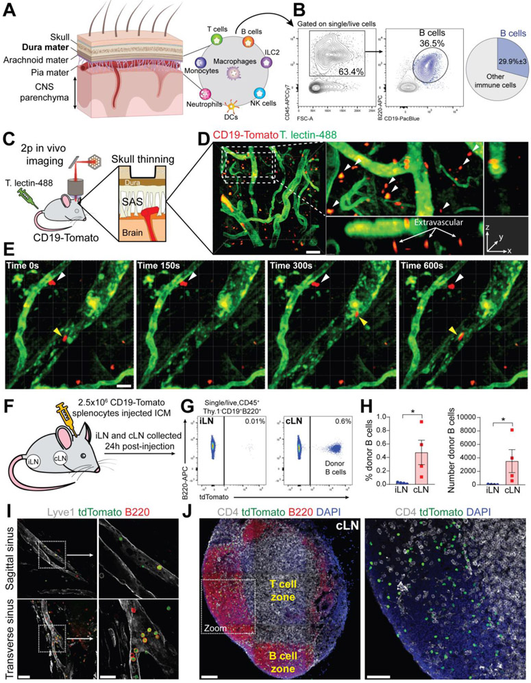
Fig. 1. B cells represent a main immune cell type in mouse meninges and are capable of trafficking through meningeal lymphatics.
(A) Cartoon representing the structural organization of the meninges. (B) Representative flow cytometry plot showing the proportion of B cells within the overall CD45+ population in mouse dura (average of n=4 mice, data generated from a single experiment). (C) Schematic depiction of the experimental approach to perform in vivo two-photon imaging in the subdural space of CD19-Tomato mice. (D) Representative two-photon image of extravascular B cells in CD19-Tomato mouse meninges (scale bar=50μm). (E) Two-photon time-lapse imaging in the meninges of a CD19-Tomato mouse. Intravascular B cell: yellow arrowhead; extravascular B cells: white arrowhead (scale bar=20μm). (F) Schematic depiction of the experimental approach (ICM: Intra-Cisterna Magna). (G) Flow cytometry analysis of donor (CD19-Tomato) derived B cells in inguinal lymph nodes (iLN) and cervical lymph nodes (cLN) 24h post-injection. (H) Frequency and absolute number of donor-derived B cells in iLN and cLN (mean ± SEM; n=4 mice; Mann Whitney U test *P<0.05; data generated from a single experiment). (I) Representative confocal image of donor (CD19-Tomato) B cells trafficking through the dura lymphatics 24h post-injection (low magnification image scale bar=50μm; high magnification image scale bar=20μm). (J) Representative confocal image of donor (CD19-Tomato) derived B cells in cLN 24h post-injection (low magnification image scale bar=100μm; high magnification image scale bar=50μm).

