. 2021 Aug 16;10:e62001. doi: 10.7554/eLife.62001

Figure 4. Synaptic inputs to dI2 neurons.

Schematic representations of the experimental design for labeling dI2::GFP or dI2::Cherry interneurons (INs; cyan) and potential sources of synaptic inputs (magenta). The soma densities of dI2 INs and the synaptic densities are illustrated in (A–E). The density values presented are 10–80%, 20–80%, 25–80%, 30–50%, and 20–80%, respectively. The laminar distributions are illustrated on the right side of (A–E). Examples of dI2 neurons contacted by axons or synaptic boutons are shown in (A′–D′), and their 3D reconstruction is shown in (A″–D″). Genetic labeling was achieved using specific enhancers (Figure 1—figure supplement 1A) introduced by electroporation at HH18. (A) Dorsal root ganglion (DRG) neurons form contacts on dI2 neurons. Inset in (A): cross-section of embryonic day (E) 17 embryos at the crural plexus level of the lumbar cord. A dorsally located dI2 neuron contacted by numerous sensory afferents, magnified in (A′) and 3D-reconstructed in (A″) (N = 18 sections, the scheme was constructed based on one representative embryo). (B) Premotor neurons (pre-MNs) form contacts on dI2 neurons. dI2 neurons were labeled at HH18. At E13, PRV virus was injected into the leg musculature, and the embryo was incubated until the infection of the pre-MNs (39 hr) (N = 34 sections, the scheme was constructed based on two representative embryos). (C) dI1 neurons form synapses on dI2 neurons. (N = 8568 synapses, 2 embryos). (C′) A representative SV2::Cherry synapse on dI2 dendrites positive for synaptotagmin. Demonstrated by horizontal and vertical optical sections in the Z-axis (see cursors and color channels). (D) V1 neurons form synapses on dI2 neurons (N = 1923 synapses, 2 embryos). (E) dI2 neurons are not contacted by 5-HT synaptic terminals (N = 1718 synapses, 1 embryo). E17 cross-sections of dI2::GFP-labeled embryos were stained for 5-HT. See Figure 4—source data 1.

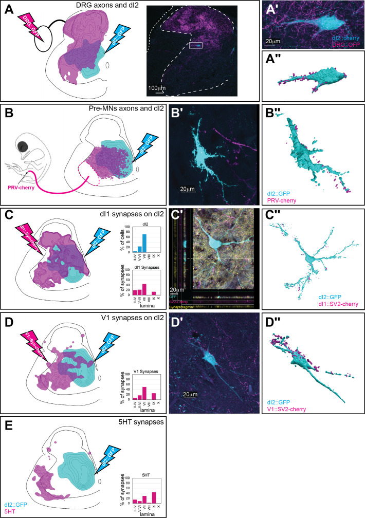
Figure 4—source data 1. Localizations of pre-dI2 terminals and synapses at the sciatic level.
The X/Y coordinates of dorsal root ganglion (DRG) axons and premotor neurons (pre-MNs) axons (Figure 4A and B). The X/Y coordinates of dI1, V1, and synapses and 5HT terminals at the sciatic level (Figure 4C–E).

Figure 4.

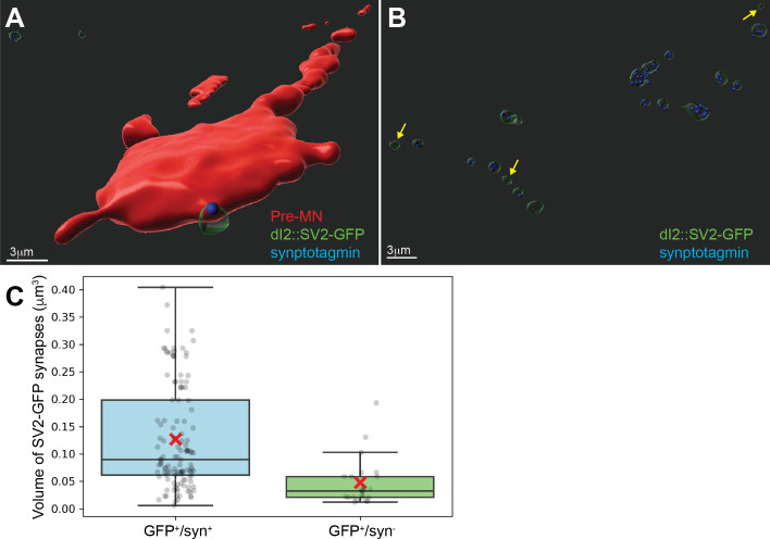
Figure 4—figure supplement 1. Validation of the synaptic reporter as an indicator of synapses.

Figure 4—figure supplement 1.

(A) Premotor neurons (red) contacted by dI2::SV2-GFP and colabeled with synaptotagmin (syn in blue) antibody. (B) A group of SV2-GFP-labeled boutons and the syn content (blue foci). Note that only the syn- boutons are small SV2-GFP boutons (yellow arrows). (C) Quantification of the volume of GFP+ boutons positive or negative for syn. The SV2-GFP+/syn+ boutons had a larger volume than the SV2-GFP+syn- boutons. (N = 121 and N = 23 for syn-positive and -negative GFP+ synapses, respectively, from one representative embryo. p<0.001, t-test).
Figure 4—figure supplement 1—source data 1. Validation of the use of SV2-GFP reporter as an indicator for synapses.
The volume of GFP+/syn+ and GFP+/syn- dI2 synapses on premotor neurons (Figure 4—figure supplement 1C).
Figure 4—figure supplement 2. dI1i neurons are premotor neurons (pre-MNs).

Figure 4—figure supplement 2.

(A, B) Schematic illustration of the strategy for studying the potential innervation of motor neurons by dI1 neurons (A). dI1 neurons were targeted by a dI1-specific enhancer (Figure 1—figure supplement 1A). At embryonic day (E) 15, synaptic boutons labeled with SV2-GFP were studied on cross-sections. A synaptic reporter (cyan) was expressed in dI1 neurons. dI1i boutons contacting Chat+ motor neurons (magenta) are apparent (B). (C, D) Schematic representation of the experimental design for colabeling dI1 and pre-MNs, supplemented by cell soma densities of dI1 neurons (cyan, N = 643 cells) and pre-MNs (magenta, N = 936 cells). dI1 neurons were labeled at HH18. At E13, PRV virus was injected into the leg musculature, and the embryo was incubated until the infection of the pre-MNs (35 hr) (C). In a cross-section taken at E13, PRV-Cherry (magenta) was detected in motor neurons (Chat+, yellow) and pre-MNs (D). An example of dI1 neurons coexpressing GFP and PRV-Cherry is shown in (D′).
Figure 4—figure supplement 2—source data 1. Localization of dI1 synapses.
The X/Y coordinates of dI1 synapses at the sciatic level (Figure 4—figure supplement 2C).
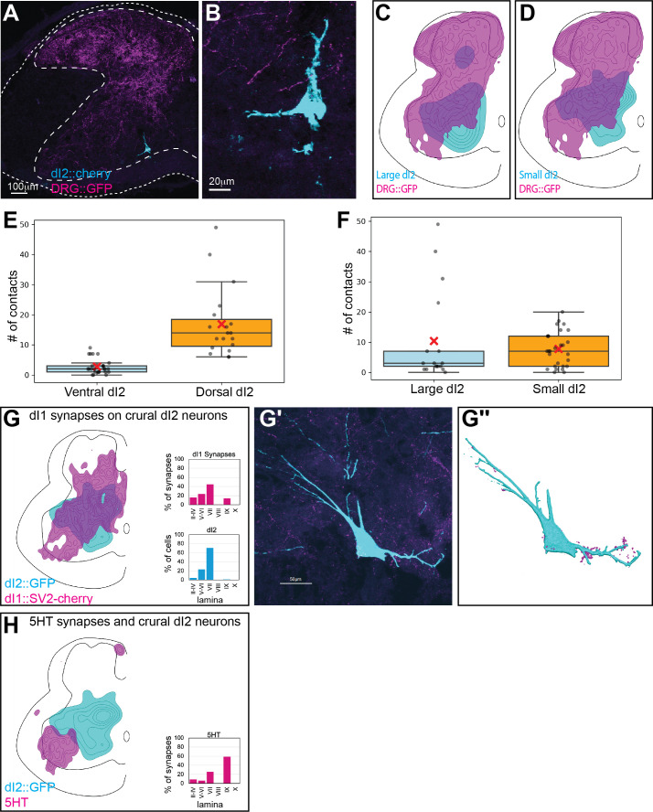
Figure 4—figure supplement 3. Input of dorsal root ganglion (DRG), dI1, and 5-HT neurons to dI2 neurons at the level of the crural plexus.

Figure 4—figure supplement 3.

(A, B) Sparse sensory innervation of ventrally located dI2 neurons. Cross-section of an embryonic day (E) 17 embryo at the lumbar spinal cord (crural plexus level). A ventrally located dI2 neuron is sparsely contacted by sensory afferents (A), magnified in (B). (C, D) Density plots of dI2large (magenta, N = 48) and dI2small (yellow, N = 502) neurons and sensory afferents (N = 18 sections, with density values 10–80%) in the crural plexus segments. (E, F) Quantification of the number of contacts between terminals of DRG axons and dI2 neurons. The number of contacts was significantly higher in dorsal than ventral dI2 neurons (E) (N = 26 and N = 19 for ventral and dorsal neurons, respectively). p<0.00001, t-test. There was no significant difference in the number of contacts on either small or large dI2 neurons (F) (N = 17 and N = 28 for large and small neurons, respectively). p=0.49, t-test. (G) dI1 neurons form synapses on dI2 neurons. Density plot and the laminar distribution of dI1 synapses (magenta) and dI2 somata (cyan) (N = 13,369 synapses with density values 50–90%). Example of dI1 boutons (magenta) on a dI2 neuron (cyan) (G′) and its 3D reconstruction in (G″). (H) dI2 neurons are not contacted by 5-HT synaptic terminals. Density plot and laminar distribution of 5-HT synapses (magenta) and dI2 somata (cyan) (N = 2754 synapses with density values 25–85%).
Figure 4—figure supplement 3—source data 1. Distribution of dorsal root ganglion (DRG) terminals on dI2 neurons.
The ventral/dorsal location and the large/small somata of dI2 neurons that are contacted by DRG terminals (Figure 4—figure supplement 3E,F).
Figure 4—figure supplement 3—source data 2. Localizations of pre-dI2 terminals and synapses at the crural level.
The X/Y coordinates of dI1 synapses and 5HT terminals at the crural level (Figure 4—figure supplement 3G,H).