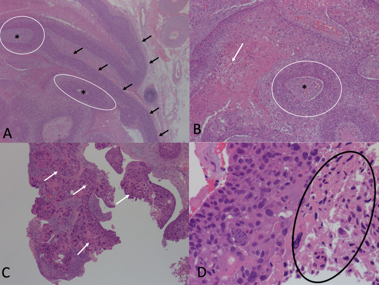
Figure 1. Histopathology of 1.5 x 1.2-centimeter biopsy of the scalp.
(A) Circles surround epithelial nests. Asterisks indicate central keratinization. Black arrows point to areas of abrupt keratinization. (B) Arrow points to the area of necrosis. Asterisk indicates central keratinization. A circle surrounds an epithelial nest. (C) Arrows point to regions of frank atypia. (D) A circle surrounds the region with abnormal mitotic figures.

