CELL BIOLOGY Correction for “Dissociating antibacterial from ototoxic effects of gentamicin C-subtypes,” by Mary E. O’Sullivan, Yohan Song, Robert Greenhouse, Randy Lin, Adela Perez, Patrick J. Atkinson, Jacob P. MacDonald, Zehra Siddiqui, Dennis Lagasca, Kate Comstock, Markus E. Huth, Alan G. Cheng, and Anthony J. Ricci, which was first published December 7, 2020; 10.1073/pnas.2013065117 (Proc. Natl. Acad. Sci. U.S.A. 117, 32423–32432).
The authors note that reference 14 is retracted and therefore should be removed from the references list.
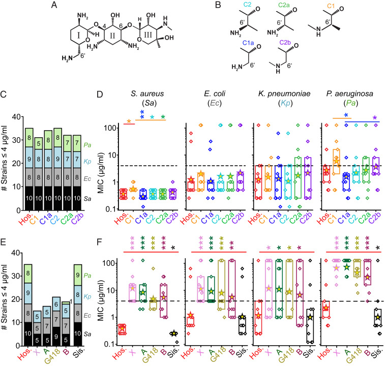
In addition, the authors note that there is an error in the “labeling of the chemical structures of gentamicin C-subtypes.” On page 32424, right column, second full paragraph, line 1, “The C-subtypes of gentamicin differ in the C5′ and C6′ positions on ring I (Fig. 1 A and B).” should instead appear as “The C-subtypes of gentamicin differ in the C6′ position on ring I (Fig. 1 A and B).”
Fig. 1.
Antimicrobial breadths and potencies of hospital gentamicin and its components. (A) Gentamicin C-subtypes are three-ringed structures, containing a central 2-deoxystreptamine with an ammonium group on either side of the deoxy carbon and added at position 4 to ring I, and at position 6 to ring III. (B) The main components of gentamicin differ at the 5′ and 6′ position on ring I. These are termed gentamicin C2 (cyan), C2a (green), C1 (orange), C1a (blue), and C2b (purple); colors used throughout. (C) The antimicrobial breadths of the C-subtypes relative to hospital gentamicin (Hos.), where breadth is defined as the number of strains inhibited by the drug at MIC value ≤ 4 μg/mL. The colors represent different species, the numbers in the columns refer to the number of individual strains inhibited, where black = S. aureus (Sa), gray = E. coli (Ec), blue = K. pneumoniae (Kp), green = P. aeruginosa (Pa); colors used throughout. Hospital gentamicin inhibits 35 of 40 strains tested, with the C-components inhibiting 31–35 strains. (D) The MIC values for all strains (diamond symbols) split by species (10 each). The box indicates the 25–75 percentiles, asterisks indicate geometric means, dashed line indicates the 4 μg/mL cutoff for susceptibility (*P < 0.05, **P < 0.01 Mann–Whitney u test). Gentamicin C1 is less potent than hospital gentamicin against S. aureus strains only (P < 0.05, Mann–Whitney u test, n = 10). There are also some differences between the C-subtypes against S. aureus and P. aeruginosa strains. (E) The antimicrobial breadths of the impurities relative to hospital gentamicin. Hospital gentamicin inhibits 35 of 40 strains tested, with the impurities gentamicin X, A, B, and G418 inhibiting 15–22 strains. The impurity sisomicin inhibited 35 of 40 strains. (F) The MIC values for all strains split by species. The impurities gentamicin X, A, B, and G418 are all significantly less potent than hospital gentamicin (P < 0.05, Mann–Whitney u test). The impurity sisomicin was the same as the hospital gentamicin against E. coli and K. pneumoniae (P > 0.05, Mann–Whitney u test), and was more potent than hospital gentamicin against S. aureus and P. aeruginosa (P < 0.05, Mann–Whitney u test). SI Appendix, Figs. S3 and S4 show individual strains for each drug. ***P < 0.001.]
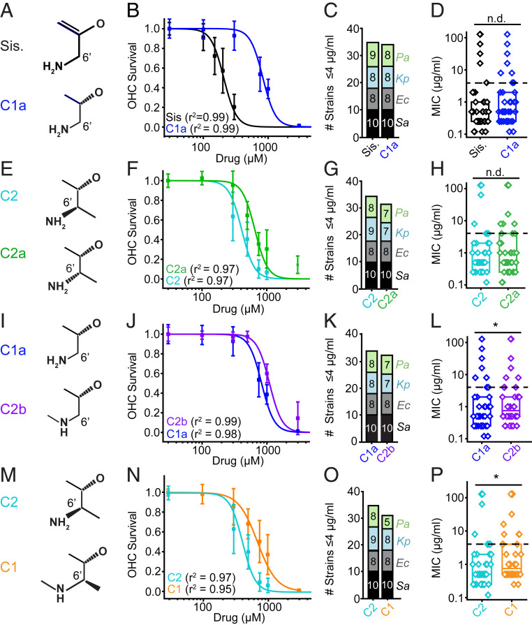
Due to the same error, Figs. 1 and 3 appeared incorrectly. The corrected figures and their legends appear below.
Fig. 3.
Structure–activity relationships between gentamicin subtypes: Ototoxicity and antimicrobial activity. (A) Sisomicin and C1a differ at the 4′ and 5′ position. (B) C4′-5′ saturation alters ototoxicity. Sisomicin has a lower EC50 than C1a (P < 0.05, F-test, n = 8–60; Sis. = 199 ± 17 μM, C1a = 821 ± 24 μM). (C). Sisomicin and C1a have similar antimicrobial breadths (35 vs. 34 stains). (D) There is no difference in potency between sisomicin and C1a (P > 0.05, Mann–Whitney u test, all strains). (E) Gentamicin C2 and C2a are epimers at the C6′ position. (F) C2 is more ototoxic than C2a (P < 0.05, F-test, n = 7–16; C2 = 403 ± 23 μM, C2a = 656 ± 36 μM). (G) C2 and C2a have similar antimicrobial breadths (35 vs. 32 strains). (H) C2 and C2a show no difference in antimicrobial potency (P > 0.05, Mann–Whitney u test, all strains). (I) Gentamicin C1a and C2b differ at the N6′ site. (J) C1a has a lower EC50 than C2b (P < 0.05, F-test, n = 8; C2b = 1,130 ± 22 μM, C1a = 821 ± 24 μM). (K) C1a and C2b have similar antimicrobial breadths (34 vs. 32 strains). (L) There is a detectable difference in potency between C1a and C2b (P < 0.05, Mann–Whitney u test, all strains). Fig. 2 shows this is related to P. aeruginosa strains. (M) Gentamicin C2 and C1 differ at the N6′ position. (N) C2 has a lower EC50 than C1 (P < 0.05, F-test, n = 8; C2 = 403 ± 23 μM, C1 = 728 ± 59 μM). (O) C2 has a slightly higher breadth than C1 (four strains). (P) C2 is more potent than C1 (P < 0.05, Mann–Whitney u test, all strains). Fig. 1 shows breakdown by species for all compounds. *P < 0.05, n.d., no difference.
Lastly, the authors note that Fig. S2 in the SI Appendix appeared incorrectly because the “labeling of rings I and III was reversed.”
The online version has been updated to include the updated reference list, the corrected text described above, the corrected Figs. 1 and 3, and the corrected SI Appendix.


