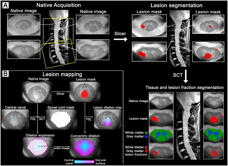
Figure 2.
Image acquisition and processing pipeline for the cervical spinal cord. (A) Spinal cord at 7 T of a 32-year-old female (RRMS, EDSS: 2.5, disease duration: 4.8 years). Acquisition with full cervical coverage aligned orthogonally to spinal cord to mitigate partial volume effects. Spinal cord lesions were manually segmented using Slicer to then be combined with the white matter and grey matter tissue segmentations from SCT to isolate the grey matter and white matter lesion fractions. (B) Spinal cord lesion mapping at 7 T of a 33-year-old female (RRMS, EDSS: 1, disease duration: 2.3 years). Manual segmentation of the central canal was used as a reference from which concentric 3D voxel-wise dilations expanded radially using scikit-image towards the subpial border, as defined by the SCT spinal cord volume. The concentric lesion dilation map was created by extracting the manual lesion segmentation from the corresponding whole spinal cord concentric dilation map using FSL. FSL = FMRIB Software Library; SCT = Spinal Cord Toolbox.

