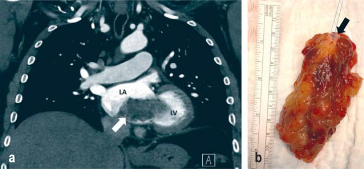
A 58-year-old patient developed dyspnea after undergoing a total hip replacement. Computed tomography revealed an intracardiac mass (arrow in Figure a). Transesophageal echocardiography was performed as part of further diagnostics. This showed an inhomogeneous structure originating in the left atrium and extending to the left ventricle. The 9 × 5 × 5-cm structure with its 1 × 1-cm “endocardial button” (endocardial attachment site—arrow in in Figure b) was successfully excised using an atrial transseptal approach under cardiopulmonary bypass. Histopathology confirmed the diagnosis of myxoma. The postoperative course was unremarkable and no recurrence was seen at 1-year postoperative follow-up. Myxoma is the most common primary cardiac tumor; it is benign, generally located in the left atrium, and usually smaller than in this case. Compared to cardiac thrombi, myxomas tend to be larger, are polypoid in shape, and are mobile structures due to the “stalk” that is usually present. The recurrence rate following myxoma excision is under 5%.
Figure.
a) Coronal section of a computed tomography angiography of the heart with contrast medium in the left ventricle and left atrium. The black filling defect of contrast medium (arrow) indicates the mass.
b) Macroscopic specimen of the surgically excised myxoma (size: 9 × 5 × 5 cm). The arrow marks the “endocardial button.”
Acknowledgments
Translated from the original German by Christine Rye.
Footnotes
Conflict of interest statement: The authors declare that no conflict of interest exists.


