. 2021 Sep 22;10:e69795. doi: 10.7554/eLife.69795

Figure 3. Genetic inactivation of agtr1a and agtr1b in DA neurons is neuroprotective.

(A) Schematic showing the procedure of FACs to isolate DA neurons for qPCR analysis of RAAS pathway gene expression. (B) qPCR data of 5 dpf larval samples show the relative expression of RAAS pathway genes normalized to the house-keeping gene eef1a1, in DA neurons (red bars) versus non-DA cells (blue bars). PRR (prorenin receptor), agtr1a (Angiotensin II receptor, type 1a), agtr1b (Angiotensin II receptor, type 1b), agtr2 (Angiotensin II receptor, type 2), ace (Angiotensin I converting enzyme), ace2 (Angiotensin I converting enzyme 2) (n = 2 biological replicates, 6 technical replicates). (C) A schematic showing the conditional CRISPR design, imaging, and analysis procedure to inactivate agtr1a and agtr1b in DA neurons. (D) Confocal images of DA neurons in 5 dpf (before MTZ treatment) and 6 dpf (24 hr after 10 mM MTZ treatment) larvae injected with either the scrambled control sgRNA construct (top) or the effective agtr1a and agtr1b sgRNA construct (bottom). Yellow cells express both NTR-mCherry and Cas9. (E) Quantification shows a significant preservation of DA neuron intensity in the agtr1a and agtr1b sgRNA construct-injected animals compared to the scrambled sgRNA control upon 10 mM MTZ treatment. (n = 15, p < 0.01, unpaired t-test).

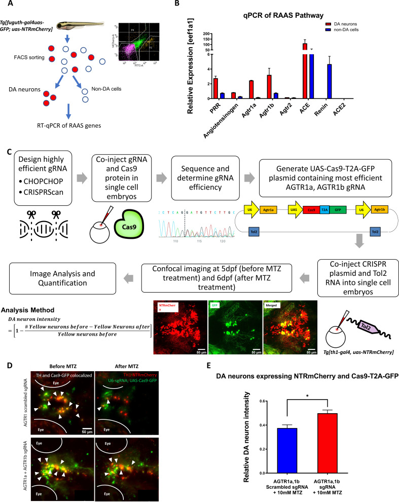
Figure 3—source data 1. DA neuron quantification for conditional CRISPR.
Figure 3—source data 2. Fluorescent activated cell sorting output file.

Figure 3.

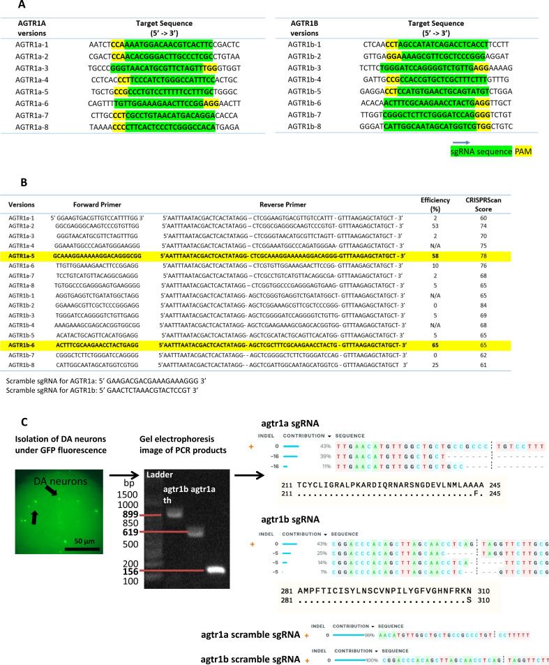
Figure 3—figure supplement 1. sgRNA design and validation for conditional CRISPR knockout of agtr1a and 1b in DA neurons.

Figure 3—figure supplement 1.

(A) sgRNA target sequences (green) with PAM sites highlighted in yellow in the agtr1a (left) and agtr1b (right) genes. 8 different targets for each gene were examined to determine the highest sgRNA KO efficiency. (B) Primers used to construct template plasmids for sgRNA synthesis. sgRNA efficiency was calculated based on sequencing followed by ICE software analysis (https://ice.synthego.com/#/) comparing the knockouts to control. CRISPRScan scores with predicted efficiency is also shown in column 5. The sgRNAs highlighted in yellow were used for conditional CRISPR KO of agtr1a and agtr1b on DA neurons. (C) Schematic showing the workflow for validating successful knockout of agtr1a and agtr1b in DA neurons. After DA neuron imaging, larval zebrafish brains were dissociated and DA neurons expressing Cas9-GFP and NTR-mCherry were collected via mouth-pipetting and pooled. PCR was performed to amplify genomic DNA regions targeted by agtr1a and agtr1b sgRNAs followed by sequencing. The samples were also amplified with th primers for QC. Sequencing results were analyzed with the Synthego ICE software. In this example dataset, at least 50% or 40% of sequenced reads carry open reading frame-shifting deletions in agtr1a and agtr1b genes, respectively. The scrambled sgRNAs for agtr1a and agtr1b showed no indel mutations when compared to controls.