Abstract
Background.
Peri-operative cardiac xenograft dysfunction (PCXD) was described by McGregor et al. to be a major barrier to the translation of heterotopic cardiac xenotransplantaton into the orthotopic position. It is characterized by graft dysfunction in the absence of rejection within 24-48 hours of transplantation. We describe our experience with PCXD at a single program.
Methods.
Orthotopic transplantation of genetically engineered pig hearts was performed in 6 healthy baboons. The immunosuppression regimen included induction by anti-CD20 mAb, Thymoglobulin, cobra venom factor and anti-CD40 mAb and maintenance with anti-CD40 mAb, MMF and tapering doses of steroids. Telemetry was used to asses graft function. Extracorporeal membrane oxygenation was used to support one recipient. A full human clinical transplant team was involved in these experiments and the procedure was performed by skilled transplant surgeons.
Results.
A maximal survival of 40 hours was achieved in these experiments. The surgical procedures were uneventful and all hearts were weaned from cardiopulmonary bypass (CPB) without issue. Support with inotropes and vasopressors was generally required after separation from CPB. The cardiac xenografts performed well immediately, but within the first several hours required increasing support and ultimately suffered arrest despite maximal interventions. All hearts were explanted immediately; histology showed no signs of rejection.
Conclusions.
Despite excellent surgical technique, uneventful weaning from CPB, and adequate initial function, orthotopic cardiac xenografts slowly fail within 24-48 hours without evidence of rejection. Modification of preservation techniques and minimizing donor organ ischemic time may be able to ameliorate PCXD.
Our group has established extremely long-term survival of the cardiac xenograft in a heterotopic position (1). This required optimization of the immunosuppressive regimen in a xenotransplant model, as well as the addition of human transgenes to porcine donors (2-4). The next step toward clinical application is applying the lessons learned in the heterotopic model to a life-supporting orthotopic model. Prior groups have attempted this and described a perioperative cardiac xenograft dysfunction (PCXD), unrelated to rejection, in the orthotopic position.
Failure in the orthotopic position can be due to a number of factors. Technical failure has certainly been a concern in early experiments, particularly with non-clinically active and inexperienced surgeons. Immunologic rejection is the major hurdle to xenotransplantation, both acute and chronic. The hallmark histopathologic feature of xenograft rejection is microthrombi formation in the microvasculature and is the primary finding used to differentiate between graft failure secondary to rejection versus technical misstep, inflammation, or progressive intrinsic graft dysfunction.
PCXD is thought to be secondary to a systemic inflammatory response associated with xenotransplantation compounded with the hemodynamic load placed on the xenograft in a life-sustaining position (5,6). Reichart et al. have attempted to overcome PCXD with changes to myocardial preservation with some recent success; however, in addition to a preservation approach that is not commonly employed in clinical practice, they have utilized a medication regimen that may not be translatable to human subjects (7). Here, we report our early preclinical experience with PCXD in a pig-to-non-human primate model.
Material and Methods
Animals
This research was approved by the Institutional Animal Care and Use Committee of the University of Maryland School of Medicine. Six healthy specific pathogen-free (SPF) juvenile baboons of either sex from University of Oklahoma (Norman, OK) underwent orthotopic xenotransplantation of genetically engineered (GE) pig hearts by clinical thoracic transplant surgeons. Donor pigs were provided by Revivicor, Inc and were all alpha α-1,3 - galactosyl transferase gene knockout (GTKO) with additional human transgene expression (shown in table 1). GTKO pigs were produced by breeding over six generations. The methods of genetic modification of these pigs has been previously described (8). Transgenes were stable at the genomic and protein level. The weights of donor pigs were matched with the baboon recipient to accommodate the heart in the baboon chest.
Table 1.
Genetic Modifications
| 1 | GTKO.hCD46 |
| 2 | GTKO.hCD46.hHLAE.B4KO |
| 3 | GTKO.hCD46.hvWF.hTBM.hEPCR.hCD47.hHO1.B4KO |
| 4 | GTKO.hCD46.hTBM.hCD47.hECPR.hHO1 |
| 5 | GTKO.hCD46.hTBM.hCD47.hECPR.hHO1 |
| 6 | TKO.hTBM.hEPCR.hCD46.hDAF.hHO1.hCD47 |
B4KO, β4galNT2 knockout; GTKO, α-1,3-galactosyltransferase gene knockout; hCD46, human CD46; hCD47, human CD47; hHLAE, human leukocyte antigen-E; human hTBM, human thrombomodulin; hvWF, human von Willebrand factor; TKO, triple knockout.
Immunosuppression
Immunosupression regimen included induction with anti-thymocyte globulin (ATG), anti-CD20 antibody (Rituximab), anti-CD40 antibody (clone 2C10R4) and cobra venom factor (CVF; Quidel, San Diego, CA). Maintenance therapy was achieved with anti-CD40 antibody, mycophenolate mofetil (MMF), and a tapered course of systemic steroids. Immunosuppression regimen is shown in table 2.
Table 2.
Immunosuppression Regimen
| Drugs | Dose | Timing |
|---|---|---|
| Induction | ||
| ATG | 4-5 mg/kg | Preop days −2 & −1 |
| CVF | 50-100 U/kg | Preop days −1, 0 & 1 |
| Anti-CD20 Ab | 19 mg/kg | Preop days −7, 0, 7 & 14 |
| Anti-CD40 Ab | 50 mg/kg | Preop days −1 & 0 |
| Maintenance | ||
| Anti-CD40 Ab | 50 mg/kg | Postop days 3, 7, 10, 14, 19, q weekly |
| MMF | 20 mg/kg/2 h IV infusion | BID daily |
| Steroids | 2 mg/kg | BID, tapered off in 7 wk |
| Aspirin | 81 mg | Daily |
| Heparin | Maintain ACT 2x baseline | Continuous infusion starting post-op day 1 |
| Supportive | ||
| Ganciclovir | 5 mg/kg/d | Daily |
| Cefazolin | 250 mg | BID for 7 d |
| Epogen | 200 U/kg | Daily from −7 to 7 then weekly |
Orthotopic Xenotransplantation
Anesthesia was provided by clinical cardiac anesthesia attendings in conjunction with senior veterinary staff. Clinical perfusionists managed cardiopulmonary bypass and, in one case, extracorporeal membrane oxygenation (ECMO). The single recipient supported with ECMO was placed on mechanical support in a planned, prophylactic fashion to attempt to overcome peri-operative xenograft dysfunction. Therefore, ECMO support was initiated immediately following cardiopulmonary bypass. Invasive monitoring was undertaken with an arterial line and a temporary implantable telemetry device. This was placed at the time of operation and monitoring was achieved utilizing a pressure line in the apex of the left ventricle and epicardial leads producing an electrocardiogram tracing (9,10). Serial laboratory values were obtained in-house, including arterial blood gas (ABG) and point-of-care iSTAT, with samples sent out for comprehensive chemistry and complete blood counts. Resuscitation with crystalloid, colloid, matched allogeneic blood, exogenous bicarbonate, magnesium, and potassium administration all followed standard clinical guidelines and were performed by an intensive care provider in conjunction with a bedside cardiac surgery nurse with supervision by the surgical team (11). External defibrillation and limited cardiopulmonary resuscitation was performed where necessary, but in each case futility was decided liberally at the discretion of the principal investigator to preserve the quality of xenograft histology.
Non-Gal Antibody Assays
Anti-pig non-Gal (IgG and IgM) antibodies were measured in serum samples collected from recipient baboons. Samples were collected 7 days prior to transplant, immediately prior to transplant, and within 24 hours of transplant. Levels were quantified by flow cytometry using GTKO porcine endothelial cells, as described previously (1).
Histology
Tissue samples were taken from each cardiac chamber of explanted xenografts for paraffin sections, hematoxylin and eosin staining and light microscopy. Sections were reviewed for the presence of cellular infiltrates, hemorrhage, thrombosis, necrosis, and myocyte damage. Percentage of necrosis and degree of microthrombi formation was determined for each section. Presence of microthrombi was defined by a scoring system as follows: 1+ = > 0; 2+ = >1-5; 3+ = >5-10; and 4+ = > 10.
Results
Xenograft Survival
All baboons underwent surgery without technical complication (Table 3). In each case, separation from cardiopulmonary bypass occurred with ease. Median epinephrine dose was 0.03mcg/kg/min; median norepinephrine dose was 0.10mcg/kg/min. Early function as assessed by inotropic requirements, mean arterial blood pressure, laboratory data including lactate, pH, and bicarbonate levels, and transesophageal echocardiography was adequate to good, with left ventricular ejection fraction greater than 40% in all measures in the first 6 hours post-operatively. Persistently worsening of lactate and bicarbonate levels was characteristic of each post-operative case.
Table 3.
Clinical courses
| Donor weight |
Recipient weight |
Survival (hours) |
Course | |
|---|---|---|---|---|
| 1 | 13kg | 13kg | 18 | Progressive biventricular dysfunction; increasing requirements to epinephrine 0.04mcg/kg/min, norepinephrine 0.28mcg/kg/min; PEA arrest. |
| 2 | 9kg | 10kg | 7 | Large volume resuscitation, bicarbonate and calcium dependence; epinephrine to 0.06mcg/kg/min, vasopressin to 0.04mcg/kg/min; bradycardic arrest. |
| 3 | 21kg | 25kg | 26 | Stable overnight; weaned off of inotropes and pressors; sudden VF arrest post-operative day 1, unable to recover. |
| 4 | 10kg | 10kg | 2 | Weaned from CPB requiring support; maximal inotropic and vasopressor support, unsustainable with rising lactate; elective termination. |
| 5 | 23kg | 21kg | 22 | Worsening diastolic function with preserved systolic function. Maximum epinephrine 0.03mcg/kg/min, norepinephrine 0.12mcg/kg/min. Progressive edema, ascites, multisystem organ dysfunction and respiratory arrest. |
| 6 | 9kg | 11kg | 40 | Epinephrine stable at 0.03mcg/kg/min, norepinephrine 0.20mcg/kg/min, consistent volume requirement for chatter/flow drops, dependence on ECMO support. |
Abbreviated clinical courses are shown in table 3. Survival was between 6 and 26 hours in this group, and 40 hours in the single recipient receiving mechanical circulatory support. All recipients ultimately suffered worsening clinical status with increasing requirements for chemical support (and, in a single case, mechanical support) prior to cardiopulmonary collapse. ECMO was utilized in a single case to prolong survival to 48 hours. Adequate flows were achieved throughout as calculated based on recipient size; despite this, the recipient underwent progressive cardiac xenograft failure and was completely ECMO-dependent by conclusion of the experiment at the time of withdrawal of support at 40 hours. All grafts were immediately explanted for histology at the time of hemodynamic collapse (following elective euthanasia by exsanguination under deep anesthesia as per institutional protocol).
Non-Gal Antibody Assays and Histology
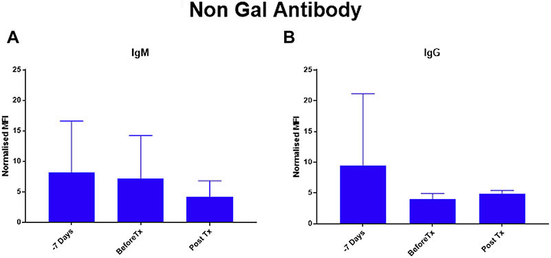
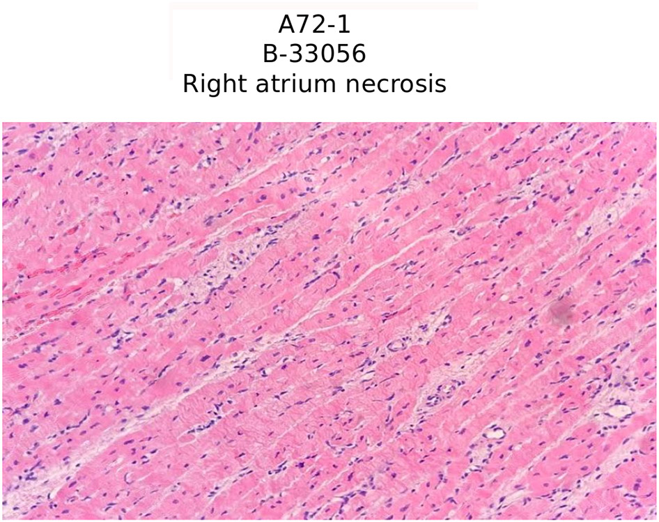
Non-Gal IgG and IgM levels decreased after induction, and remained at levels lower than naive baseline in both pre- and post-transplant serum measures (figure 1). Representative histology is shown in figure 2. In H & E staining, cardiac myocyte death and necrosis was seen in all samples. Microthrombi, markers of rejection, were completely absent from 5 explanted grafts (score 0+). A single graft demonstrated 1+ microthrombi isolated to the left ventricle and apex.
Figure 1.
Non-Gal Antibody Assay: Anti pig non-Gal (A) IgG and (B) IgM antibody levels in mean fluorescence intensity (MFI) were measured in cardiac xenograft baboon recipient’s serum by flow cytometry using GTKO porcine endothelial cells. MFI was normalized with respect to positive control serum as one hundred.
Figure 2.
Hematoxylin and eosin staining from right atrium (representative) of xenograft with necrosis and without any sign of rejection or microthrombosis.
Comment
PCXD is a real phenomenon contributing to the loss of orthotopic cardiac xenografts in the first 48 hours of transplantation in the absence of technical failure and acute rejection. In our experience and that of other groups, meticulous post-operative care with avoidance of inotropes and correction of metabolic derangements can decrease the incidence to as low as 40-60% in the orthotopic model (5,12). In addition, new experience with myocardial preservation may facilitate a decrease in this rate to even lower levels (7). In fact, with changes in our own preservation strategy, we have decreased our incidence to 33% in early studies and significantly prolonged our survival (unpublished data). By all indications, including histopathology of the explanted grafts, rejection plays a very insignificant role in graft failure. As xenotransplantation progresses, it’s important to recognize this as a real entity representing an alternative pathway of graft failure, independent from rejection (7,13). It will be particularly important to consider the implications of PCXD and develop methods to overcome it in early clinical trials. In our single experience attempting to overcome this with prophylactic intiation of ECMO support, we found that this was insufficient. This makes it even more important to recognize ahead of clinical applications as this common “bailout” method may not be sufficient. In the absence of technical failure or rejection, we advocate for early retransplantation in these patients. As noted above, with significant changes to our procurement and preservation method, we are seeing early sustainable success in this model. This has greatly reduced, but not completely eliminated, the risk of PCXD in our program. The integral next steps to advancing the field of cardiac xenotransplantation will include the refinement and optimization of the preservation approach in these GE porcine donors, through experiments involving preservation of the GE pig heart alone and in concert with transplantation into a baboon recipient.
Conclusion
Cardiac xenotransplantation is continuing its slow march toward a clinical reality, but substantial hurdles still remain. Current experiences in both heterotopic and orthotopic models are encouraging. However, PCXD remains a real threat to xenograft survival in the orthotopic position and occurs independently from mechanisms of rejection. As xenotransplantation moves closer to clinical trials, establishing approaches to both reduce the risk of PCXD and rescue the patient when it does occur is crucial.
Footnotes
Publisher's Disclaimer: This is a PDF file of an article that has undergone enhancements after acceptance, such as the addition of a cover page and metadata, and formatting for readability, but it is not yet the definitive version of record. This version will undergo additional copyediting, typesetting and review before it is published in its final form, but we are providing this version to give early visibility of the article. Please note that, during the production process, errors may be discovered which could affect the content, and all legal disclaimers that apply to the journal pertain.
References
- 1.Mohiuddin MM, Singh AK, Corcoran PC, Thomas ML 3rd, Clark T, Lewis BG, Hoyt RF, Eckhaus M, Pierson RN 3rd, Belli AJ, Wolf E, Klymiuk N, Phelps C, Reimann KA, Ayares D, Horvath KA. Chimeric 2C10R4 anti-CD40 antibody therapy is critical for long-term survival of GTKO.hCD46.hTBM pig-to-primate cardiac xenograft. Nat Commun. 2016April5;7:11138. [DOI] [PMC free article] [PubMed] [Google Scholar]
- 2.Thompson P, Cardona K, Russell M, Badell IR, Shaffer V, Korbutt G, Rayat GR, Cano J, Song M, Jiang W, Strobert E, Rajotte R, Pearson T, Kirk AD, Larsen CP. CD40-specific costimulation blockade enhances neonatal porcine islet survival in nonhuman primates. Am J Transplant. 2011;11:947–957. 3. [DOI] [PMC free article] [PubMed] [Google Scholar]
- 3.Mohiuddin MM, Corcoran PC, Singh AK, Azimzadeh A, Hoyt RF Jr, Thomas ML, Eckhaus MA, Seavey C, Ayares D, Pierson RN 3rd, Horvath KA. B cell depletion ex tends the survival of GTKO.hCD46Tg pig heart xenografts in baboons for up to 8 months. Am J Transplant. 2012;12:763–771. [DOI] [PMC free article] [PubMed] [Google Scholar]
- 4.Singh AK, Chan JL, DiChiacchio L, Hardy NL, Corcoran PC, Lewis BGT, Thomas ML, Burke AP, Ayares D, Horvath KA, Mohiuddin MM. Cardiac xenografts show reduced survival in the absence of transgenic human thrombomodulin expression in donor pigs. Xenotransplantation. 2018October5:e12465. [DOI] [PMC free article] [PubMed] [Google Scholar]
- 5.Byrne GW, McGregor CG. Cardiac xenotransplantation: progress and challenges. Curr Opin Organ Transplant. 2012April;17(2):148–54. doi: 10.1097/MOT.0b013e3283509120. [DOI] [PMC free article] [PubMed] [Google Scholar]
- 6.Byrne GW, Du Z, Sun Z, Asmann YW, McGregor CG. Changes in cardiac gene expression after pig-to-primate orthotopic xenotransplantation. Xenotransplantation. 2011Jan-Feb;18(1):14–27. [DOI] [PMC free article] [PubMed] [Google Scholar]
- 7.Längin M, Mayr T, Reichart B, Michel S, Buchholz S, Guethoff S, Dashkevich A, Baehr A, Egerer S, Bauer A, Mihalj M, Panelli A, Issl L, Ying J, Fresch AK, Buttgereit I, Mokelke M, Radan J, Werner F, Lutzmann I, Steen S, Sjöberg T, Paskevicius A, Qiuming L, Sfriso R, Rieben R, Dahlhoff M, Kessler B, Kemter E, Klett K, Hinkel R, Kupatt C, Falkenau A, Reu S, Ellgass R, Herzog R, Binder U, Wich G, Skerra A, Ayares D, Kind A, Schönmann U, Kaup FJ, Hagl C, Wolf E, Klymiuk N, Brenner P, Abicht JM. Consistent success in life-supporting porcine cardiac xenotransplantation. Nature. 2018December;564(7736):430–433. [DOI] [PubMed] [Google Scholar]
- 8.Phelps CJ, Koike C, Vaught TD, Boone J, Wells KD, Chen SH, Ball S, Specht SM, Polejaeva IA, Monahan JA, Jobst PM, Sharma SB, Lamborn AE, Garst AS, Moore M, Demetris AJ, Rudert WA, Bottino R, Bertera S, Trucco M, Starzl TE, Dai Y, Ayares DL. Production of alpha 1,3 galactosyltransferase deficient pigs. Science. 2003;299:411–414. [DOI] [PMC free article] [PubMed] [Google Scholar]
- 9.Längin M, Panelli A, Reichart B, Kind A, Brenner P, Mayr T, Abicht JM. Perioperative Telemetric Monitoring in Pig-to-Baboon Heterotopic Thoracic Cardiac Xenotransplantation. Ann Transplant. 2018July20;23:491–499. [DOI] [PMC free article] [PubMed] [Google Scholar]
- 10.Horvath KA, Corcoran PC, Singh AK, Hoyt RF, Carrier C, Thomas ML 3rd, Mohiuddin MM. Left ventricular pressure measurement by telemetry is an effective means to evaluate transplanted heart function in experimental heterotopic cardiac xenotransplantation. Transplant Proc. 2010Jul-Aug;42(6):2152–5. [DOI] [PMC free article] [PubMed] [Google Scholar]
- 11.Marino PL. The ICU Book. 4th edition. 2014. [Google Scholar]
- 12.Byrne GW, Du Z, Sun Z, Asmann YW, McGregor CG. Changes in cardiac gene expression after pig-to-primate orthotopic xenotransplantation. Xenotransplantation. 2011Jan-Feb;18(1):14–27 [DOI] [PMC free article] [PubMed] [Google Scholar]
- 13.Mohiuddin MM, DiChiacchio L, Singh AK, Griffith BP. Xenotransplantation: A step closer to clinical reality? Transplantation. In press. [DOI] [PubMed] [Google Scholar]


