Abstract
Chronic thromboembolic pulmonary hypertension is an underdiagnosed and potentially fatal subgroup of pulmonary hypertension, if left untreated. Clinical signs include exertional dyspnea and non-specific symptoms. Diagnosis requires multimodality imaging and heart catheterization. Pulmonary endarterectomy, an open heart surgery, is the gold standard treatment of choice in selected patients in specialized centers. Targeted medical therapy and balloon pulmonary angioplasty can be effective in high-risk patients with significant comorbidities, distal pulmonary vascular obstructions, or recurrent/persistent pulmonary hypertension after pulmonary endarterectomy. Currently, there is a limited number of data regarding novel coronavirus-2019 infection in patients with chronic thromboembolic pulmonary hypertension and the changing spectrum of the disease during the pandemic. Challenging times during this outbreak due to healthcare crisis and relatively higher case-fatality rates require convergence; that is an ultradisciplinary collaboration, which crosses disciplinary and sectorial boundaries to develop integrated knowledge and new paradigms. Management strategies for the "new normal" such as virtual care, preparedness for further threats, redesigned standards and working conditions, reevaluation of specific recommendations, and online collaborations for optimal decisions for chronic thromboembolic pulmonary hypertension patients may change the poor outcomes.
Keywords: Balloon pulmonary angioplasty, chronic thromboembolic pulmonary hypertension, pulmonary endarterectomy, pulmonary thromboembolism, targeted medical therapy
Introduction
Pulmonary hypertension (PH) is a group of diseases that may interfere, involve, and contribute to various cardiovascular and respiratory diseases.[1-4] The clinical classification is consisted of five groups according to clinical presentations, pathological findings, pathogenetic and hemodynamic characteristics, and treatment strategies.[1,2,5-9]
Chronic thromboembolic pulmonary hypertension (CTEPH) is a progressive, potentially life-threatening obstructing vasculopathy considered a long-term complication of pulmonary thromboembolism (PTE), leading to significant mortality, morbidity, and poor quality of life.[1,2,6,10-17] It is a potentially curable form of PH and classified as Group 4 in the PH classification and shown in Table 1 and abbreviations in Table 2.[1,2,4,7,8,11,12,18-20]
Table 1. Pulmonary hypertension classification.
| Group 1 | Group 2 | Group 3 | Group 4 | Group 5 |
| Idiopathic PAH | PH due to left heart disease | PH due to lung diseases and/or hypoxia | PH due to pulmonary artery obstructions | PH with unclear and/or multifactorial mechanisms |
| PAH related with connective tissue disease | PH with preserved LVEF | Obstructive and restrictive lung disease | CTEPH | Hematological disorders |
| HIV infection | PH with reduced LVEF | Other lung disease with mixed restrictive/ obstructive pattern Hypoxia without lung disease | Other obstructive reasons (parasites, sarcoma) | Systemic and metabolic disorders |
| Portal hypertension | Valvular heart disease | Developmental lung disorders | Other reasons | |
| Congenital heart diseases | Congenital/ acquired cardiovascular conditions leading to post-capillary PH | |||
| Drug- and toxin-induced PAH | ||||
| PAH long-term responders to calcium channel blockers | ||||
| Persistent PH of the newborn syndrome | ||||
| PAH: Pulmonary arterial hypertension; PH: Pulmonary hypertension; LVEF: Left ventricular ejection fraction; CTEPH: Chronic thromboembolic pulmonary hypertension; HIV: Human immunodeficiency virus. | ||||
Table 2. Abbreviations.
| BPA | Balloon pulmonary angioplasty |
| CTED | Chronic thromboembolic disease |
| CTEPH | Chronic thromboembolic pulmonary hypertension |
| DHCA | Deep hypothermic circulatory arrest |
| DSA | Digital subtraction angiography |
| DVT | Deep vein thrombosis |
| ECMO | Extracorporeal membrane oxygenation |
| IVC | Inferior vena cava |
| mPAP | Mean pulmonary artery pressure |
| MRA | Magnetic resonance angiography |
| MRI | Magnetic resonance imaging |
| NT-proBNP | N-terminal pro-B-type natriuretic peptide |
| PAP | Pulmonary artery pressure |
| PEA | Pulmonary endarterectomy |
| PH | Pulmonary hypertension |
| PVR | Pulmonary vascular resistance |
| PTE | Pulmonary thromboembolism |
| RCT | Randomized controlled trial |
| SVC | Superior vena cava |
| VA-ECMO | Veno-arterial extracorporeal membrane oxygenation |
| VKA | Vitamin K antagonist |
| V/Q | Ventilation/perfusion |
| VV-ECMO | Veno-venous extracorporeal membrane oxygenation |
| VTE | Venous thromboembolism |
In this disease, pulmonary vascular resistance (PVR) and pulmonary artery pressure (PAP) increases, resulting in the gradual progress of the disease, followed by right ventricular (RV) failure and death.[10-17] Mechanism in CTEPH depends on segmental obstruction of pulmonary blood flow, high shear stress in non-occluded areas, and progressive PH due to vascular remodeling in distal vessels.[16,17] Non-operable CTEPH has been associated with a five-year survival of 30%.[15]
Early diagnosis is challenging.[8,20] Late diagnosis have a negative impact on prognosis.[8] The diagnosis is made in the presence of the following criteria: (i) (with or without a prior episode of PTE), at least segmental perfusion defects in ventilationperfusion (V/Q) scintigraphy should be obtained with intraluminal filling defects in computed tomography pulmonary angiography (CTPA) and/or magnetic resonance angiography (MRA), and/or pulmonary angiography; (ii) finalizing the diagnosis of PH with a right heart catheterization, resting mean PAP (mPAP) should be equal or greater than 25 mmHg, and pulmonary capillary wedge pressure should be lower than 15 mmHg; and (iii) these findings should be obtained after effective anticoagulant therapy for at least three months.[1-3,21-23]
Sometimes the thrombi in the pulmonary arteries may not resolve, but the patient may still not suffer from PH at rest either. This disease has similar symptoms and perfusion defects with CTEPH, but without PH at rest and defined as chronic thromboembolic pulmonary disease (CTED).[1,2,24] There are robust data regarding satisfactory results with pulmonary endarterectomy (PEA) for CTEPH. However, currently, we do not have sufficient data to conclude about a definitive evolution of CTED. There are no clear guideline recommendations for CTED,[1,2,4,6,22,23] unlike the precise guidelines for PH and PTE.[1,22]
Currently, limited data from surveys are available regarding COVID-19 infection in patients with PH and CTEPH.[24] Patients with CTEPH having multiple comorbidities are distinctively at a higher risk for severe novel coronavirus-2019 (COVID-19) infection. Relatively higher case-fatality rates in comparison with the general population are alarming and require reevaluation of specific recommendations. Convergence (ultradisciplinary collaboration), which crosses disciplinary and sectorial boundaries to develop integrated knowledge and new paradigms, may help to solve too many gray zones to be considered.[25-27]
EPIDEMIOLOGY
Studies have proven that CTEPH is an underdiagnosed disease.[28-39] In a prospective follow-up study, the cumulative incidence of symptomatic CTEPH after an acute episode of PTE was 1% at six months, 3.1% at one year, and 3.8% at two years.[28] A recent study showed that approximately 3.2% of all survivors who had an acute episode of PTE would develop CTEPH.[30] The International Prospective Registry data including 679 newly diagnosed consecutive patients with CTEPH from Europe and Canada showed that 75% of CTEPH patients had a history of PTE, and 56% of them had a history of deep vein thrombosis (DVT).[20]
It is estimated from the registries that the incidence of CTEPH is thought to be 3 to 30 per million in the general population.[4,31] The incidence of CTEPH in the United States and Europe ranges from 3 to 5/100,000.[29] While the incidence of CTEPH increases in the fourth to sixth decade, it is rare in childhood.[2,20,28] A lack of awareness and ambiguousness of symptoms results in misdiagnosis. Furthermore, a significant number of cases may be asymptomatic with a honeymoon period after PE.[21,32] Screening is recommended within two years following a PE episode.[33]
In Turkey, it is estimated that 800 individuals per year are suffering from CTEPH, but only 200 of them can have a chance for an optimal treatment. In the Registry on Clinical Outcome and Survival in Pulmonary Hypertension Groups (SIMURG), CTEPH accounted for 19% of the enrolled 1,501 patients with PH from 20 adult cardiology PH centers.[34]
The International Prospective Registry data showed that 32% of CTEPH patients had thrombophilic disorders, including elevated Factor VIII and lupus anticoagulant/antiphospholipid antibodies.[20,35] Moreover, this group of patients is more likely to have blood groups A, B, and AB, which may be associated with an increased level of Factor VIII. The relationship between thrombophilia and PTE is well known. However, the association of CTEPH and several disorders, including protein C and protein S, the G20210A mutation of prothrombin, and hyperhomocysteinemia, has not been fully understood, yet.[40] Despite that, routine thrombophilia screening, including antiphospholipid antibodies, may be useful.
Other clinical risk factors are genetic predisposition, a history of splenectomy, infected ventriculoatrial shunts for the treatment of hydrocephalus, hypothyroidism and thyroid hormone replacement, chronic inflammatory bowel disease, chronic osteomyelitis, malignancy, and infected pacemaker lines.[28,36] The prognosis is poor with these conditions.[28,36-38]
The CTEPH incidence after the COVID-19 pandemic and coronavirus challenge in patients with existing CTEPH remains to be resolved. A recent prospective study during the pandemic reported a high prevalence of PE in patients with COVID-19 at the time of hospital admission.[39] In the near future, the impact of COVID-19 on the epidemiology of CTEPH would be clearer.
PATHOPHYSIOLOGY
The pathophysiology includes intraluminal thrombus organization, fibrotic scar tissue-like stenosis resulting from incomplete thrombus resolution following PE, and subsequent vascular remodeling of unaffected pulmonary vessels.[40-44] Previous studies have described that the organized thrombi are tightly attached to the pulmonary arterial medial layer and can form complete occlusion or form different grades of stenosis, webs and bands.[45,46] Chronic intravascular scar formation, high shear stress in non-occluded areas, and progressive PH due to microvascular remodeling in distal pulmonary vessels lead to progressive PH and finally RV dysfunction.[17,42,47]
Catheter studies in healthy individuals have demonstrated a normal mPAP at rest of about 14.0±3.3 mmHg.[48] The task force has proposed the inclusion of an mPAP of >20 mmHg as the threshold and PVR of ≥3 WU in the definition of pre-capillary PH.[30] Hemodynamically increased mPAP results in increased RV wall tension, RV hypertrophy and dilatation and, at later stages, RV failure in untreated patients. The main cause of vascular disease in patients with CTEPH can be considered the remodeling of the vascular wall after thromboembolism and the increased vascular resistance in the following period. Thus, RV failure is the main cause of death in CTEPH.
CLINICAL PRESENTATION
The cardinal symptom is progressive exertional dyspnea.[1,2,3,9] However, the diagnosis is often delayed or missed due to non-specific symptoms and a lack of awareness. Non-specific symptoms, such as exercise intolerance, chest discomfort, fatigue, and depression can be potentially misleading.[10,11] As the disease progresses, signs of RV failure, including jugular venous distension, hepatomegaly, ascites, and peripheral edema, may be seen.[7,49] Occasionally, hypoxemia, syncopal episodes during effort, lightheadedness, stress angina, hemoptysis, or chest pain may be present.
Chronic thromboembolic pulmonary hypertension should be kept in mind for patients with a history of VTE in the presence of progressive exertional dyspnea.[9,10,21] Typically, clinical history reveals a suspicion of previous PE or DVT of the lower limbs in more than half of the patients with CTEPH.[7,9] With effective anticoagulation, resolution is expected within three to six months.[7] The persistence of symptoms more than six months should alert the physicians.
DIAGNOSIS
Any patient with a history of PTE and progressive dyspnea is a potential candidate for CTEPH. A high index of suspicion is essential for early diagnosis.[48,49,50]
Physical examination may reveal prominent RV impulse, a split of the second heart sound, a RV, S4 gallop, a right-sided S3, and varying degrees of tricuspid regurgitation.
The International Prospective Registry data reported a median time interval of 14.1 months between the first symptoms and CTEPH diagnosis.[20] However, early detection of CTEPH has a paramount importance on survival rates.[8,28] Furthermore, an accurate diagnosis requires multimodality imaging.[21,50]
Chest radiography and pulmonary function tests may help in excluding possible airway or parenchymal lung diseases.[7] Right ventricular enlargement, a prominent pulmonary trunk, and hilar pulmonary arteries can be detected on chest X-ray.
Electrocardiographic (ECG) findings may show RV strain and hypertrophy, including P-pulmonale, right bundle branch block, T-wave abnormalities, and rightaxis deviation.[1,51,52]
Elevated brain natriuretic peptide (BNP) and N-terminal prob-type natriuretic peptide (NT-proBNP) correlate with the severity of RV dysfunction and hemodynamic outcome.[53] A simple, non-invasive diagnostic model was developed by Klok et al.[52] based on ECG assessment and NT-proBNP measurements with a sensitivity of 94% (95% confidence interval [CI]: 86-98%) and a specificity of 65% (95% CI: 56-72%).
ECHOCARDIOGRAPHY
The current guidelines recommend echocardiography as the first-line non-invasive screening tool to detect PH and RV dysfunction.[1,2,16] Echocardiographic evaluations include estimation of pulmonary arterial systolic, diastolic, and mean pressures, quantifiable tricuspid regurgitant signals, RV pressure size, and function, septal shift, right atrial size, inferior vena cava (IVC) diameter, and pericardial effusion. Echocardiographic imaging can also rule out left-sided valvular lesions, left ventricular systolic and diastolic dysfunction, or intracardiac shunts. Echocardiographic evaluation in symptomatic patients with a suspicion of PH aims to estimate probability of PH, and has been based on two-step assessment comprising measurement of tricuspid regurgitation peak velocity by Doppler, and assessment for supportive findings suggestive of PH. Following the assessment for PH probability and the presence or absence of risk factors for PAH and CTEPH determines the decision for heart catheterization to confirm PH diagnosis.[1,2,9,18,20,23,53,54]
LUNG V/Q SCINTIGRAPHY
The V/Q scintigraphy is the first-line screening imaging method to rule out CTEPH.[1,2,7,50,55] A normal V/Q scan effectively eliminates CTEPH diagnosis with a sensitivity of 90 to 100% and a specificity of 94 to 100%.[1,50,55]
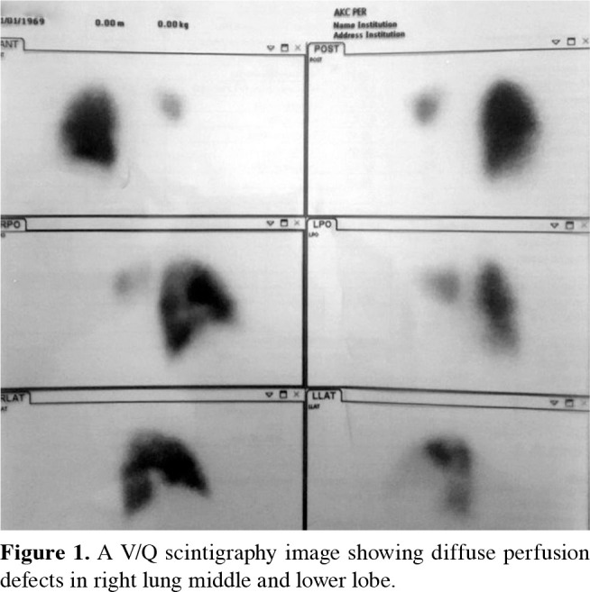
Lobar and segmental perfusion defects are typical in CTEPH patients. An example of V/Q scintigraphy is shown in Figure 1 demonstrating diffuse perfusion defects in the right lung middle and lower lobe. False-positive V/Q scans may be misleading with pulmonary artery sarcoma, large-vessel pulmonary vasculitis, extrinsic compressive lesions, pulmonary microvasculopathy.
Figure 1. A V/Q scintigraphy image showing diffuse perfusion defects in right lung middle and lower lobe.
COMPUTED TOMOGRAPHY (CT)
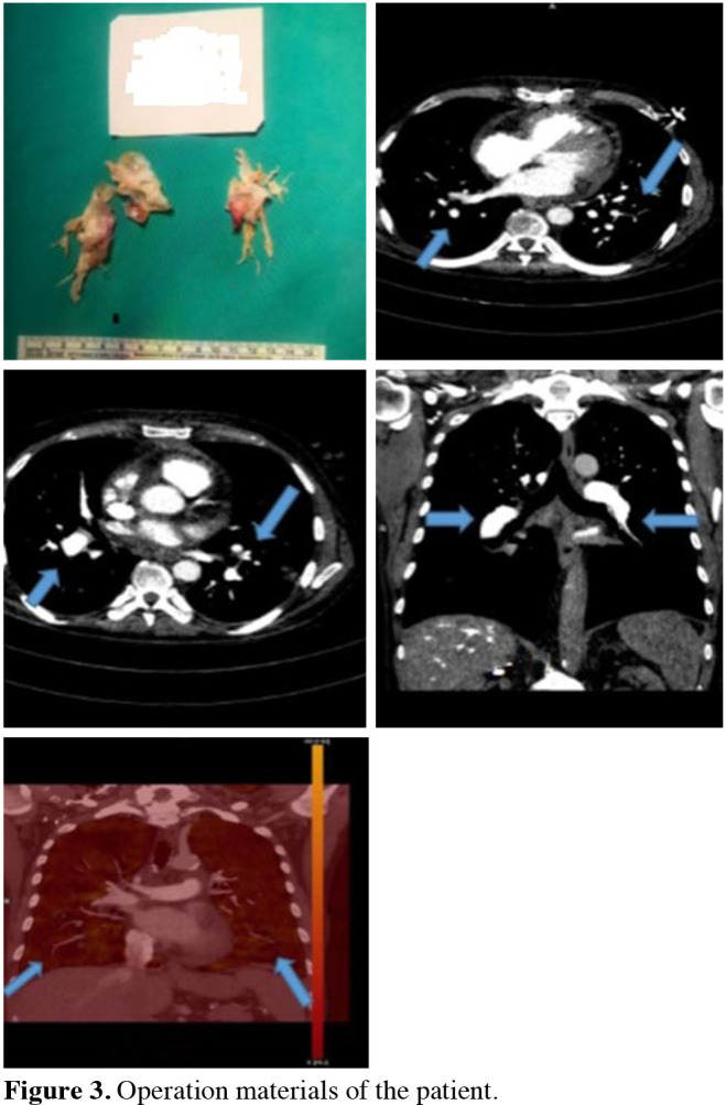
Spiral CT is an excellent imaging modality with a sensitivity of 85 to 90% to visualize occlusion of pulmonary arteries, eccentric filling defects compatible with thrombi, recanalization, webs, or bands. Moreover, post-stenotic dilatation or aneurysms and calcification in the thrombus can be identified using this method. Dual-energy CT, which has been successfully applied in recent years, provides valuable information regarding perfusion defects.[9,19,55] Pre- and postoperative CT scans and the operation material of a demonstrative patient are shown in Figures 2 and 3.
Figure 2. Pre and postoperative dual energy computed tomography scans showing preoperative occlusions of the pulmonary artery branches, the endarterectomy material and the significant improvement after surgical treatment.
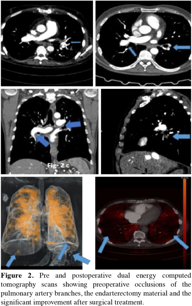
Figure 3. Operation materials of the patient.
MAGNETIC RESONANCE IMAGING (MRI)
The MRI is a promising and evolving modality without radiation exposure in CTEPH diagnosis and evaluating cardiac remodeling both before and after PEA.[56] It not only allows diagnosis of both acute and chronic thromboembolism, but also enables the qualitative and quantitative determination of right and left ventricular function.[57]
DIGITAL SUBTRACTION PULMONARY ANGIOGRAPHY
Digital subtraction pulmonary angiography is considered the gold standard for characterizing vessel morphology, surgical operability or applicability of interventional treatments in CTEPH.[2,9,23,24,44,48]
If the V/Q scan is suggestive of CTEPH, pulmonary angiography is required for accurate diagnosis and anatomical guidance for surgery. Selective pulmonary angiography is helpful for visualization of filling defects, web, ring-like or total obstructive lesions in pulmonary artery branches. It is also quite accurate in the identification of distal obstructions.
HEART CATHETERIZATION
Cardiac catheterization has a critical role in the diagnostic algorithm of PH and CTEPH to establish the specific diagnosis and determine the severity of PH. It includes measurement of systolic, diastolic, and mean arterial pressures, pulmonary capillary wedge pressure, RV and right atrial pressures, serial blood sampling for oxygen saturation, and calculation of cardiac output and index, and PVR. Intracardiac shunting should be also ruled out.[1,2,9]
Furthermore, coronary angiography for obstructive lesions and extrinsic compression of left main coronary artery by the aneurysmal or dilated pulmonary artery, and selective bronchial angiography should be considered during heart catheterization. The cardiologist's perspective prefers simultaneous pressure tracing for right and left-sided circulatory units to evaluate the changes in systemic pressure on the pulmonary arterial wedge and mean pressures. Therefore, heart catheterization rather than right heart catheterization seems to be the more appropriate definition for this procedure.
EXERCISE TESTING
The 6-Minute Walking Test (6-MWT) is another diagnostic modality that has become widely used in both follow-up and clinical studies, and is a simple and repeatable test to measure the exercise capacity of patients.[1-4,11]
DIFFERENTIAL DIAGNOSIS
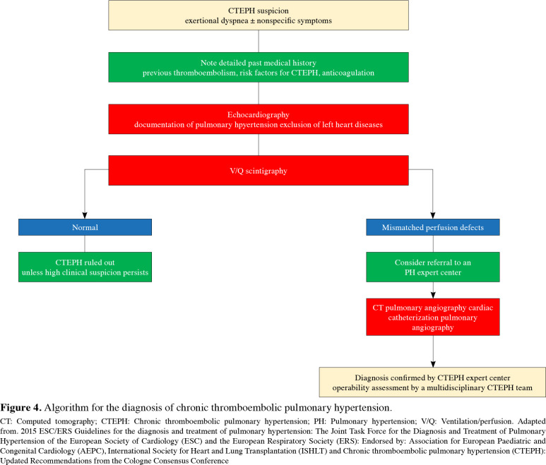
If perfusion disorder is detected when there is no problem in respiratory function test, PH should be considered in the differential diagnosis, and if the patient has a history of PTE, CTEPH should be considered.[5] The diagnostic algorithm is summarized in Figure 4.[1,2,3,58,59]
Figure 4. Algorithm for the diagnosis of chronic thromboembolic pulmonary hypertension. CT: Computed tomography; CTEPH: Chronic thromboembolic pulmonary hypertension; PH: Pulmonary hypertension; V/Q: Ventilation/perfusion. Adapted from. 2015 ESC/ERS Guidelines for the diagnosis and treatment of pulmonary hypertension: The Joint Task Force for the Diagnosis and Treatment of Pulmonary Hypertension of the European Society of Cardiology (ESC) and the European Respiratory Society (ERS): Endorsed by: Association for European Paediatric and Congenital Cardiology (AEPC), International Society for Heart and Lung Transplantation (ISHLT) and Chronic thromboembolic pulmonary hypertension (CTEPH): Updated Recommendations from the Cologne Consensus Conference.
Non-thrombotic pulmonary embolism (i.e., tumors, parasites, foreign materials) should be excluded in the differential diagnosis. Rare sarcomas and Echinococcosis-related hydatidosis should be considered, and these entities can be distinguished clinically from CTEPH by specific features.[60]
TREATMENT
A critical point is to make the decision based on an interdisciplinary approach consisting of different disciplines such as pulmonology, cardiology, cardiovascular surgery (cardiothoracic surgery in certain locations), radiology, nuclear medicine, and anesthesia and intensive care.[9] Curative treatment is PEA[1,2,3,9-12,42-44] performed under cardiopulmonary bypass (CPB) and deep hypothermic circulatory arrest (DHCA).[1-3,9-12,42-44,61-66] This surgery has become an accepted and curative treatment modality worldwide with the increase of awareness and can be applied with low mortality with the help of recent advances in imaging and diagnostic methods, advances in myocardial protection techniques in cardiovascular surgery, advances in supporting systems for postoperative care in intensive care unit settings, and advances in medical treatment.[67-74] It is well known that the learning curve for diagnosis and management of CTEPH is an essential factor such as the commitment and the technical background of the center.[74-83] As cardiovascular surgeons, we must focus not only on how to perform a proper endarterectomy, but also on fundamental training and management of the problems in cardiac surgery principles, such as the management of CPB and DHCA, weaning from CPB in the presence of RV failure due to PH, and also the use of extracorporeal membrane oxygenation (ECMO) support in challenging cases.[61,67] Therefore, the importance of experienced and committed inter/ultradisciplinary teams who can provide a second or third opinion should not be neglected. This fact has emerged as a reality, particularly in the COVID-19 pandemic and pandemic-related travel restrictions.[26]
The severity of the patients" symptoms, correlation between accessibility of thrombus and the degree of PH and RV failure are important criteria to consider while deciding on the operation. The level and accessibility of obstruction are the most important surgical criteria. An "haute couture" approach has paramount importance similar to all areas of cardiovascular surgery. Life expectancy and individual expectations may vary depending on several factors.[1,2,9,20,23]
Although increased PVR or advanced RV failure is not an absolute contraindication to surgery, these patients may have some challenges in the postoperative period. In some cases, additional medications or mechanical circulatory support systems may be needed. Patients with PVR values of 1,000 dyn·s/cm5 or ≤12 Wood units are defined as relatively low-risk patients.[9-11] If the thrombus extends to the main branches of the pulmonary artery, lobar arteries and even segmental arteries, surgical removal of chronic thrombus material is considered safe. Removal of chronic clot material from the distal segmental or sub-segmental arteries may be difficult and, in some centers, this group of patients may be considered inoperable. In this case, a second opinion should be taken from another experienced center.
Patient-specific comorbidities and risk factors, long-term risk analysis, technical difficulties (hostile thorax such as previous sternotomy, chest anomalies, previous coronary artery bypass grafting) are the other factors that can play a role in the decision-making process.[15,31,83] However, two issues need to be considered with a special caution. First, patients with parenchymal lung damage (patients with severe emphysema or lung parenchymal damage) would experience severe respiratory failure in the postoperative period and, therefore, surgery may not be the right choice. Hence, it may not be beneficial to perform surgery to perfuse a non-ventilated lung. Second, surgery for patients with severe comorbid conditions such as end-stage lung disease or malignancy should be considered non-beneficial.[1,2,3,9,20,23] Several risk models are described for PH patients (e.g., COMPERA), and we, as the authors of this paper, prefer the risk-benefit ratio described by Jenkins et al.[43]
SURGERY
Convergence (Ultradisciplinary) Approach
Chronic thromboembolic pulmonary hypertension with RV failure may cause hemodynamic instability during anesthetic induction and in the pre-CPB period, and associated comorbidities (pulmonary, hepatic) may affect the actions and metabolism of anesthetic drugs. Therefore, anesthetic care for patients needs special attention in every step. During the CPB period, the anesthesia team must ensure that adequate perfusion, cerebral oxygenation, and hypothermia for DHCA are employed. Very close collaboration with the anesthesiology team has a vital importance to manage either specific problems of the operation (i.e., residual PH, pulmonary edema, pulmonary bleeding, and RV failure) or various metabolic and cardiovascular issues related to hypothermic circulatory arrest.
Surgical details
A median sternotomy is required for CPB and surgical accessibility to both lungs. The IVC and superior vena cava (SVC) are encircled. The CPB is established with standard aorta-bicaval cannulation and a left ventricular vent through the right upper pulmonary vein. A second vent can be placed into the pulmonary artery, if necessary.[9,20,23]
The cardioplegia cannula is inserted into the aortic root, and the circulation should be discontinued by cooling to 20ºC. The DHCA is probably the most important part of the operation to provide a clear, bloodless field of vision. Another crucial point is the identification of the correct plane of dissection in the segmental and sub-segmental branches of the pulmonary artery.
It is important to stay in the same plane circumferentially to avoid losing the plane of dissection, as it is advanced into the segmental and sub-segmental branches.
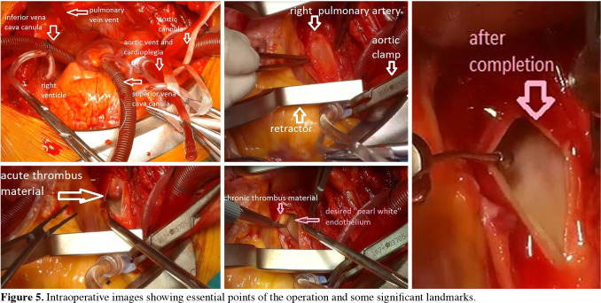
The critical point is to be able to perform a complete endarterectomy without any residue.[43,44,54,68] Particularly in the distal part, it is vital that the endarterectomy material should not be cut, not be ripped off, and again with a meticulous dissection, removing the material as en bloc w ith i ts t ail h as a vital importance to prevent residual PH.[43,44,54,58] If the disease has a very distal localization, another DHCA period can be necessary. After completing endarterectomy, the arteriotomy is closed with 5-0 or 6-0 Prolene, usually with a double running fashion. Concomitant cardiac pathologies such as coronary artery disease or valvular heart disease can be treated surgically during the warming period after total circulatory arrest.[1,2,20,23] The most common procedure is closure of patent foramen ovale.[1,2,3,9] Some essential points and significant landmarks of the operation are shown in Figure 5.
Figure 5. Intraoperative images showing essential points of the operation and some significant landmarks.
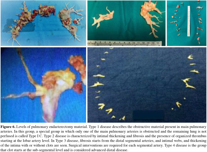
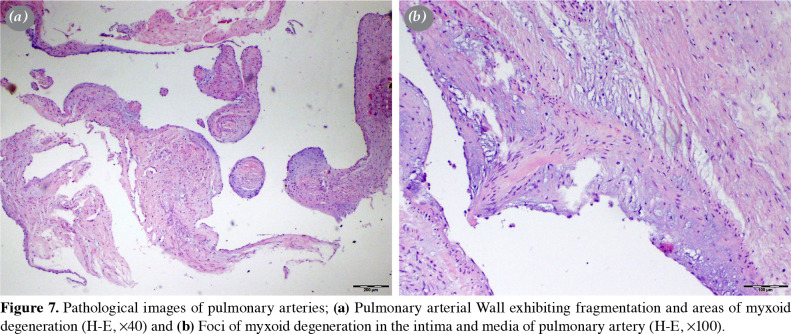
The surgical classification was made according to materials extracted and there are five levels.[9,20,23] The types of diseases are summarized in Figure 6 and the pathological images are demonstrated in Figure 7.
Figure 6. Levels of pulmonary endarterectomy material. Type 1 disease describes the obstructive material present in main pulmonary arteries. In this group, a special group in which only one of the main pulmonary arteries is obstructed and the remaining lung is not perfused is called Type I C. Type 2 disease is characterized by intimal thickening and fibrosis and the presence of organized thrombus starting at the lobar artery level. In Type 3 disease, fibrosis starts from the distal segmental arteries, and intimal webs, and thickening of the intima with or without clots are seen. Surgical interventions are required for each segmental artery. Type 4 disease is the group that clot starts at the sub-segmental level and is considered advanced distal disease.
Figure 7. Pathological images of pulmonary arteries; (a) Pulmonary arterial Wall exhibiting fragmentation and areas of myxoid degeneration (H-E, x40) and (b) Foci of myxoid degeneration in the intima and media of pulmonary artery (H-E, x100).
Another important question has emerged about brain protection during DHCA. This question was answered after Pulmonary Endarterectomy Cognitive (PEACOG) study, neurocognitive functions of patients who underwent DHCA and selective perfusion were compared, and no significant difference was found between the two groups.[68]
Weaning from CPB
The snares are released from SVC and IVC. Air is removed from the cardiac chambers and the aorta is unclamped. The patient is warmed to 37ºC and vent cannulas are removed followed by a mandatory meticulous hemostasis, particularly focused on the suture lines of both pulmonary arteries. Pericardial patch can be sutured over the bleeding suture lines along the pulmonary arteries to control the bleeding. Reinforcing stitches along the suture lines should be avoided to prevent tears on pulmonary arteries, particularly in patients with postoperative PH.[1,2,4,9,20,23]
Transesophageal echocardiography (TEE) during surgery, which is a non-invasive monitoring method, provides extremely important information regarding the hemodynamic follow-up and treatment processes. After PEA, RV parameters reflecting RV EF such as global functional area change (FAC) and tricuspid annular plane systolic excursion (TAPSE) can be quantitatively measured.
Complications related to open heart surgery such as postoperative bleeding, mediastinitis, atrial arrhythmias, central nervous system complications, renal problems, recurrence, laryngeal, and phrenic nerve injuries can be seen after the operation. Another important complication is massive pulmonary hemorrhage which can be potentially fatal.[9,20,23] Gentle dissection in the right plane is the main principle in preventing such an injury; however, the damage may occur, particularly in fragile arteries. The entire operating team needs to be aware of this complication and treatment options. Flexible bronchoscopy devices, surgical adhesives, and bronchial blockers must be ready in the operating theatre. After finishing the endarterectomy, a bubble test should be performed before weaning off CPB. The identification of bleeding before restarting the circulation is important to repair the disruption easily; otherwise, it may be difficult to repair and a pulmonary resection option may be also considered.[69]
After PEA, patients with residual PH are natural candidates for reperfusion injury, pulmonary edema, and RV failure. Fluid replacement should be done carefully in the postoperative period. Nitric oxide may be a good option to decrease PVR. Milrinone can be another option for RV support. Pressure control, positive end-expiratory pressure, and inverse ratio ventilation are helpful strategies to maintain an acceptable ventilation-perfusion matching and decreasing the risk of further damage. However, if pulmonary edema and RV failure persist and the strategies mentioned earlier fail, veno-arterial ECMO (VA-ECMO) is a useful treatment modality to decrease the RV volume overload and improve cardiac output, leading to better oxygen transport. It must be always remembered that ECMO is one of the devices that should be found in CTEPH centers.[9,61,70]
What about the tricuspid valve? Repair or not to repair? Other concomitant procedures
Tricuspid valve regurgitation in various degrees may be detected in CTEPH patients undergoing PEA. As cardiovascular surgeons, our intention is usually to perform a tricuspid valve repair. However, tricuspid valve repair is unnecessary, unless there is a serious structural problem involving the leaflets or chorda tendinea. After PEA, RV remodeling resolves and, therefore, we believe that tricuspid regurgitation secondary to annular dilatation should be omitted.[2,31,32,56,70]
TARGETED MEDICAL THERAPY
Traditionally, diuretic agents, oxygen therapy, and lifelong anticoagulant therapy are important parts of classical medical therapy. In anticoagulant therapy, the target international normalized ratio (INR) should be between 2.0 and 3.0. Anticoagulant therapy prevents pulmonary artery thrombosis and recurrent thromboembolism. We do not have enough data on the usage of direct-acting oral anticoagulants, despite the accumulated information that has increased in the medical treatment of DVT in recent years. Specific treatment for PH in CTEPH should be considered mainly in three patient groups:[1-3,7,9] (i) patients who are anatomically unsuitable for endarterectomy, since the lesion is placed very distally. This decision should be confirmed by a second opinion; (ii) patients with persistent PH after PEA; and (iii) p atients w ho a re deemed to be very risky as a result of the decision of the Multidisciplinary Council due to accompanying comorbid conditions (severe pulmonary parenchymal disease, morbid obesity, hepatic or renal dysfunction, diabetes mellitus, coronary artery disease, hostile thorax due to previous mediastinal surgery). Patients who are unwilling to undergo surgery can be considered in this group.[2,9,71,72]
Riociguat treatment has emerged as an effective therapeutic option additionally to classical medical therapy in CTEPH.[71-73] It is a soluble guanylate cyclase stimulator, acts on nitric oxide receptor, used orally, and the only approved drug for inoperable cases or treatment of recurrent/persistent PH after surgery. The starting dose is 0.5 mg t.i.d. By controlling the side effects, titrating and increasing the dose, the optimum dose of 2.5 mg t.i.d. should be reached.[2,9,71-73]
In the literature, there are studies conducted with other agents, although they have not been approved yet.[74,75] Discussions on bridging treatment are still in the gray zone, and the results of multi-center studies are waiting.[9]
BALLOON PULMONARY ANGIOPLASTY (BPA)
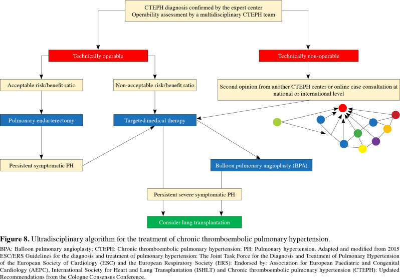
Balloon pulmonary angioplasty has been considered an emerging treatment modality in small, segmental, and sub-segmental vessels in high-risk surgical patients due to multiple comorbidities or unwillingness of patients for PEA or residual CTEPH following PEA. The rationale beneath this percutaneous treatment is based on the presence of web, ring-like narrowing, or total occlusion amenable to balloon dilations. The role of BPA in CTEPH and treatment algorithm is summarized in Figure 8.
Figure 8. Ultradisciplinary algorithm for the treatment of chronic thromboembolic pulmonary hypertension. BPA: Balloon pulmonary angioplasty; CTEPH: Chronic thromboembolic pulmonary hypertension; PH: Pulmonary hypertension. Adapted and modified from 2015 ESC/ERS Guidelines for the diagnosis and treatment of pulmonary hypertension: The Joint Task Force for the Diagnosis and Treatment of Pulmonary Hypertension of the European Society of Cardiology (ESC) and the European Respiratory Society (ERS): Endorsed by: Association for European Paediatric and Congenital Cardiology (AEPC), International Society for Heart and Lung Transplantation (ISHLT) and Chronic thromboembolic pulmonary hypertension (CTEPH): Updated Recommendations from the Cologne Consensus Conference.
The Japanese experience for BPA seems to provide robust data for efficacy and safety issues of this method in a selected subgroup of cases with CTEPH. The fundamental principles of different BPA strategies are to initiate balloon dilations with smaller balloon sizes than vessel diameters and multiple dilations for target lesions and target branches in each session, and to increase the balloon size at sequential sessions until achieving a satisfactory drop in PA pressures and PVR.
The risks for reperfusion pulmonary edema and vessel perforation are the most dreadful complications.[76-80] The National Hospital Organization, Okayama Medical Center reported that mPAP reduced to below 25 mmHg threshold in 85% of patients with sequential BPA sessions within six months. After BPA, the 10-year survival rate was reported as 89.7%.[80] Following these encouraging results of the Japanese experience, BPA programs have been also initiated in many countries.[77]
The Turkish BPA program was launched under the leadership of Dr. Matsubara in 2017, and was followed by at least five PH/CTEPH centers capable of performing BPA with satisfactory efficacy and safety results. Despite this enthusiasm, BPA should not be regarded as a treatment strategy that is an alternative to PEA, and decisions for eligibility to BPA should be based on the consensus of the multidisciplinary CTEPH teams.[2,16,24]
A recent study examined the changes in NT-proBNP in patients undergoing BPA for inoperable CTEPH and showed that NT-proBNP levels decreased after BPA, providing valuable evidence for the efficiency of this treatment modality.[81]
As a late-breaking abstract, the Riociguat versus Balloon Pulmonary Angioplasty in Non-operable Chronic Thromboembolic Pulmonary Hypertension (RACE) study was an open-label randomized, parallel-group study evaluating the efficacy and safety of riociguat versus BPA in newly diagnosed and treatment-naïve patients with inoperable CTEPH in 20 centers.[82] Patients aged 18 to 80 years, with inoperable CTEPH and eligible for BPA, without a history of specific PH therapies were enrolled in the study. Change in PVR from baseline to Week 26 was set as the primary endpoint, whereas changes in the 6MWT, functional class (FC), NT-pro BNP, and Borg Dyspnea Scale scores were secondary endpoints. The BPA significantly improved PVR compared to riociguat (BPA vs. r iociguat: P VR r eduction 4 0%, ratio of geometric means, 95% CI: 0.60, 0.53 to 0.69, p<0.0001), FC and NT-proBNP. There was no significant difference in the 6MWT between the two groups. However, BPA was associated with a higher number of serious adverse events. It seems that further randomized-controlled trials and prospective, longitudinal CTEPH registries including inoperable CTEPH are required to draw a firm conclusion on this subject.[83]
In conclusion, chronic thromboembolic pulmonary hypertension is critical, but a curable disease with pulmonary endarterectomy and there are medical options for inoperable patients. Collaboration with other disciplines has of utmost importance. Increasing awareness and establishing new expert and committed centers would provide benefits to this patient population.
Acknowledgments.
We are grateful for the mentorship in starting new CTEPH programs in Turkey, provided by Dr. Madani, Dr. Mayer, Dr. Jenkins, and Dr. Matsubara. This review article is intended to merge the knowledge generated by multiple disciplines, and we thank the members of all fields for their outstanding work.
Footnotes
Conflict of Interest: The authors declared no conflicts of interest with respect to the authorship and/or publication of this article.
Financial Disclosure: The authors received no financial support for the research and/or authorship of this article.
References
- 1.Galiè N, Humbert M, Vachiery JL, Gibbs S, Lang I, Torbicki A, et al. 2015 ESC/ERS Guidelines for the diagnosis and treatment of pulmonary hypertension: The Joint Task Force for the Diagnosis and Treatment of Pulmonary Hypertension of the European Society of Cardiology (ESC) and the European Respiratory Society (ERS): Endorsed by: Association for European Paediatric and Congenital Cardiology (AEPC), International Society for Heart and Lung Transplantation (ISHLT) Eur Respir J. 2015;46:903–975. doi: 10.1183/13993003.01032-2015. [DOI] [PubMed] [Google Scholar]
- 2.Wilkens H, Konstantinides S, Lang IM, Bunck AC, Gerges M, Gerhardt F, et al. Chronic thromboembolic pulmonary hypertension (CTEPH): Updated Recommendations from the Cologne Consensus Conference 2018. Int J Cardiol. 2018;272:69–78. doi: 10.1016/j.ijcard.2018.08.079. [DOI] [PubMed] [Google Scholar]
- 3.Taş SG. In: Damar. Polat A, Akay HT, Köksal C, Bozkurt AK, editors. İstanbul: Bayçınar Tıbbi Yayıncılık; 2019. Pulmoner hipertansiyon; pp. 663–676. [Google Scholar]
- 4.Kim NH, Delcroix M, Jais X, Madani MM, Matsubara H, Mayer E, et al. Chronic thromboembolic pulmonary hypertension. Eur Respir J. 2019;53:1801915–1801915. doi: 10.1183/13993003.01915-2018. [DOI] [PMC free article] [PubMed] [Google Scholar]
- 5.Madani M, Ogo T, Simonneau G. The changing landscape of chronic thromboembolic pulmonary hypertension management. Eur Respir Rev. 2017;26:170105–170105. doi: 10.1183/16000617.0105-2017. [DOI] [PMC free article] [PubMed] [Google Scholar]
- 6.Lang IM, Madani M. Update on chronic thromboembolic pulmonary hypertension. Circulation. 2014;130:508–518. doi: 10.1161/CIRCULATIONAHA.114.009309. [DOI] [PubMed] [Google Scholar]
- 7.Mahmud E, Madani MM, Kim NH, Poch D, Ang L, Behnamfar O, et al. Chronic thromboembolic pulmonary hypertension: Evolving therapeutic approaches for operable and inoperable disease. J Am Coll Cardiol. 2018;71:2468–2486. doi: 10.1016/j.jacc.2018.04.009. [DOI] [PubMed] [Google Scholar]
- 8.Klok FA, Barco S, Konstantinides SV, Dartevelle P, Fadel E, Jenkins D, et al. Determinants of diagnostic delay in chronic thromboembolic pulmonary hypertension: Results from the European CTEPH Registry. Eur Respir J. 2018;52:1801687–1801687. doi: 10.1183/13993003.01687-2018. [DOI] [PubMed] [Google Scholar]
- 9.Akay HT, Hatipoğlu A. In: Damar. Polat A, Akay HT, Köksal C, Bozkurt AK, editors. İstanbul: Bayçınar Tıbbi Yayıncılık; 2019. Kronik tromboembolik pulmoner hipertansiyon; pp. 677–687. [Google Scholar]
- 10.Uçanok K, Akar AR. In: Pulmoner hipertansiyon. Karabıyıkoğlu G, editor. Ankara: Antıb Basımevi; 2007. Septostomi ve pulmoner tromboendarterektomi; pp. 244–256. [Google Scholar]
- 11.Okumuş G, Önen ZP, editörler , editors. Pulmoner hipertansiyon Tanı ve Tedavi Uzlaşı Raporu. İstanbul: Logos Yayıncılık; 2020. [Google Scholar]
- 12.Fedullo P, Kerr KM, Kim NH, Auger WR. Chronic thromboembolic pulmonary hypertension. Am J Respir Crit Care Med. 2011;183:1605–1613. doi: 10.1164/rccm.201011-1854CI. [DOI] [PubMed] [Google Scholar]
- 13.Khangoora VS, Shlobin OA. Evolving spectrum of treatment for CTEPH. Curr Opin Pulm Med. 2020;26:406–413. doi: 10.1097/MCP.0000000000000696. [DOI] [PubMed] [Google Scholar]
- 14.Auger WR, Fedullo PF. Chronic thromboembolic pulmonary hypertension. Semin Respir Crit Care Med. 2009;30:471–483. doi: 10.1055/s-0029-1233316. [DOI] [PubMed] [Google Scholar]
- 15.Nishimura R, Tanabe N, Sugiura T, Shigeta A, Jujo T, Sekine A, et al. Improved survival in medically treated chronic thromboembolic pulmonary hypertension. Circ J. 2013;77:2110–2117. doi: 10.1253/circj.cj-12-1391. [DOI] [PubMed] [Google Scholar]
- 16.Delcroix M, Torbicki A, Gopalan D, Sitbon O, Klok FA, Lang I, et al. ERS Statement on chronic thromboembolic pulmonary hypertension. 2002828Eur Respir J. 2020 doi: 10.1183/13993003.02828-2020. [DOI] [PubMed] [Google Scholar]
- 17.Peacock A, Simonneau G, Rubin L. Controversies, uncertainties and future research on the treatment of chronic thromboembolic pulmonary hypertension. Proc Am Thorac Soc. 2006;3:608–614. doi: 10.1513/pats.200605-114LR. [DOI] [PubMed] [Google Scholar]
- 18.Madani MM, Wittine LM, Auger WR, Fedullo PF, Kerr KM, Kim NH, et al. Chronic thromboembolic pulmonary hypertension in pediatric patients. J Thorac Cardiovasc Surg. 2011;141:624–630. doi: 10.1016/j.jtcvs.2010.07.010. [DOI] [PubMed] [Google Scholar]
- 19.Thistlethwaite PA, Kemp A, Du L, Madani MM, Jamieson SW. Outcomes of pulmonary endarterectomy for treatment of extreme thromboembolic pulmonary hypertension. J Thorac Cardiovasc Surg. 2006;131:307–313. doi: 10.1016/j.jtcvs.2005.07.033. [DOI] [PubMed] [Google Scholar]
- 20.Pepke-Zaba J, Delcroix M, Lang I, Mayer E, Jansa P, Ambroz D, et al. Chronic thromboembolic pulmonary hypertension (CTEPH): Results from an international prospective registry. Circulation. 2011;124:1973–1981. doi: 10.1161/CIRCULATIONAHA.110.015008. [DOI] [PubMed] [Google Scholar]
- 21.Piazza G, Goldhaber SZ. Chronic thromboembolic pulmonary hypertension. N Engl J Med. 2011;364:351–360. doi: 10.1056/NEJMra0910203. [DOI] [PubMed] [Google Scholar]
- 22.Konstantinides SV, Meyer G, Becattini C, Bueno H, Geersing GJ, Harjola VP, et al. 2019 ESC Guidelines for the diagnosis and management of acute pulmonary embolism developed in collaboration with the European Respiratory Society (ERS) Eur Heart J. 2020;41:543–603. doi: 10.1093/eurheartj/ehz405. [DOI] [PubMed] [Google Scholar]
- 23.Taboada D, Pepke-Zaba J, Jenkins DP, Berman M, Treacy CM, Cannon JE, et al. Outcome of pulmonary endarterectomy in symptomatic chronic thromboembolic disease. Eur Respir J. 2014;44:1635–1645. doi: 10.1183/09031936.00050114. [DOI] [PubMed] [Google Scholar]
- 24.Galiè N, McLaughlin VV, Rubin LJ, Simonneau G. An overview of the 6th World Symposium on Pulmonary Hypertension. Eur Respir J. 2019;53:1802148–1802148. doi: 10.1183/13993003.02148-2018. [DOI] [PMC free article] [PubMed] [Google Scholar]
- 25.Belge C, Quarck R, Godinas L, Montani D, Escribano Subias P, Vachiéry JL, et al. COVID-19 in pulmonary arterial hypertension and chronic thromboembolic pulmonary hypertension: A reference centre survey. ERJ Open Res. 2020;6:00520–2020. doi: 10.1183/23120541.00520-2020. [DOI] [PMC free article] [PubMed] [Google Scholar]
- 26.Akay T, Sade LE, Bozbaş Ş, Hekimoğlu K, Okyay K, Gültekin B, et al. How to deal with chronic thromboembolic pulmonary arterial hypertension (CTEPH) during the COVID-19 pandemic: Too many gray zones to be considered. Turk J Vasc Surg. 2021;30:70–76. [Google Scholar]
- 27.Akar AR, Ertugay S, Kervan Ü, İnan MB, Sargın M, Engin Ç, et al. Turkish Society of Cardiovascular Surgery (TSCVS) Proposal for use of ECMO in respiratory and circulatory failure in COVID-19 pandemic era. Turk Gogus Kalp Dama. 2020;28:229–235. doi: 10.5606/tgkdc.dergisi.2020.09293. [DOI] [PMC free article] [PubMed] [Google Scholar]
- 28.Pengo V, Lensing AW, Prins MH, Marchiori A, Davidson BL, Tiozzo F, et al. Incidence of chronic thromboembolic pulmonary hypertension after pulmonary embolism. N Engl J Med. 2004;350:2257–2264. doi: 10.1056/NEJMoa032274. [DOI] [PubMed] [Google Scholar]
- 29.Gall H, Hoeper MM, Richter MJ, Cacheris W, Hinzmann B, Mayer E. An epidemiological analysis of the burden of chronic thromboembolic pulmonary hypertension in the USA, Europe and Japan. Eur Respir Rev. 2017;26:160121–160121. doi: 10.1183/16000617.0121-2016. [DOI] [PMC free article] [PubMed] [Google Scholar]
- 30.Ende-Verhaar YM, Cannegieter SC, Vonk Noordegraaf A, Delcroix M, Pruszczyk P, Mairuhu AT, et al. Incidence of chronic thromboembolic pulmonary hypertension after acute pulmonary embolism: A contemporary view of the published literature. Eur Respir J. 2017;49:1601792–1601792. doi: 10.1183/13993003.01792-2016. [DOI] [PubMed] [Google Scholar]
- 31.Lang IM, Pesavento R, Bonderman D, Yuan JX. Risk factors and basic mechanisms of chronic thromboembolic pulmonary hypertension: A current understanding. Eur Respir J. 2013;41:462–468. doi: 10.1183/09031936.00049312. [DOI] [PubMed] [Google Scholar]
- 32.Lang IM. Chronic thromboembolic pulmonary hypertension- -not so rare after all. N Engl J Med. 2004;350:2236–2238. doi: 10.1056/NEJMp048088. [DOI] [PubMed] [Google Scholar]
- 33.Coquoz N, Weilenmann D, Stolz D, Popov V, Azzola A, Fellrath JM, et al. Multicentre observational screening survey for the detection of CTEPH following pulmonary embolism. Eur Respir J. 2018;51:1702505–1702505. doi: 10.1183/13993003.02505-2017. [DOI] [PubMed] [Google Scholar]
- 34.Kaymaz C, Mutlu B, Küçükoğlu MS, Kaya B, Akdeniz B, Kılıçkıran Avcı B, et al. Preliminary results from a nationwide adult cardiology perspective for pulmonary hypertension: RegiStry on clInical outcoMe and sUrvival in pulmonaRy hypertension Groups (SIMURG) Anatol J Cardiol. 2017;18:242–250. doi: 10.14744/AnatolJCardiol.2017.7549. [DOI] [PMC free article] [PubMed] [Google Scholar]
- 35.Wolf M, Boyer-Neumann C, Parent F, Eschwege V, Jaillet H, Meyer D, et al. Thrombotic risk factors in pulmonary hypertension. Eur Respir J. 2000;15:395–399. doi: 10.1034/j.1399-3003.2000.15b28.x. [DOI] [PubMed] [Google Scholar]
- 36.Bonderman D, Skoro-Sajer N, Jakowitsch J, Adlbrecht C, Dunkler D, Taghavi S, et al. Predictors of outcome in chronic thromboembolic pulmonary hypertension. Circulation. 2007;115:2153–2158. doi: 10.1161/CIRCULATIONAHA.106.661041. [DOI] [PubMed] [Google Scholar]
- 37.Bonderman D, Jakowitsch J, Adlbrecht C, Schemper M, Kyrle PA, Schönauer V, et al. Medical conditions increasing the risk of chronic thromboembolic pulmonary hypertension. Thromb Haemost. 2005;93:512–516. doi: 10.1160/TH04-10-0657. [DOI] [PubMed] [Google Scholar]
- 38.Fernandes T, Auger W, Fedullo P. Epidemiology and risk factors for chronic thromboembolic pulmonary hypertension. Thromb Res. 2018;164:145–149. doi: 10.1016/j.thromres.2018.01.012. [DOI] [PubMed] [Google Scholar]
- 39.Jevnikar M, Sanchez O, Chocron R, Andronikof M, Raphael M, Meyrignac O, et al. Prevalence of pulmonary embolism in patients with COVID 19 at the time of hospital admission. 2100116Eur Respir J. 2021 doi: 10.1183/13993003.00116-2021. [DOI] [PMC free article] [PubMed] [Google Scholar]
- 40.Simonneau G, Torbicki A, Dorfmüller P, Kim N. The pathophysiology of chronic thromboembolic pulmonary hypertension. Eur Respir Rev. 2017;26:160112–160112. doi: 10.1183/16000617.0112-2016. [DOI] [PMC free article] [PubMed] [Google Scholar]
- 41.Yan L, Li X, Liu Z, Zhao Z, Luo Q, Zhao Q, et al. Research progress on the pathogenesis of CTEPH. Heart Fail Rev. 2019;24:1031–1040. doi: 10.1007/s10741-019-09802-4. [DOI] [PubMed] [Google Scholar]
- 42.Matthews DT, Hemnes AR. Current concepts in the pathogenesis of chronic thromboembolic pulmonary hypertension. Pulm Circ. 2016;6:145–154. doi: 10.1086/686011. [DOI] [PMC free article] [PubMed] [Google Scholar]
- 43.Jenkins D, Madani M, Fadel E, D Armini AM, Mayer E. Pulmonary endarterectomy in the management of chronic thromboembolic pulmonary hypertension. Eur Respir Rev. 2017;26:160111–160111. doi: 10.1183/16000617.0111-2016. [DOI] [PMC free article] [PubMed] [Google Scholar]
- 44.Madani MM, Jamieson SW. Technical advances of pulmonary endarterectomy for chronic thromboembolic pulmonary hypertension. Semin Thorac Cardiovasc Surg. 2006;18:243–249. doi: 10.1053/j.semtcvs.2006.09.003. [DOI] [PubMed] [Google Scholar]
- 45.Korn D, Gore I, Blenke A, Collins DP. Pulmonary arterial bands and webs: An unrecognized manifestation of organized pulmonary emboli. Am J Pathol. 1962;40:129–151. [PMC free article] [PubMed] [Google Scholar]
- 46.Moser KM, Bloor CM. Pulmonary vascular lesions occurring in patients with chronic major vessel thromboembolic pulmonary hypertension. Chest. 1993;103:685–692. doi: 10.1378/chest.103.3.685. [DOI] [PubMed] [Google Scholar]
- 47.Dorfmüller P, Günther S, Ghigna MR, Thomas de Montpréville V, Boulate D, Paul JF, et al. Microvascular disease in chronic thromboembolic pulmonary hypertension: A role for pulmonary veins and systemic vasculature. Eur Respir J. 2014;44:1275–1288. doi: 10.1183/09031936.00169113. [DOI] [PubMed] [Google Scholar]
- 48.Kovacs G, Berghold A, Scheidl S, Olschewski H. Pulmonary arterial pressure during rest and exercise in healthy subjects: A systematic review. Eur Respir J. 2009;34:888–894. doi: 10.1183/09031936.00145608. [DOI] [PubMed] [Google Scholar]
- 49.Kim NH, Delcroix M, Jenkins DP, Channick R, Dartevelle P, Jansa P, et al. Chronic thromboembolic pulmonary hypertension. D92-9J Am Coll Cardiol. 2013;62(25 Suppl) doi: 10.1016/j.jacc.2013.10.024. [DOI] [PubMed] [Google Scholar]
- 50.Gopalan D, Delcroix M, Held M. Diagnosis of chronic thromboembolic pulmonary hypertension. Eur Respir Rev. 2017;26:160108–160108. doi: 10.1183/16000617.0108-2016. [DOI] [PMC free article] [PubMed] [Google Scholar]
- 51.Al-Naamani K, Hijal T, Nguyen V, Andrew S, Nguyen T, Huynh T. Predictive values of the electrocardiogram in diagnosing pulmonary hypertension. Int J Cardiol. 2008;127:214–218. doi: 10.1016/j.ijcard.2007.06.005. [DOI] [PubMed] [Google Scholar]
- 52.Klok FA, Surie S, Kempf T, Eikenboom J, van Straalen JP, van Kralingen KW, et al. A simple non-invasive diagnostic algorithm for ruling out chronic thromboembolic pulmonary hypertension in patients after acute pulmonary embolism. Thromb Res. 2011;128:21–26. doi: 10.1016/j.thromres.2011.03.004. [DOI] [PubMed] [Google Scholar]
- 53.Surie S, Reesink HJ, van der Plas MN, Hardziyenka M, Kloek JJ, Zwinderman AH, et al. Plasma brain natriuretic peptide as a biomarker for haemodynamic outcome and mortality following pulmonary endarterectomy for chronic thromboembolic pulmonary hypertension. Interact Cardiovasc Thorac Surg. 2012;15:973–978. doi: 10.1093/icvts/ivs415. [DOI] [PMC free article] [PubMed] [Google Scholar]
- 54.Thistlethwaite PA, Kaneko K, Madani MM, Jamieson SW. Technique and outcomes of pulmonary endarterectomy surgery. Ann Thorac Cardiovasc Surg. 2008;14:274–282. [PubMed] [Google Scholar]
- 55.Tunariu N, Gibbs SJ, Win Z, Gin-Sing W, Graham A, Gishen P, et al. Ventilation-perfusion scintigraphy is more sensitive than multidetector CTPA in detecting chronic thromboembolic pulmonary disease as a treatable cause of pulmonary hypertension. J Nucl Med. 2007;48:680–684. doi: 10.2967/jnumed.106.039438. [DOI] [PubMed] [Google Scholar]
- 56.Reesink HJ, Marcus JT, Tulevski II, Jamieson S, Kloek JJ, Vonk Noordegraaf A, et al. Reverse right ventricular remodeling after pulmonary endarterectomy in patients with chronic thromboembolic pulmonary hypertension: Utility of magnetic resonance imaging to demonstrate restoration of the right ventricle. J Thorac Cardiovasc Surg. 2007;133:58–64. doi: 10.1016/j.jtcvs.2006.09.032. [DOI] [PubMed] [Google Scholar]
- 57.Memon HA, Lin CH, Guha A. Chronic thromboembolic pulmonary hypertension: Pearls and pitfalls of diagnosis. Methodist Debakey Cardiovasc J. 2016;12:199–204. doi: 10.14797/mdcj-12-4-199. [DOI] [PMC free article] [PubMed] [Google Scholar]
- 58.Madani M, Mayer E, Fadel E, Jenkins DP. Pulmonary endarterectomy. Patient selection, technical challenges, and outcomes. S240-7Ann Am Thorac Soc. 2016;13 Suppl 3 doi: 10.1513/AnnalsATS.201601-014AS. [DOI] [PubMed] [Google Scholar]
- 59.Klok FA, Tesche C, Rappold L, Dellas C, Hasenfuß G, Huisman MV, et al. External validation of a simple noninvasive algorithm to rule out chronic thromboembolic pulmonary hypertension after acute pulmonary embolism. Thromb Res. 2015;135:796–801. doi: 10.1016/j.thromres.2014.12.009. [DOI] [PubMed] [Google Scholar]
- 60.Orhan G, Bastopcu M, Aydemir B, Ersoz MS. Intracardiac and pulmonary artery hydatidosis causing thromboembolic pulmonary hypertension. Eur J Cardiothorac Surg. 2018;53:689–690. doi: 10.1093/ejcts/ezx330. [DOI] [PubMed] [Google Scholar]
- 61.Thistlethwaite PA, Madani MM, Kemp AD, Hartley M, Auger WR, Jamieson SW. Venovenous extracorporeal life support after pulmonary endarterectomy: Indications, techniques, and outcomes. Ann Thorac Surg. 2006;82:2139–2145. doi: 10.1016/j.athoracsur.2006.07.020. [DOI] [PubMed] [Google Scholar]
- 62.Thistlethwaite PA, Mo M, Madani MM, Deutsch R, Blanchard D, Kapelanski DP, et al. Operative classification of thromboembolic disease determines outcome after pulmonary endarterectomy. J Thorac Cardiovasc Surg. 2002;124:1203–1211. doi: 10.1067/mtc.2002.127313. [DOI] [PubMed] [Google Scholar]
- 63.Moser KM, Braunwald NS. Successful surgical intervention in severe chronic thromboembolic pulmonary hypertension. Chest. 1973;64:29–35. doi: 10.1378/chest.64.1.29. [DOI] [PubMed] [Google Scholar]
- 64.Hazan E, Şişli E, Uğurlu ŞB, Akdeniz B, Barış N, Silistreli E. Pulmonary embolectomy and thromboendarterectomy in seven cases. Turk Gogus Kalp Dama. 2012;20:442–449. [Google Scholar]
- 65.Kumbasar U, Aypar E, Karagöz T, Demircin M, Doğan R. Pulmonary thromboendarterectomy in pediatric patients: Report of three cases. Turk J Pediatr. 2018;60:604–607. doi: 10.24953/turkjped.2018.05.023. [DOI] [PubMed] [Google Scholar]
- 66.Yağdı T, Engin Ç. Kronik tromboembolik pulmoner hipertansiyonda pulmoner tromboendarterektomi. Turkiye Klinikleri J Cardiovasc Surg-Special Topics. 2015;7:87–93. [Google Scholar]
- 67.Berman M, Tsui S, Vuylsteke A, Snell A, Colah S, Latimer R, et al. Successful extracorporeal membrane oxygenation support after pulmonary thromboendarterectomy. Ann Thorac Surg. 2008;86:1261–1267. doi: 10.1016/j.athoracsur.2008.06.037. [DOI] [PubMed] [Google Scholar]
- 68.Vuylsteke A, Sharples L, Charman G, Kneeshaw J, Tsui S, Dunning J, et al. Circulatory arrest versus cerebral perfusion during pulmonary endarterectomy surgery (PEACOG): A randomised controlled trial. Lancet. 2011;378:1379–1387. doi: 10.1016/S0140-6736(11)61144-6. [DOI] [PubMed] [Google Scholar]
- 69.Morsolini M, Azzaretti A, Orlandoni G, D'Armini AM. Airway bleeding during pulmonary endarterectomy: The "bubbles" technique. J Thorac Cardiovasc Surg. 2013;145:1409–1410. doi: 10.1016/j.jtcvs.2012.12.045. [DOI] [PubMed] [Google Scholar]
- 70.Karahan M, Sert DE, Yavaş S, Kervan Ü. A successful surgical treatment of pulmonary thromboembolism after endovenous radiofrequency ablation with extracorporeal membrane oxygenation bridging. Turk Gogus Kalp Dama. 2020;28:369–371. doi: 10.5606/tgkdc.dergisi.2020.17565. [DOI] [PMC free article] [PubMed] [Google Scholar]
- 71.Ghofrani HA, D"Armini AM, Grimminger F, Hoeper MM, Jansa P, Kim NH, et al. Riociguat for the treatment of chronic thromboembolic pulmonary hypertension. N Engl J Med. 2013;369:319–329. doi: 10.1056/NEJMoa1209657. [DOI] [PubMed] [Google Scholar]
- 72.Rubin LJ, Galiè N, Grimminger F, Grünig E, Humbert M, Jing ZC, et al. Riociguat for the treatment of pulmonary arterial hypertension: A long-term extension study (PATENT-2) Eur Respir J. 2015;45:1303–1313. doi: 10.1183/09031936.00090614. [DOI] [PubMed] [Google Scholar]
- 73.Ghofrani HA, Galiè N, Grimminger F, Grünig E, Humbert M, Jing ZC, et al. Riociguat for the treatment of pulmonary arterial hypertension. N Engl J Med. 2013;369:330–340. doi: 10.1056/NEJMoa1209655. [DOI] [PubMed] [Google Scholar]
- 74.Jaïs X, D"Armini AM, Jansa P, Torbicki A, Delcroix M, Ghofrani HA, et al. Bosentan for treatment of inoperable chronic thromboembolic pulmonary hypertension: BENEFiT (Bosentan Effects in iNopErable Forms of chronIc Thromboembolic pulmonary hypertension), a randomized, placebo-controlled trial. J Am Coll Cardiol. 2008;52:2127–2134. doi: 10.1016/j.jacc.2008.08.059. [DOI] [PubMed] [Google Scholar]
- 75.Ghofrani HA, Simonneau G, D'Armini AM, Fedullo P, Howard LS, Jaïs X, et al. Macitentan for the treatment of inoperable chronic thromboembolic pulmonary hypertension (MERIT-1): Results from the multicentre, phase 2, randomised, double-blind, placebo-controlled study. Lancet Respir Med. 2017;5:785–794. doi: 10.1016/S2213-2600(17)30305-3. [DOI] [PubMed] [Google Scholar]
- 76.Inami T, Kataoka M, Yanagisawa R, Ishiguro H, Shimura N, Fukuda K, et al. Long-term outcomes after percutaneous transluminal pulmonary angioplasty for chronic thromboembolic pulmonary hypertension. Circulation. 2016;134:2030–2032. doi: 10.1161/CIRCULATIONAHA.116.024201. [DOI] [PubMed] [Google Scholar]
- 77.Lang IM, Matsubara H. Balloon pulmonary angioplasty for the treatment of chronic thromboembolic pulmonary hypertension: Is Europe behind. Eur Respir J. 2019;54:1901639–1901639. doi: 10.1183/13993003.01639-2019. [DOI] [PubMed] [Google Scholar]
- 78.Räber L, Ueki Y, Lang IM. Balloon pulmonary angioplasty for the treatment of chronic thromboembolic pulmonary hypertension. e814-e815EuroIntervention. 2019;15 doi: 10.4244/EIJ-D-18-01212. [DOI] [PubMed] [Google Scholar]
- 79.Tanabe N, Kawakami T, Satoh T, Matsubara H, Nakanishi N, Ogino H, et al. Balloon pulmonary angioplasty for chronic thromboembolic pulmonary hypertension: A systematic review. Respir Investig. 2018;56:332–341. doi: 10.1016/j.resinv.2018.03.004. [DOI] [PubMed] [Google Scholar]
- 80.Ogawa A, Satoh T, Fukuda T, Sugimura K, Fukumoto Y, Emoto N, et al. Balloon pulmonary angioplasty for chronic thromboembolic pulmonary hypertension: Results of a multicenter registry. e004029Circ Cardiovasc Qual Outcomes. 2017;10 doi: 10.1161/CIRCOUTCOMES.117.004029. [DOI] [PubMed] [Google Scholar]
- 81.Kriechbaum SD, Wiedenroth CB, Wolter JS, Hütz R, Haas M, Breithecker A, et al. N-terminal pro-B-type natriuretic peptide for monitoring after balloon pulmonary angioplasty for chronic thromboembolic pulmonary hypertension. J Heart Lung Transplant. 2018;37:639–646. doi: 10.1016/j.healun.2017.12.006. [DOI] [PubMed] [Google Scholar]
- 82.Jais X, Brenot P, Bouvaist H, Canuet M, Chabanne C, Chaouat A, et al. Late Breaking Abstract - Balloon pulmonary angioplasty versus riociguat for the treatment of inoperable chronic thromboembolic pulmonary hypertension: Results from the randomised controlled RACE study. RCT1885European Respiratory Journal. 2019;54(suppl 63) [Google Scholar]
- 83.Kerr KM, Elliott CG, Benza RL, Channick RN, Chin KM, Davis RD, et al. The United States chronic thromboembolic pulmonary hypertension registry: Protocol for a prospective, longitudinal study. e25397JMIR Res Protoc. 2021;10 doi: 10.2196/25397. [DOI] [PMC free article] [PubMed] [Google Scholar]


