Figure 9.
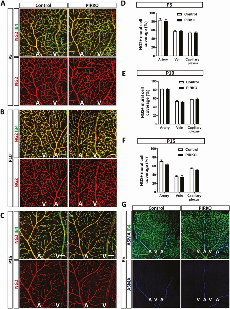
Mural cell coverage is not affected in PIRKO. Mural cell coverage was assessed by NG2 staining at (A) P5, (B) P10, and (C) P15, scale bar 100 µm. NG2 + mural cell coverage is unchanged in arteries, veins and the capillary plexus at (D) P5, (E) P10, and (F) P15. (G) P5 retinas were stained for α-smooth muscle actin (ASMA) to locate vascular smooth muscle cells (VSMCs). Arteries, but not the capillary plexus or veins are covered by ASMA-expressing VSMCs. Data presented as mean ± SEM, 2-way ANOVA with Sidak’s multiple comparison test, not significant, n = 9, 9 at P5, n = 8, 8 at P10, and n = 7, 8 at P15; A, artery; V, vein.

