Figure 2.
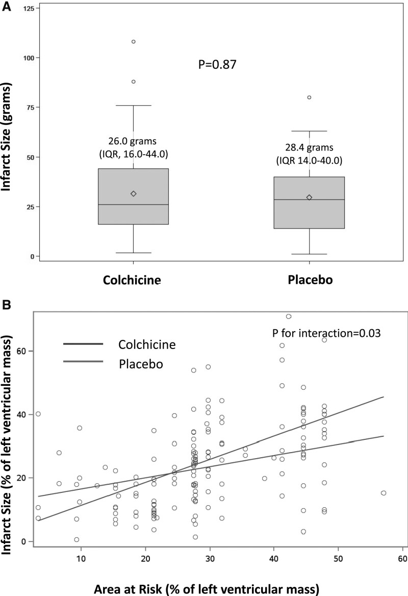
Assessment of infarct size by late gadolinium enhancement cardiac magnetic resonance and as a function of the area at risk (A). Infarct size (IS) was measured in a centralized core laboratory by quantification of the area of late gadolinium enhancement (LGE) by cardiac magnetic resonance at 5 days. IS Colchicine administration did not result in a significant reduction in IS in comparison with placebo (estimate: 0.99 [95% CI, –2.64 to 4.61]; P=0.59). The IS measured by LGE was expressed as a function of the APPROACH angiographic score,16 an estimate of the area at risk, as shown in B. To assess the relationship between the area at risk and IS, we performed a prespecified analysis of regression plots of IS by LGE at 5 days on angiographically estimated area at risk. There was a significant association between the 2 variables in the colchicine group (β=0.73; P<0.001) and the placebo group (β=0.35; P=0.003). There was a significant positive relationship between the 2 variables in both groups, and significantly larger in the colchicine group (P interaction=0.03). These data suggest that, for the largest areas at risk, colchicine administration was associated with an increase in the resulting infarct size as measured by LGE. This difference was confirmed to be significant by analysis of covariance (P for interaction= 0.03). APPROACH indicates Alberta Provincial Project for Outcome Assessment in Coronary Heart Disease; and IQR, interquartile range.

