1. Case report
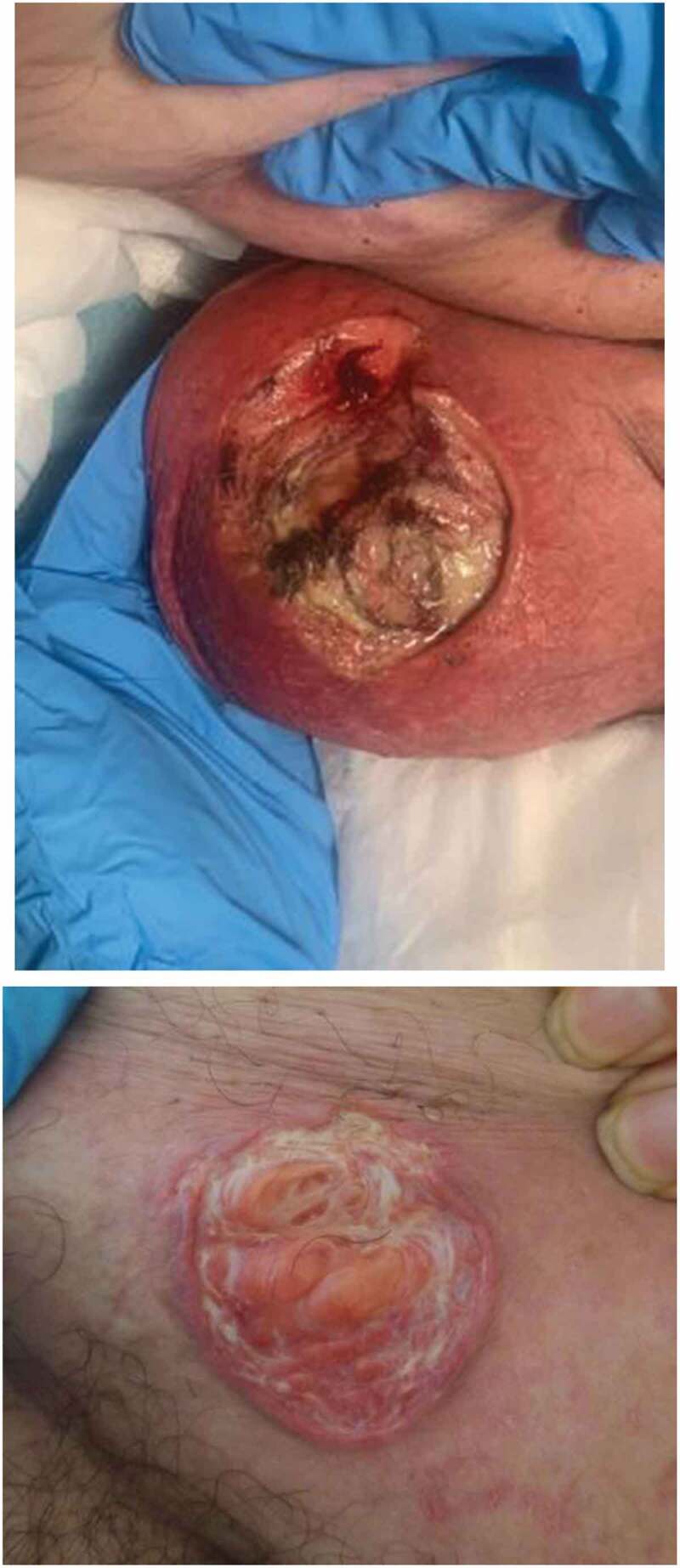
A 71-year-old-male with hypertension, type 2 diabetes mellitus, morbid obesity, and diastolic heart failure presented to the hospital with mild symptoms of sore throat, fevers, and shortness of breath and tested positive for COVID-19 by RT-PCR. He did not require admission and was discharged home with instructions to self-quarantine for 14 days. About 10 days after diagnosis, he developed painful and pruritic pustules on his left scrotum that quickly ulcerated within a few days. After self-treatment with over-the-counter antifungal and antibiotic creams, the patient was treated at an urgent care facility with cephalexin 500 mg three times daily and valacyclovir 1000 mg twice daily due to concern for cellulitis and shingles. Over the next few weeks, the small ulcers coalesced to form a large painful ulcer (Figure 1(a)), followed by the appearance of a similar lesion in the lower abdomen (Figure 1(b)).
Figure 1.

Ulcerated lesions on the scrotum (a) and lower abdomen (b) of the patient
He was prescribed topical triamcinolone by his physician and referred to a wound care center where he was diagnosed with pressure ulcers and treated with topical collagenase, a debriding agent, which worsened the ulcers. The patient gradually developed more ulcers on the penis, groin, buttocks, and abdomen over a span of two to three months. He eventually underwent a lesional biopsy revealing neutrophilic dermatosis with perivascular and interstital neutrophilic infiltrates in the dermis and was diagnosed with pyoderma gangrenosum (PG) based on the clinical presentation and histopathology. The patient was started on 60 mg prednisone daily and topical corticosteroids with prompt improvement of ulcers and he is currently being transitioned to infliximab for long-term treatment. Work up for PG-associated disorders including rheumatoid arthritis and other major autoimmune diseases, MGUS, inflammatory bowel disease, and hematological malignancies were all negative.
2. Discussion
A little more than a year after being termed a pandemic, our knowledge about COVID-19 symptoms, presentation, pathogenesis, and treatment continues to evolve rapidly. Several cutaneous manifestations of COVID-19 have been described but the major ones include chilblain-like or pseudo-chilblain lesions, urticarial lesions, morbilliform lesions, varicella-like vesicular lesions, purpuric lesions, and livedoid lesions [1]. The mechanisms behind cutaneous manifestations in COVID-19 are still under investigation, but likely involve the indirect effects of immune system hyperactivity and hypercoagulability.
Several immunological similarities exist between COVID-19 and PG. Proinflammatory cytokines and neutrophilic abnormalities play a significant role in the pathogenesis of both diseases. A high neutrophil-to-lymphocyte ratio is an independent risk factor for severe COVID-19 and neutrophilia is an indicator of poor outcomes in COVID-19 patients [2]. PG’s histological finding of neutrophilic abscesses and suppurative inflammation in the dermis is indicative of neutrophilic dysfunction [3].
Biologics targeting the proinflammatory cytokines TNF-α, Interleukin-12 (IL-12), and IL-23 have been very effective in treating PG [4]. IL-6 plays a key role in the activation and accumulation of neutrophils and potentially plays a significant role in hyperinflammation in both COVID-19 and PG. The use of IL-6 antagonist, tocilizumab, along with systemic corticosteroids is currently recommended by the National Institutes of Health (NIH) COVID-19 treatment guidelines panel for severe or rapidly deteriorating patients [5] and was shown to be effective for PG treatment in one case study [6].
3. Conclusion
Awareness about the various cutaneous manifestations of COVID-19 is an important step in increasing our overall understanding of the pathophysiology and clinical presentation of this novel disease. With considerable overlap in disease pathogenesis, COVID-19 patients may also be at increased risk for developing PG. An early diagnosis will help facilitate timely treatment and prevent PG-associated morbidity.
Disclosure statement
No potential conflict of interest was reported by the author(s).
References
- [1].Genovese G, Moltrasio C, Berti E, et al. Skin manifestations associated with COVID-19: current knowledge and future perspectives. Dermatology. 2021;237(1):1–12.Epub 2020 Nov 24. [DOI] [PMC free article] [PubMed] [Google Scholar]
- [2].Borges L, Pithon-Curi TC, Curi R, et al. COVID-19 and neutrophils. The relationship between hyperinflammation and neutrophil extracellular traps. Mediators Inflamm. 2020Dec2;2020:8829674. [DOI] [PMC free article] [PubMed] [Google Scholar]
- [3].Patel F, Fitzmaurice S, Duong C, et al. Effective strategies for the management of pyoderma gangrenosum: a comprehensive review. Acta Derm Venereol. 2015May;95(5):525–531. [DOI] [PubMed] [Google Scholar]
- [4].Fletcher J, Alhusayen R, Alavi A.. Recent advances in managing and understanding pyoderma gangrenosum. F1000Res. 2019Dec12;8:F1000Faculty Rev–2092. [DOI] [PMC free article] [PubMed] [Google Scholar]
- [5].Lee WS, Choi YJ, Yoo WH. Use of tocilizumab in a patient with pyoderma gangrenosum and rheumatoid arthritis. J Eur Acad Dermatol Venereol. 2017Feb;31(2):e75–e77. Epub 2016 Jun 21. [DOI] [PubMed] [Google Scholar]
- [6].COVID-19 Treatment Guidelines Panel . Coronavirus Disease 2019 (COVID-19) Treatment Guidelines. National Institutes of Health. [cited 2021 Apr 26]. Available from: https://www.covid19treatmentguidelines.nih.gov/. [PubMed] [Google Scholar]


