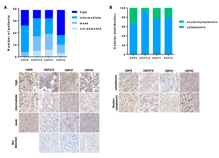
Figure 2.
Expression of USP43, USP41, USP27x and USP6 in OS TMA OS. Tissue microarray (TMA) glass slides containing 40 OS specimens were stained with anti USP6, USP27x, USP41 and USP43 antibodies. Samples were analyzed using standard light microscopy by two different pathologists blinded to the experiment. Expression (A) and distribution (B) of USPs was assessed within the biopsies as described in the Materials and Methods section. USPs expression was assessed as follows: no detected expression, weak expression, intermediate expression and high expression.

