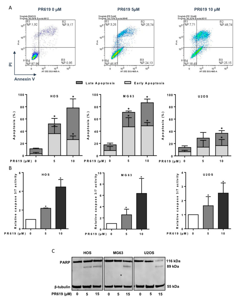
Figure 6.
PR619 induces in vitro cell death in OS cell lines. (A) Upper panels: Representative dot plots of HOS cells treated or not with 5 or 10 μM PR619 for 24 h are shown (representative graphs of three independent experiments). Lower panels: OS cells were treated or not with 5 or 10 μM PR619 for 24 h. Bars indicate the means ± SD of the relative number of cells in early- or late-phase apoptosis (3 independent experiments) (* p < 0.05). (B) OS cells were treated with 5 or 10 μM PR619 for 14 h. Relative caspase-3/7 activity was measured as described in the Materials and Methods section. Bars indicate the caspase-3/7 activity (mean ± SD) of three independent experiments, each performed in triplicate (* p < 0.05). (C) OS cells were treated or not treated with 5 or 15 μM PR619, as indicated, for 24 h. After incubation, PARP cleavage levels were detected by Western blot analysis as described in the Materials and Methods section. Representative blots of three experiments are shown.

