Abstract
The aim of this systematic review was to summarize concentrations of human milk oligosaccharides (HMOs) in the Chinese population. We searched articles originally published in both Chinese and English. When compiling data, lactation was categorized into five stages. We found that 6′-sialyllactose, lacto-N-tetraose, and lacto-N-neotetraose decreased over lactation. Conversely, 3′-fucosyllactose increased over lactation. Our study represents the first systematic review to summarize HMO concentrations in Chinese population. Our findings not only provide data on HMO profiles in Chinese population but suggest future directions in the study of the metabolism of HMOs.
Keywords: breast milk, lactation, longitudinal changes, sialic acid, fucose
1. Introduction
Human milk represents the optimal source of nutrition for neonatal growth, development, and health. Currently, there are gaps between breastfed and formula-fed infants. For example, when compared with breastfed infants, formula-fed infants gain excessive weight, possess lower abundance of Bifidobacteria in the gut, have higher incidences of infections, etc. [1]. Furthermore, the differences between breastfeeding and formula feeding could prolong to later life, especially in cognitive development and risks of metabolic disorders [2]. The advantages of breastfeeding are mainly attributed to human milk constituents.
Human milk oligosaccharides (HMOs) are the fourth largest group of nutrients in human milk after water, lactose, and lipids [3]. Concentrations of total HMOs range from 5–25 g/L in human milk [4,5,6]. Although HMOs are not a significant source of metabolic energy or building blocks of tissues such as lactose, lipids, and protein, there are several possible mechanisms via which HMOs play vital roles in infant health. First, HMOs nourish probiotics in the gut as a carbon source [7]. Second, HMOs block the adhesion of bacterial and viral pathogens to the surface of intestinal epithelial cells [8,9]. Third, HMOs directly interact with cells of the body and modulate cellular functions [10]. Because intact HMOs can enter the circulation, HMOs can theoretically interact with all cells in the body [1]. Fourth, HMOs can donate fucose and sialic acid (N-acetylneuraminic acid) moieties in the glycosylation of proteins and lipids [11].
HMOs are highly diverse in structures. Currently, more than 200 HMOs have been characterized [6]. Nevertheless, structures of HMOs generally share a common blueprint [12]: (1) the backbones of HMOs all contain lactose at the reducing end; (2) lactose at the reducing end can be elongated by adding glucose (Glc), galactose (Gal), and N-acetylglucosamine (GlcNAc); (3) backbones of HMOs can be modified by adding fucose or sialic acid moieties.
Interindividual differences in HMO profiles due to genetic factors have been demonstrated. The α1-2-fucosyltransferase (FUT2; encoded by Se gene) is involved in the fucosylation in the synthesis of α1-2-fucosylated HMOs such as 2’-fucosyllactose (2′-FL) and lacto-N-fucopentaose I (LNPF I) [4,13]. Single-nucleotide polymorphisms (SNPs) in Se may cause dramatically lower concentrations of α1-2-fucosylated glycans in human milk [14]. Similarly, α1-3/4-fucosyltransferase (FUT3; encoded by Le gene) participates in the synthesis of α1-4-fucosylated HMOs such as lacto-N-fucopentaose II (LNFP II) [4,13]. Accordingly, SNPs in Le may lead to lower levels of α1-4-fucosylated HMOs in human milk [15]. It should be noted that more than 200 glycosyltransferases have been identified in the human genome [16]. Moreover, other families of enzymes are involved in the biogenesis of HMOs [17]. Therefore, it is possible that HMOs in addition to α1-2-fucosylated and α1-4-fucosylated glycans may display interindividual variations.
Human milk from different populations may exhibit different typical HMO profiles due to genetic factors [18]. For example, frequencies of SNPs in Se gene were found different among populations [19,20]. Previously, Thurl et al. attempted to establish representative HMO profiles by conducting a systematic review and compiling data from 15 countries and regions (except for the Mainland of China) [3]. Additionally, they identified that genetic factors, as well as lactation stage, affect HMO concentrations. To our best knowledge, the study by Thurl et al. remains the only systematic review on HMO concentrations to date. Unfortunately, none of the included studies reported HMO data from the mainland of China.
China has more than 20% of the world population. In recent years, HMO studies in the Chinese population have been published in both English and Chinese, which are not readily available to non-Chinese scholars and policymakers. In our initial literature search, we found that small sample sizes and limited locations of sample collection were common in the original studies that reported HMOs in Chinese human milk, making it impossible to conclude typical HMO profile in Chinese population from any of the studies. Accordingly, the aim of this study was to compile HMO concentrations in Chinese population by a systematic review. We also attempted to interrogate the influences of lactation stages and genetic factors on HMO concentrations. The findings of our study could potentially enhance the knowledge of lactation biology, improve infant feeding practices, and shed light on the metabolism of HMOs in the infant body.
2. Methods
2.1. Literature Selection
The guidelines in Preferred Reporting Items for Systematic Review and Meta-Analysis Protocols (PRISMA-p) were followed [21]. Briefly, for the literature published in English, the databases Web of Science, PubMed, and ScienceDirect were searched using the formula “(human OR breast) AND (milk) AND (oligosaccharide*) AND (composition OR concentration OR content* OR quantification OR profile* OR amount) AND (China OR Chinese)”. For the literature published in Chinese, the databases China National Knowledge Infrastructure (CNKI) (http://cnki.net, accessed on 25 May 2020), Wanfang (http://www.wanfangdata.com.cn/index.html, accessed on 25 May 2020), and Chongqing VIP Information (http://qikan.cqvip.com, accessed on 25 May 2020) were searched, and the formula was optimized to “(human milk OR breast milk) AND (oligosaccharide)” to adapt to the style of the Chinese language. Literature searching was completed in May 2020. The titles and abstracts of all hits were checked, and the obviously unrelated articles were removed. The full texts of the remaining articles were examined by applying the inclusion and exclusion criteria listed in the PICOS table (Supplementary Table S1). Quality of included studies was assessed as previously described [22]. The literature selection was performed independently by two investigators (Y.Z. and H.S.). Discrepancies were addressed in the presence of a third investigator (K.L.).
2.2. Data Mining and Processing
To compile data, lactation was divided into five stages: 1–7, 8–14, 15–60, 61–120, and beyond 121 postnatal days. Means and standard deviations (SDs) of HMO concentrations were extracted. Units were all converted to mg/L. The human milk density was 1.03 g/mL in the conversions [23]. Data mining and processing was performed independently by two investigators (Y.Z. and H.S.). Discrepancies were addressed in the presence of a third investigator (K.L.). Nomenclatures were adapted from Bode (2012) [1].
2.3. Statistical Analysis
Weighted means, SDs, and standard errors (SEs) of HMOs in each lactation stage were calculated as described [24]. The 95% CIs of each HMO across lactation stages were estimated as weighted mean ± t × SE, where t follows a t-distribution with degrees of freedom equal to the group sample size minus 1. Cochran’s Q statistic of a fixed effect model, which, under the null hypothesis of no subgroup (namely, lactation stage), heterogeneity follows a Χ2-distribution with degrees of freedom equal to number of subgroup minus 1, was calculated [25]. A random effect model was used when significant heterogeneity was observed using the fixed model. Higgins and Thompson’s I2 was derived according to the Q statistic [26]. Figures were generated by using ggplot2 R package [27]. All the statistical analyses were performed using R (version 4.0.3) [28]. Forrest plots were generated to compare HMO concentrations of included studies using Stata/SE 14.0 (StataCorp LLC, College station, TX, USA). Publication biases were estimated by plotting mean concentration of HMO against its corresponding SE of each study included.
3. Results
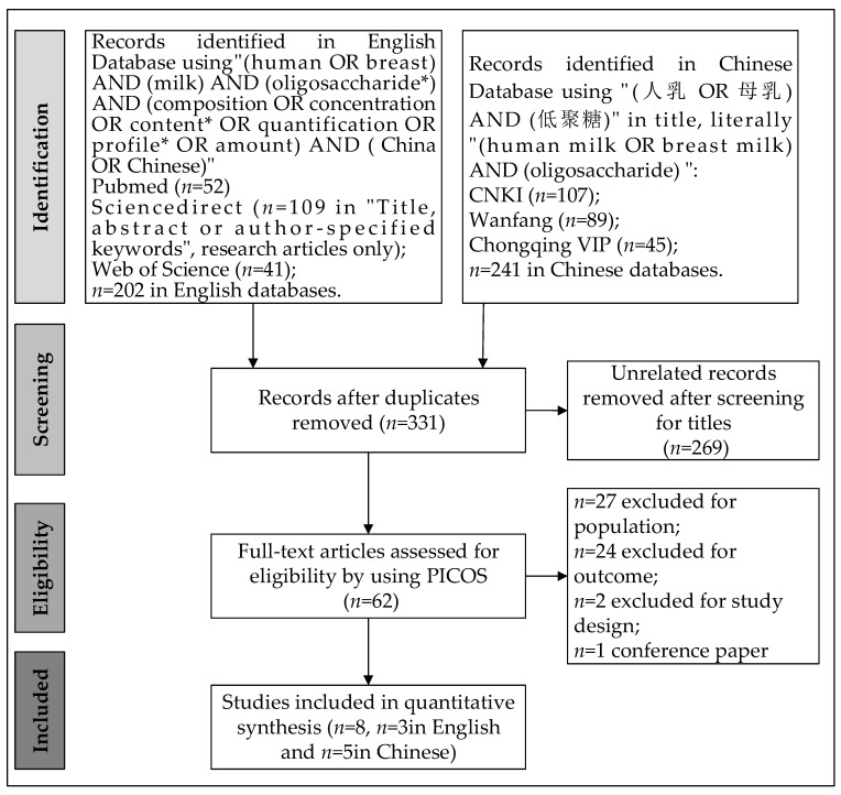
The article selection process is summarized in Figure 1. Included studies [4,14,29,30,31,32,33,34] were listed (Table 1) and assessed (Supplementary Table S2). Articles were published from 2009 to 2020. Publication biases of included studies were visualized by the funnel plots (Supplementary Figure S2). Concentrations of 14 structures of HMOs were reported in the included articles (Supplementary Table S3). Among the investigated HMOs, eight were fucosylated, four were sialylated, and two were not modified by fucose or sialic acid. Lengths of the included glycans range from 3–6 sugar residues with four trisaccharides, two tetrasaccharides, five pentasaccharides, and three hexasaccharide. One study reported SNPs in Se. None of the included studies directly investigated Lewis types or SNPs in Le.
Figure 1.
Flow chart of literature searching and screening.
Table 1.
Inclusion and exclusion criteria for article selection.
| Reference | Period of Investigation | Site | Collection Time (Days) | Number of Mothers | Milk Samples Collected | Term/Preterm | Secretor Status | HMOs Determination Method | Language | Measurement Unit |
|---|---|---|---|---|---|---|---|---|---|---|
| [4] | October 2011–February 2012 | Beijing Guangzhou Suzhou |
5–11 12–30 31–60 61–120 121–240 |
446 | 88 88 90 90 90 |
Full term | NR 1 | UHPLC | English | mg/L |
| [14] | March–December 2018 | Shanghai | 8–14 | 30 | 27 | Full term | FUT2(AA) FUT2(AT) FUT2(TT) |
HPLC | Chinese | mg/L |
| [29] | NR | Guangzhou | 14, 30, 60, 90, 120, 180, 240 | NR | 20 | Full term | Non-secretor phenotype (37%) | HPLC–MS | English | mg/L |
| [30] | NR | Beijing | 3 and 20 | 5 | 5 5 |
NR | NR | UPLC–QqQ-MS | Chinese | mg/L |
| [31] | NR | Nanjing Qiqihar |
0–7; 8–15; 16–180 | NR | 102 | NR | NR | UHPLC–FLD | Chinese | μg/g |
| [32] | Fedruary–May 2018 | Beijing, Guangzhou, Nanjing, Zhengzhou, Mudanjiang, Chengdu |
90 ± 15 | 96 | 10 6 20 20 20 20 |
Full term | NR | UHPLC–FLD | Chinese | μg/mL |
| [33] | March–July 2008 | Shanghai | 8–14 15–21 22–28 |
51 | 51 44 11 |
Full term | Le(a−, b+): 56.8% Le(a−, b−): 19.6%; Le(a+, b−): 23.6% |
HPLC | Chinese | mg/L |
| [34] | NR | Beijing | 31–180 | 61 | 61 | Full term | NR | UPLC–tandem MS | English | mg/mL |
1 NR, not reported.
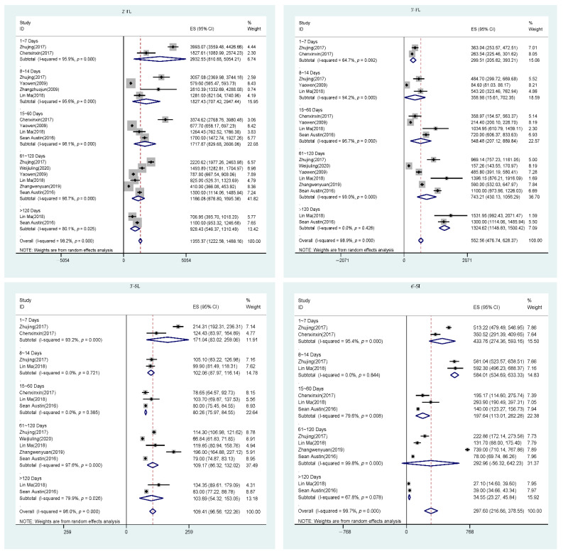
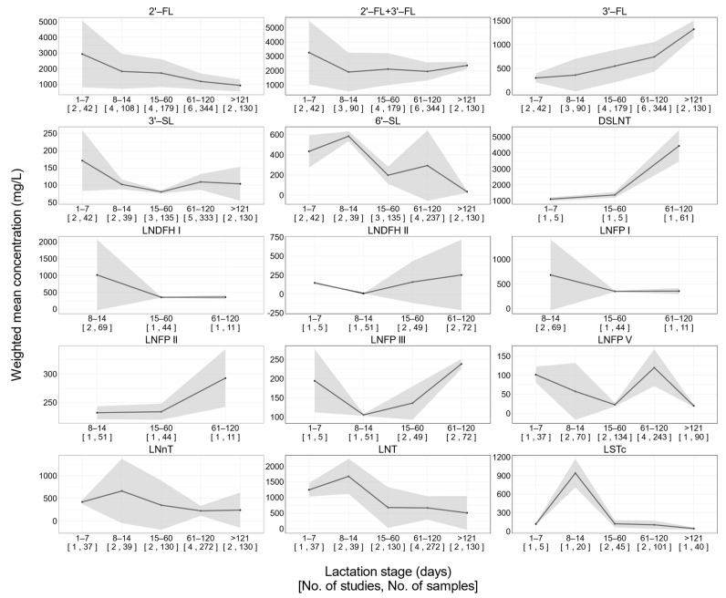
Significant between-study heterogeneities were observed for 2‘-FL, 3′-FL, 6′-SL, and 3′-SL (Figure 2). This remained true for other HMOs included in our study (Supplementary Figure S1). To characterize the longitudinal pattern of each HMO during lactation stages, subgroup analysis was carried out using random effect model considering substantial between-study homogeneities being observed (Table 2 and Figure 3).
Figure 2.
Forest plot of comparison of HMO concentrations.
Table 2.
Results of subgroup heterogeneity analysis.
| 1–7 Days | 8–14 Days | 15–60 Days | 61–120 Days | >121 Days | Q | df | p | I 2 | ||||||
|---|---|---|---|---|---|---|---|---|---|---|---|---|---|---|
| Mean | SE | Mean | SE | Mean | SE | Mean | SE | Mean | SE | |||||
| 2′-FL | 2932.5 | 1082.5 | 1827.4 | 571.4 | 1717.9 | 453.2 | 1186.1 | 259.8 | 928.4 | 194.9 | 7.0 | 4 | 0.137 | 42.7 |
| 3′-FL | 299.5 | 47.8 | 359.0 | 175.2 | 548.5 | 174.2 | 743.2 | 159.7 | 1324.6 | 89.7 | 104.5 | 4 | 1.1 × 10²¹ | 96.2 |
| 2′-FL + 3′-FL | 3265.1 | 1131.7 | 1905.0 | 691.7 | 2111.0 | 567.6 | 1947.5 | 322.2 | 2365.4 | 135.7 | 2.6 | 4 | 0.629 | 0.0 |
| 3′-SL | 171.0 | 44.9 | 102.1 | 7.2 | 80.3 | 2.2 | 109.2 | 11.7 | 103.7 | 25.2 | 18.1 | 4 | 0.001 | 78.0 |
| 6′-SL | 433.8 | 81.3 | 584.0 | 25.2 | 197.6 | 43.2 | 293.0 | 178.2 | 34.6 | 5.8 | 484.9 | 4 | 1.2 ×10103 | 99.2 |
| DSLNT | 1098.7 | 53.9 | NA | NA | 1363.8 | 92.7 | 4443.0 | 501.6 | NA | NA | 48.4 | 2 | 3.1 × 10¹¹ | 95.9 |
| LNDFH I | NA | NA | 1015.9 | 535.1 | 352.2 | 6.6 | 356.8 | 31.5 | NA | NA | 1.6 | 2 | 0.459 | 0.0 |
| LNDFH II | 147.3 | 8.1 | 8.9 | 0.3 | 158.6 | 140.0 | 251.8 | 236.6 | NA | NA | 290.7 | 3 | 1.0 × 1062 | 99.0 |
| LNFP I | NA | NA | 684.4 | 365.3 | 352.2 | 6.6 | 356.8 | 31.5 | NA | NA | 0.8 | 2 | 0.655 | 0.0 |
| LNFP II | NA | NA | 232.4 | 5.8 | 234.0 | 7.2 | 292.7 | 25.7 | NA | NA | 5.3 | 2 | 0.072 | 62.0 |
| LNFP III | 194.3 | 41.9 | 105.6 | 1.3 | 136.2 | 22.2 | 238.1 | 6.6 | NA | NA | 393.2 | 3 | 6.7 × 1085 | 99.2 |
| LNFP V | 101.6 | 10.7 | 57.7 | 38.0 | 22.8 | 2.4 | 120.0 | 24.5 | 20.0 | 1.6 | 73.5 | 4 | 4.1 × 10¹⁵ | 94.6 |
| LNnT | 421.1 | 25.2 | 663.6 | 363.2 | 349.6 | 277.4 | 224.9 | 56.3 | 240.6 | 198.2 | 11.3 | 4 | 0.024 | 64.5 |
| LNT | 1247.6 | 110.4 | 1678.9 | 287.1 | 679.4 | 333.1 | 667.4 | 190.5 | 507.7 | 276.1 | 17.1 | 4 | 0.002 | 76.6 |
| LSTc | 118.9 | 7.8 | 940.9 | 118.1 | 122.5 | 29.9 | 105.7 | 34.4 | 44.8 | 9.9 | 87.3 | 4 | 4.9 × 10¹⁸ | 95.4 |
Means are presented as weighted means calculated from included studies. Q: Cochran’s Q statistic which follows a chi-square distribution with df degrees of freedom; df: degrees of freedom; I2: Higgins and Thompson’s I2. NA: data not available.
Figure 3.
Longitudinal changes in individual HMOs.
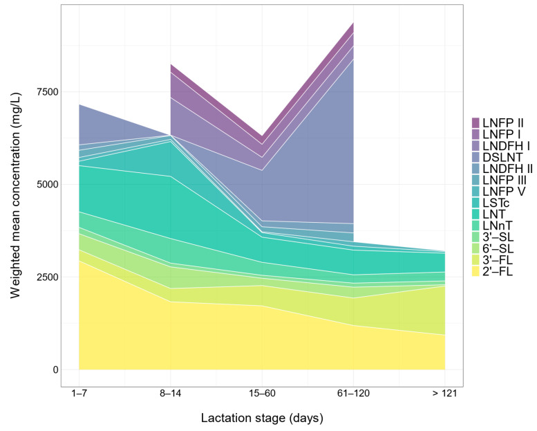
Fucosylated HMOs exhibited distinct patterns of changes over lactation. The concentration of 2′-FL in the 1–7 postnatal days was higher than that at all following lactation stages (Figure 3). The concentration of 2′-FL in the 8–14 postnatal days was about 60% of that in the 1–7 postnatal days (Table 2). Nevertheless, longitudinal changes of 2′-FL over lactation was not significant according to subgroup heterogeneity analysis using a random effect model (Table 2). (Q = 7.0, df = 4, p = 0.137). By contrast, the concentrations of 3′-fucosyllactose (3′-FL) significantly increased (Q = 104.5, df = 4, p < 0.001) with the progression of lactation according to the subgroup heterogeneity analysis (Figure 3 and Table 2). The sum of 2′-FL and 3′-FL remained constant over lactation (p = 0.538). Additionally, the concentration of lacto-N-difucohexaose II (LNDFH II) in the 61–120 postnatal days was significantly (Q = 290.7, df = 3, p < 0.001) higher than that at the preceding stages (Figure 3 and Table 2). There was no clear pattern of longitudinal changes in LNFP I, LNFP II, lacto-N-fucopentaose III (LNFP III), and lacto-N-fucopentaose V (LNFP V) (Figure 3). In the first 120 postnatal days, 2′-FL was the dominant fucosylated HMO (Figure 4). In the postnatal days beyond 121 days, 3′-FL was the most abundant fucosylated HMO (Figure 4). The concentration of 2′-FL was higher than 900 mg/L during the entire lactation (Table 2).
Figure 4.
Longitudinal changes in HMO composition.
4. Discussion
Our study provides a summary of 14 HMOs in Chinese population by using a defined literature search and selection process. We compiled data from original research articles that were published in both English and Chinese. Our study demonstrates that HMO concentrations exhibit dynamic changes during lactation. The concentrations of 6′-SL, LNT, and LNnT decline over lactation. Conversely, the concentration of 3′-FL increases with the progression of lactation. The longitudinal changes in LNFP I, LNFP II, LNFP V, and 3′-SL are not significant. Our study is the first systematic review to establish reference HMO profile in Chinese population.
Unlike previous studies on HMO content from varied populations in distinct countries, our study focused on Chinese mothers. Although variations in detection methods and targeted population from studies to studies limit the comparability of the data, the dynamic change in HMOs along the lactation period may comply with a general trend.
We calculated the total concentration of 14 kinds of HMOs and observed the downward trend with the progression of lactation from the holistic perspective (Figure 4). The findings were supported by previous studies. Many studies demonstrated that total concentrations of interest HMOs were the highest in the first week of lactation and decreased thereafter [4,5]. This result is in accordance with the overall dynamic changes in total concentration of all HMOs along the lactation period. Although it is not possible to measure all the HMOs in human milk, it has been reported that the total level of HMOs in colostrum is estimated to be the highest at 9–22 g/L and declines slightly in transitional milk (8–19g/L at postnatal 8–15 days), followed by a gradual decrease in mature milk [35]. However, it is worth noting that the total content of 14 HMOs showed a sharp increase in the 61–120 postnatal days, which is likely attributed to the dramatic rise in the concentration of DSLNT. This may be explained by the scant number of studies on DSLNT and the limited sample population involved. There are insufficient basic data available on the HMO content of lactating mothers in China, implying that sufficient caution should be taken when drawing conclusions.
2′-FL is the most abundant HMO whose biological significance includes modulating immune functions, nourishing gut prebiotics, and providing fucose moieties for the glycosylation of biomolecules such as proteins and lipids [1]. Although not significantly, the concentration of 2′-FL declined gradually along with the lactation course, which is consistent with almost all studies [11,35]. 3′-FL is another neutral fucosylated HMO. Differing from 2′-FL, the level of 3′-FL rose significantly and may have compensated for 2′-FL. It is possible that the secretion of 3′-FL, an isomer of 2′-FL, is upregulated with the progression of lactation to compensate for 2′-FL. In our study, although 2′-FL and 3′-FL concentrations displayed opposite trends, the sum of 2′-FL and 3′-FL remained constant after the first week of lactation. In another study, researchers reviewed the HMO concentration among populations from different countries and observed an increasing trend of 3′-FL and a negative correlation between the production of 2′-FL and 3′-FL [35]. These observations implied the possibility that 3′-FL may compensate for 2′-FL in human milk from non-secretor mothers whose 2′-FL is dramatically lower than that from secretor mothers. Subsequently, another question was raised regarding the potential compensatory mechanisms of 2′-FL in non-secretor mothers. Therefore, in the future, studies that directly compare 2′-FL with 3′-FL in their functions and metabolism in infants are necessary to formally evaluate the compensatory mechanism of 2′-FL by 3′-FL.
Despite no significance, other neutral fucosylated HMOs such as LNFP I and LNFP V decreased during the course of lactation, which is in accordance with findings reported in previous studies [3,11,35]. LNnT and LNT, neutral non-fucosylated HMOs, declined throughout the lactation, and the changes reached the significant level. Sumiyoshi [36] also reported the similar findings among Japanese population. Additionally, LNnT and LNT showed parallel dynamic changes among population from North America, South America, and Europe reported by McGuire [11].
In our study, the concentrations of two major sialylated HMOs (6′-SL and 3′-SL) showed a downward trend across lactation, while the decrease in 3′-SL did not reach a significant level, unlike findings reported in a previous study [37]. 6′-SL predominated in the early period of lactation (<15 days) and declined with the progression of lactation. Beyond 120 days, the concentrations of 6′-SL were comparable to those of 3′-SL. In line with our findings, it was proposed that 6′-SL occupies the main position among sialylated HMOs at early stages of lactation (<3 months), and the concentration of 3′-SL is higher at 4–8 months [3,35,37].
DSLNT is another major acidic HMO. In several previous studies, the level of DSLNT declined from colostrum to transitional milk to mature milk, but the longitudinal changes were not significant [37,38]. Conversely, unlike the findings reported, we found that the concentration of DSLNT in the 61–120 day stage rose dramatically to at least 1.5 times that at preceding stages. We acknowledge that the results are debatable because of the scant literature and small sample size included. It is advisable for us to draw a conclusion with an abundance of caution. The sialylated oligosaccharides may have an important role in early postnatal cognitive development. It should be highlighted that each DSLNT molecule contains two sialic acid moieties, whereas 6′-SL and 3′-SL include one moiety. In addition, existing evidence underpins a significant relationship between fucosylated/sialylated HMOs in maternal milk and offspring blood [39,40]. Although sialic acid moieties widely exist in body tissues, their concentration in the gray matter of the central nervous system is more than 10 times higher than that in all other organs [41]. Sialic acid is linked with the interface between neurons (synapse), where it engages in the neuron signal transduction [42]. Although infants are born with almost all neurons, the development of synapses continues into postnatal life [43]. The velocity of synaptogenesis peaks at the age of 3 months [44]. Interestingly, 3 months is also the time when the concentration of 3′-SL becomes comparable to 6′-SL. Together, it is likely that the predominant sialylated HMOs depend on the lactation stages and play varied roles in cognitive development by providing sialic acid moieties.
The content and composition of HMOs in milk are affected by multiple factors including environmental and maternal factors such as secretor status, lactation stages, maternal age, physical status, and diet. Some previous systematic reviewed data from different countries all around the world and investigated the influencing factors related to the concentration of HMOs. For example, there was a systematic review on data from 15 countries and reporting 33 structures of HMOs [3]. The study had the advantage of separating milk from secretor and non-secretor mothers, as well as analyzing the differences between milk from mothers delivered at term and preterm. Regretfully, in our study, only one included article directly sequenced SNPs in Se, and none of the studies sequenced SNPs in Le [14]. It is also possible to predict secretor status by calculating the ratios of 2′-FL to 3′-FL [40]. However, the prediction of secretor status is dependent on the availability of HMO concentrations from individual mothers, which was available only in the study by Chen et al. [30]. In that study, three out of five mothers were secretors [30]. Mean concentrations of 2′-FL and 3′-6′-FL were 3770.75 mg/L and 227.98 mg/L, respectively. In non-secretor mothers, mean concentrations of 2′-FL and 3′-FL were 2781.02 mg/L and 555.46 mg/L, respectively. In addition, another systematic review summarized the influencing factors including secretor status and Lewis, the country of origin, and maternal physical status [35]. However, our study had several advantages. Firstly, focusing on a single population from China meant relatively smaller individual variations. Secondly, we allocated data available into five lactation stages and analyzed the dynamic changes throughout the lactation period. Lastly, the meta-analysis made it possible to offer the pooled individual concentrations at different lactation stages.
The longitudinal changes in HMOs revealed in our study should be further validated. Inter-study variations are inherited weaknesses when combining data from different original research articles, as confirmed by our results where significant between-study heterogeneities were observed for most of the HMOs. Instruments, quantification methods, sample storage and preparation, and performance of experiments may all contribute to the variations. To further establish reference values of HMOs, standardized quantification methods should be developed. In addition, substantial publication biases were observed for some of the HMOs. Those HMOs are generally under-studied in Chinese population, which calls for future investigation.
5. Conclusions
Our study represents the first systematic review that summarizes HMOs in Chinese population. We demonstrated that the levels of 3′-FL increase throughout lactation, whereas the levels of 6′-SL, LNT, and LNnT decrease with the progression of lactation. The longitudinal changes in HMOs during lactation suggest future directions investigating the functions and metabolism of HMOs, such as a direct comparison of 2′-FL and 3′-FL in their metabolism in infant bodies and formally validating the donation of sialic acid moieties by sialylated HMOs such as 6′-SL, 3′-SL, and DSLNT to the synaptogenesis process in brain development.
Supplementary Materials
The following are available online at https://www.mdpi.com/article/10.3390/nu13092912/s1: Table S1. Inclusion and exclusion criteria of article selection; Table S2. Quality assessment of included studies; Table S3. Blueprint of HMOs; Figure S1. Forest plots of HMO concentrations of included studies; Figure S2. Publication biases of included studies.
Author Contributions
Conceptualization, Y.X. and S.J.; methodology, Y.Z. and H.S.; investigation, Y.Z., H.S., K.L. and C.Z.; writing—original draft preparation, W.Z.; writing—review and editing, M.J., Y.L., R.Z. and W.W.; visualization, H.S., K.L. and C.Z.; supervision, Y.X., S.J. and W.Z.; project administration, Y.X., S.J. and W.Z.; funding acquisition, K.L., W.Z. and S.J. All authors read and agreed to the published version of the manuscript.
Funding
This study was supported by the Chinese Human Milk Project (CHMP) Study grant (funded by Heilongjiang Feihe Co., Ltd., Tsitsihar, China to K.L., W.Z., and S.J.) and the “Bai-Qian-Wan Engineering and Technology Master Project” (Grant #2019ZX07B01, funded by the Government of Heilongjiang Province of the People’s Republic of China, to W.Z. and S.J.).
Institutional Review Board Statement
Not applicable.
Informed Consent Statement
Not applicable.
Data Availability Statement
Not applicable.
Conflicts of Interest
H.S., K.L., C.Z., M.J., W.W., W.Z. and S.J are employed by Feihe Dairy Co., Ltd., Tsitsihar, China. The other authors declare no conflicts of interest. Feihe Dairy Co., Ltd., Tsitsihar, China provided the funding.
Footnotes
Publisher’s Note: MDPI stays neutral with regard to jurisdictional claims in published maps and institutional affiliations.
References
- 1.Bode L. Human milk oligosaccharides: Every baby needs a sugar mama. Narnia. 2012;22:1147–1162. doi: 10.1093/glycob/cws074. [DOI] [PMC free article] [PubMed] [Google Scholar]
- 2.Anderson J.W., Johnstone B.M., Remley D.T. Breast-feeding and cognitive development: A meta-analysis. Am. J. Clin. Nutr. 1999;70:525–535. doi: 10.1093/ajcn/70.4.525. [DOI] [PubMed] [Google Scholar]
- 3.Thurl S., Munzert M., Boehm G., Matthews C., Stahl B. Systematic review of the concentrations of oligosaccharides in human milk. Nutr. Rev. 2017;75:920–933. doi: 10.1093/nutrit/nux044. [DOI] [PMC free article] [PubMed] [Google Scholar]
- 4.Austin S., De Castro C.A., Benet T., Hou Y., Sun H., Thakkar S.K., Vinyes-Pares G., Zhang Y., Wang P. Temporal change of the content of 10 oligosaccharides in the milk of Chinese urban mothers. Nutrients. 2016;8:346. doi: 10.3390/nu8060346. [DOI] [PMC free article] [PubMed] [Google Scholar]
- 5.Elwakiel M., Hageman J.A., Wang W., Szeto I.M., van Goudoever J.B., Hettinga K.A., Schols H.A. Human milk oligosaccharides in colostrum and mature milk of Chinese mothers: Lewis positive secretor subgroups. J. Agric. Food Chem. 2018;66:7036–7043. doi: 10.1021/acs.jafc.8b02021. [DOI] [PMC free article] [PubMed] [Google Scholar]
- 6.Wang Y., Zhou X., Gong P., Chen Y., Feng Z., Liu P., Zhang P., Wang X., Zhang L., Song L. Comparative major oligosaccharides and lactose between Chinese human and animal milk. Int. Dairy J. 2020;108:104727. doi: 10.1016/j.idairyj.2020.104727. [DOI] [Google Scholar]
- 7.Asakuma S., Hatakeyama E., Urashima T., Yoshida E., Katayama T., Yamamoto K., Kumagai H., Ashida H., Hirose J., Kitaoka M. Physiology of consumption of human milk oligosaccharides by infant gut-associated bifidobacteria. J. Biol. Chem. 2011;286:34583–34592. doi: 10.1074/jbc.M111.248138. [DOI] [PMC free article] [PubMed] [Google Scholar]
- 8.Kunz C., Rudloff S., Baier W., Klein N., Strobel S. Oligosaccharides in human milk: Structural, functional, and metabolic aspects. Annu. Rev. Nutr. 2000;20:699–722. doi: 10.1146/annurev.nutr.20.1.699. [DOI] [PubMed] [Google Scholar]
- 9.Newburg D.S., Ruiz Palacios G.M., Morrow A.L. Human milk glycans protect infants against enteric pathogens. Annu. Rev. Nutr. 2005;25:37–58. doi: 10.1146/annurev.nutr.25.050304.092553. [DOI] [PubMed] [Google Scholar]
- 10.Plaza-Díaz J., Fontana L., Gil A. Human milk oligosaccharides and immune system development. Nutrients. 2018;10:1038. doi: 10.3390/nu10081038. [DOI] [PMC free article] [PubMed] [Google Scholar]
- 11.McGuire M.K., Meehan C.L., McGuire M.A., Williams J.E., Foster J., Sellen D.W., Kamau-Mbuthia E.W., Kamundia E.W., Mbugua S., Moore S.E., et al. What’s normal? Oligosaccharide concentrations and profiles in milk produced by healthy women vary geographically. Am. J. Clin. Nutr. 2017;105:1086–1100. doi: 10.3945/ajcn.116.139980. [DOI] [PMC free article] [PubMed] [Google Scholar]
- 12.Cheng K., Zhou Y., Neelamegham S. DrawGlycan-SNFG: A robust tool to render glycans and glycopeptides with fragmentation information. Glycobiology. 2017;27:200–205. doi: 10.1093/glycob/cww115. [DOI] [PMC free article] [PubMed] [Google Scholar]
- 13.Wang M., Zhao Z., Zhao A., Zhang J., Wu W., Ren Z., Wang P., Zhang Y. Neutral human milk oligosaccharides are associated with multiple fixed and modifiable maternal and infant characteristics. Nutrients. 2020;12:826. doi: 10.3390/nu12030826. [DOI] [PMC free article] [PubMed] [Google Scholar]
- 14.Zhang Z. Master’s Thesis. Fudan University; Shanghai, China: 2010. Study on the Relationship between the Concentration of Neutral Oligosaccharides in Breast Milk and FUT2 Gene Polymorphism and the Early Growth of Offspring. [Google Scholar]
- 15.Lefebvre G., Shevlyakova M., Charpagne A., Marquis J., Vogel M., Kirsten T., Wieland K., Sean A., Sprenger N., Binia A. Time of lactation and maternal fucosyltransferase genetic polymorphisms determine the variability in human milk oligosaccharides. Front. Nutr. 2020;7:225. doi: 10.3389/fnut.2020.574459. [DOI] [PMC free article] [PubMed] [Google Scholar]
- 16.Narimatsu Y., Joshi H.J., Yang Z., Gomes C., Chen Y.-H., Lorenzetti F.C., Furukawa S., Schjoldager K.T., Hansen L., Clausen H., et al. A validated gRNA library for CRISPR/Cas9 targeting of the human glycosyltransferase genome. Glycobiology. 2018;28:295–305. doi: 10.1093/glycob/cwx101. [DOI] [PubMed] [Google Scholar]
- 17.Newburg D.S., Grave G. Recent advances in human milk glycobiology. Pediatric Res. 2014;75:675–679. doi: 10.1038/pr.2014.24. [DOI] [PMC free article] [PubMed] [Google Scholar]
- 18.Leo F., Asakuma S., Kukuda K., Senda A., Urashima T. Determination of sialyl and neutral oligosaccharide levels in transition and mature milks of samoan women, using anthranilic derivatization followed by reverse phase high performance liquid chromatography. Biosci. Biotechnol. Biochem. 2010;74:912261789. doi: 10.1271/bbb.90614. [DOI] [PubMed] [Google Scholar]
- 19.Van Trang N., Vu Hau T., Le Nhung T., Huang P., Jiang X., Anh Dang D. Association between norovirus and rotavirus infection and histo-blood group antigen types in Vietnamese children. J. Clin. Microbiol. 2014;52:1366–1374. doi: 10.1128/JCM.02927-13. [DOI] [PMC free article] [PubMed] [Google Scholar]
- 20.Soejima M., Koda Y. FUT2 polymorphism in Latin American populations. Clin. Chim. Acta. 2020;505:1–5. doi: 10.1016/j.cca.2020.02.011. [DOI] [PubMed] [Google Scholar]
- 21.Moher D., Shamseer L., Clarke M., Ghersi D., Liberati A., Petticrew M., Shekelle P., Stewart L.A., Group P.-P. Preferred reporting items for systematic review and meta-analysis protocols (PRISMA-P) 2015 statement. Syst. Rev. 2015;4:1–9. doi: 10.1186/2046-4053-4-1. [DOI] [PMC free article] [PubMed] [Google Scholar]
- 22.Ren Q., Sun H., Zhao M., Xu Y., Xie Q., Jiang S., Zhao X., Zhang W. Longitudinal changes in crude protein and amino acids in human milk in Chinese population: A systematic review. J. Pediatric Gastroenterol. Nutr. 2020;70:555–561. doi: 10.1097/MPG.0000000000002612. [DOI] [PubMed] [Google Scholar]
- 23.Yin S. Human Milk Ingredients: Existence Form, Content, Function, and Detection Method. Chemical Industry Press; Beijing, China: 2016. [Google Scholar]
- 24.Higgins J.P., Thomas J., Chandler J., Cumpston M., Li T., Page M.J., Welch V.A. Cochrane Handbook for Systematic Reviews of Interventions. John Wiley & Sons; Hoboken, NJ, USA: 2019. [Google Scholar]
- 25.Cochran W.G. The combination of estimates from different experiments. Biometrics. 1954;10:101–129. doi: 10.2307/3001666. [DOI] [Google Scholar]
- 26.Higgins J.P., Thompson S.G. Quantifying heterogeneity in a meta-analysis. Stat. Med. 2002;21:1539–1558. doi: 10.1002/sim.1186. [DOI] [PubMed] [Google Scholar]
- 27.R Core Team . R: A Language and Environment for Statistical Computing. Version 4.0. 2 (Taking off Again) R Foundation for Statistical Computing; Vienna, Austria: 2020. [Google Scholar]
- 28.Wickham H., Navarro D., Pedersen T.L. Ggplot2: Elegant Graphics for Data Analysis (Use R!) Springer; New York, NY, USA: 2009. [Google Scholar]
- 29.Ma L., McJarrow P., Jan Mohamed H.J.B., Liu X., Welman A., Fong B.Y. Lactational changes in the human milk oligosaccharide concentration in Chinese and Malaysian mothers’ milk. Int. Dairy J. 2018;87:1–10. doi: 10.1016/j.idairyj.2018.07.015. [DOI] [Google Scholar]
- 30.Chen X. Master’s Thesis. Sichuan Agricultural University; Ya’an, China: 2017. Study on Determination Methods of Oligosaccharides in Human Milk and Bovine Milk and the Changes of Oligosaccharides in Different Lactation Periods. [Google Scholar]
- 31.Zhu J., Shi Y., Wu L., Sun H., Wang X., Wang Z., Guan L., Yu K., Yang Y. Detection and content analysis of 10 free oligosaccharides in breast milk at different stages. Chin. J. Food Hyg. 2017;29:417–422. [Google Scholar]
- 32.Wei J., Ren X., Wang X., Wu L., Han J., Yang Y. Study on the macronutrient composition of human milk in six regions of China. Acta Nutr. SINICA. 2020;42:7–11. [Google Scholar]
- 33.Yao W., Zhang Z., Zhou T., Newburg D.S., Peng Y. Changes in the concentration of neutral oligosaccharides in breast milk of Chinese mothers. Chin. J. Child Health Care. 2009;17:251–253. [Google Scholar]
- 34.Zhang W., Wang T., Chen X., Pang X., Zhang S., Obaroakpo J.U., Shilong J., Lu J., Lv J. Absolute quantification of twelve oligosaccharides in human milk using a targeted mass spectrometry-based approach. Carbohydr. Polym. 2019;219:328–333. doi: 10.1016/j.carbpol.2019.04.092. [DOI] [PubMed] [Google Scholar]
- 35.Thum C., Wall C.R., Weiss G.A., Wang W., Szeto I.M., Day L. Changes in HMO Concentrations throughout Lactation: Influencing Factors, Health Effects and Opportunities. Nutrients. 2021;13:2272. doi: 10.3390/nu13072272. [DOI] [PMC free article] [PubMed] [Google Scholar]
- 36.Sumiyoshi W., Urashima T., Nakamura T., Arai I., Saito T., Tsumura N., Wang B., Brand-Miller J., Watanabe Y., Kimura K. Determination of each neutral oligosaccharide in the milk of Japanese women during the course of lactation. Br. J. Nutr. 2003;89:61–69. doi: 10.1079/BJN2002746. [DOI] [PubMed] [Google Scholar]
- 37.Plows J.F., Berger P.K., Jones R.B., Alderete T.L., Yonemitsu C., Najera J.A., Khwajazada S., Bode L., Goran M.I. Longitudinal Changes in Human Milk Oligosaccharides (HMOs) over the Course of 24 Months of Lactation. J. Nutr. 2021;151:876–882. doi: 10.1093/jn/nxaa427. [DOI] [PMC free article] [PubMed] [Google Scholar]
- 38.Siziba L.P., Mank M., Stahl B., Gonsalves J., Blijenberg B., Rothenbacher D., Genuneit J. Human Milk Oligosaccharide Profiles over 12 Months of Lactation: The Ulm SPATZ Health Study. Nutrients. 2021;13:1973. doi: 10.3390/nu13061973. [DOI] [PMC free article] [PubMed] [Google Scholar]
- 39.Radzanowski G.G., Garrett P.N., Xi L., Anita M.J.T.F.J. Short-chain milk oligosaccharide levels in human milk and infant plasma. J. Fed. Am. Soc. Exp. Biol. 2013;27:629.16. [Google Scholar]
- 40.Goehring K.C., Kennedy A.D., Prieto P.A., Buck R.H. Direct evidence for the presence of human milk oligosaccharides in the circulation of breastfed infants. PLoS ONE. 2014;9:e101692. doi: 10.1371/journal.pone.0101692. [DOI] [PMC free article] [PubMed] [Google Scholar]
- 41.Svennerholm L. Cholera and Related Diarrheas. Karger Publishers; Basel, Switzerland: 1980. Structure and biology of cell membrane gangliosides; pp. 80–87. [DOI] [Google Scholar]
- 42.Wang B., Brand-Miller J. The role and potential of sialic acid in human nutrition. Eur. J. Clin. Nutr. 2003;57:1351–1369. doi: 10.1038/sj.ejcn.1601704. [DOI] [PubMed] [Google Scholar]
- 43.Uauy R., Mena P., Peirano P. Mechanisms for nutrient effects on brain development and cognition. Nestlé Nutr. Workshop Ser. Clin. Perform. Programme. 2001;5:41–72. doi: 10.1159/000061845. [DOI] [PubMed] [Google Scholar]
- 44.Huttenlocher P.R., Dabholkar A.S. Regional differences in synaptogenesis in human cerebral cortex. J. Comp. Neurol. 1997;387:167–178. doi: 10.1002/(SICI)1096-9861(19971020)387:2<167::AID-CNE1>3.0.CO;2-Z. [DOI] [PubMed] [Google Scholar]
Associated Data
This section collects any data citations, data availability statements, or supplementary materials included in this article.
Supplementary Materials
Data Availability Statement
Not applicable.


