Abstract
Primary ciliary dyskinesia (PCD) is a rare genetic disease characterized by dyskinetic cilia. Respiratory symptoms usually start at birth. The lack of diagnostic gold standard tests is challenging, as PCD diagnostics requires different methods with high expertise. We founded PCD-UNIBE as the first comprehensive PCD diagnostic center in Switzerland. Our diagnostic approach includes nasal brushing and cell culture with analysis of ciliary motility via high-speed-videomicroscopy (HSVM) and immunofluorescence labeling (IF) of structural proteins. Selected patients undergo electron microscopy (TEM) of ciliary ultrastructure and genetics. We report here on the first 100 patients assessed by PCD-UNIBE. All patients received HSVM fresh, IF, and cell culture (success rate of 90%). We repeated the HSVM with cell cultures and conducted TEM in 30 patients and genetics in 31 patients. Results from cell cultures were much clearer compared to fresh samples. For 80 patients, we found no evidence of PCD, 17 were diagnosed with PCD, two remained inconclusive, and one case is ongoing. HSVM was diagnostic in 12, IF in 14, TEM in five and genetics in 11 cases. None of the methods was able to diagnose all 17 PCD cases, highlighting that a comprehensive approach is essential for an accurate diagnosis of PCD.
Keywords: airways, ciliopathy, air-liquid interface cell culture, high-speed videomicroscopy, immunofluorescence, transmission electron microscopy
1. Introduction
Primary ciliary dyskinesia (PCD) is a rare genetic disorder (prevalence between 1:10,000 and 1:20,000 [1,2,3]) that manifests with chronic respiratory symptoms caused by impaired mucociliary clearance [4,5,6]. Symptoms include chronic wet cough, chronic rhinitis, recurrent otitis media, and subsequent hearing impairment, recurrent respiratory infections, and may start early in life with neonatal respiratory distress [6,7,8,9,10]. Pulmonary long-term morbidity includes function impairment and structural changes (e.g., lung atelectasis, bronchiectasis) [8,11,12,13]. The impaired ciliary function with inefficient or absent beating is based on defects in a large number of genes. Those genes primarily involve the ciliary motor protein family, the dyneins, but also other components of the ciliary structure [9,14]. Despite recent diagnostic guidelines of the European Respiratory Society (ERS) [15] and the American Thoracic Society (ATS) [16], the diagnosis of PCD remains challenging. Difficulties are related to several issues: (i) clinical presentation shows a wide phenotypical spectrum and is not specific for PCD; (ii) there is no single test that is diagnostic for PCD as a stand-alone test; current diagnostic guidelines therefore include a combination of different functional, structural and molecular methods; (iii) all diagnostic methods require a high level of expertise; (iv) over ¼ of the genetic defects causing PCD are unknown [17]; (v) analytical methods have not been standardized sufficiently [18] and have unsatisfactory sensitivity and/or specificity if considered individually; and (vi) PCD is a rare disease and satisfactory diagnostic experience is available in only a few specialized centers. Hence, PCD is often diagnosed late in life or not at all, resulting in an overall underdiagnosis and undertreatment [1,2]. Even if causal therapy is not yet available, early diagnosis and interdisciplinary management increases quality of life and prognosis of affected patients [19,20].
The current diagnostic guidelines include a combination of different functional, structural, and molecular methods. All require a high level of expertise. Currently, the following methods are used routinely for the diagnosis of PCD: (i) measurement of nasal nitric oxide (nNO); (ii) high-speed-videomicroscopy (HSVM) to assess ciliary motility of viable cells (fresh samples or air-liquid interface (ALI) cell cultures); (iii) immunofluorescence staining (IF) of different structural proteins; (iv) assessment of the axonemal ultrastructure by transmission electron microscopy (TEM); and (v) genetic testing for the identification of pathogenic or likely pathogenic variants in PCD-associated genes.
Based on 30 years of experience in structural PCD diagnosis, we founded the interdisciplinary center for comprehensive PCD diagnostics (PCD-UNIBE) at the University Children’s Hospital, Inselspital, Bern University Hospital and the Institute of Anatomy, University of Bern, in January 2018. This is the first comprehensive PCD diagnostic center in Switzerland and is led by an experienced team of clinicians and biomedical researchers.
The aim of this study is to summarize the experiences of the first 100 patients assessed by PCD-UNIBE and to report on the workflow, benefits, and challenges of our center and the diagnostic algorithm used by us.
2. Materials and Methods
2.1. Study Design and Study Population
This observational study includes the first 100 patients referred to our PCD-UNIBE diagnostic center. The study was approved by the ethics committees of the University Children’s Hospital Bern (Ethikkommission der Kinderkliniken) and of the Canton Bern (Kantonale Ethikkomission Bern), Switzerland (project identification code 2018-02155, 01/2019). We obtained written informed consents from all participants or their legal guardians.
2.2. Diagnostic Workflow
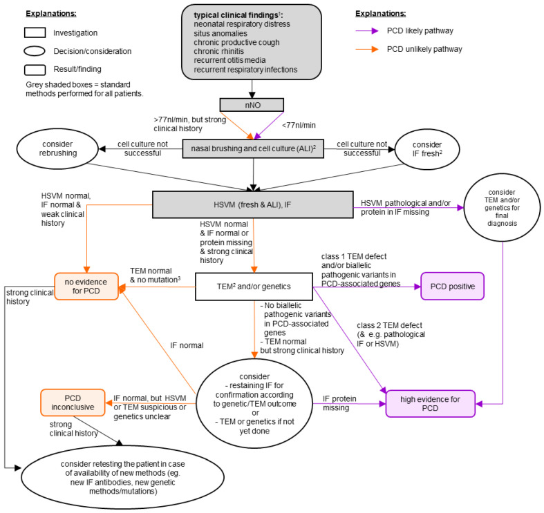
The diagnostic workflow used at PCD-UNIBE implements the ERS guidelines [15], but adds (for all patients) ALI cell culture of brushed cells and IF of structural proteins of the ciliary axoneme. In brief, we perform HSVM and IF for all patients, but execute TEM and genetics analysis only for selected patients. Our diagnostic workflow is shown in Figure 1 and our detailed diagnostic algorithm has been recently published [21]. An important part of PCD-UNIBE are interdisciplinary meetings with clinicians (pneumologists) and diagnostic research specialists to discuss cases, decide on further investigations and confirm diagnosis. nNO measurements are not performed by PCD-UNIBE, but by the referring centers themselves (since some patients are not physically present at PCD-UNIBE, but only their nasal brushes are sent to us).
Figure 1.
Diagnostic workflow of our comprehensive PCD-UNIBE diagnostic center. “PCD positive” and “high evidence for PCD” are considered as a diagnosis of PCD. For patients with a high clinical suspicion, we usually perform all available methods. 1 As a clinical screening, the PICADAR-Score [22] may be useful. 2 Further investigations (HSVM, IF, and TEM) are preferably conducted by analyzing the material of the ALI cell cultures. A re-brushing is considered if cell culture was not successful. When re-brushing was not possible, fresh material was used. 3 We recommend further investigations (e.g., RNA-analysis or array-CGH) if other results suggest PCD. Abbreviations: ALI: air-liquid interface, HSVM: high-speed-videomicroscopy, IF: immunofluorescence staining, nNO: nasal nitric oxide, PCD: primary ciliary dyskinesia, TEM: transmission electron microscopy.
2.3. Nasal Brushing and Further Processing
Nasal epithelial cells (NECs) are obtained by brushings with adapted interdental brushes from both nostrils. The cells are then removed from the brushes and used for different investigations: (i) HSVM of the fresh sample; (ii) preparation of slides for IF staining; (iii) cultivation of cells; and (iv) fixation for TEM (if the sample contains sufficiently large groups of ciliated cells). Detailed descriptions of the nasal brushing itself, its processing and the further procedures are provided in the Appendix A—Supplementary Methods’ Description.
2.4. Cell Culture
Primary NECs are cultured using the PneumaCult Media Kits (Stemcell Technologies), according to the manufacturer’s protocol with minor changes. A detailed description of our protocol is available in Appendix A.
2.5. High-Speed Videomicroscopy (HSVM)
Ciliary motility of fresh or ALI cells is analyzed via HSVM. To summarize, we use a silicon spacer to build an imaging chamber and record videos on an inverted bright field microscope. These videos are then analyzed using our own analysis software (termed “Cilialyzer” [23]) by assessing the ciliary beating pattern (CBP), beating frequency (CBF), coordination of cilia movement, and particle transport. A detailed description of the methods is provided in Appendix A. The Excel analysis template is available online under the Supplementary Material.
2.6. Immunofluorescence Staining (IF)
Different structural proteins of the ciliary axoneme are labeled using standard protocols for IF. Our standard panel includes dynein axonemal heavy chain 5 (DNAH5), growth arrest specific 8 (GAS8), and radial-spoke-head 9 (RSPH9) (and dynein axonemal light intermediate chain 1 (DNALI1) for earlier samples). Based on findings from the IF and the HSVM, further proteins are stained. A detailed description of the methods and a list of available proteins are provided in Appendix A.
2.7. Transmission Electron Microscopy (TEM)
We perform an ultrastructural analysis of cilia via TEM for patients with high clinical suspicion and/or suspicious results from the standard tests (HSVM and IF). In short, cells (fresh or from ALI cultures) are fixed and processed, and 100–200 well assessable cilia cross sections imaged. The axonemal structures are systematically evaluated and scored according to the international consensus guidelines on TEM in PCD diagnosis [24]. Additionally, we discriminate between proximal and distal localization. A detailed method’s description is provided in Appendix A and the Excel analysis template is available online under the Supplementary Material.
2.8. Genetical Analysis
Analysis of pathogenic or likely pathogenic variants in all currently known PCD-associated genes is performed via next-generation sequencing of the whole exome by specialized centers; for more details see Appendix A.
3. Results
3.1. Study Population
The first 100 patients referred to the new PCD-UNIBE center for comprehensive PCD diagnostics came from various hospitals all over Switzerland, mostly children’s clinics. Details on the study population are presented in Table 1.
Table 1.
Characteristics of the study population, summary of the referring centers, and overview of the diagnostic status and outcome. Percentages are related to the total number of cases in the respective column.
| Characteristic of the Study Population | All Cases | PCD Cases | Non-PCD Cases |
|---|---|---|---|
| Mean age (Standard deviation), years | 12.9 (18.0) | 29.2 (26.4) | 9.1 (12.8) |
| Median age (range), years | 5.6 (0.04–68.9) | 16.9 (1.4–68.9) | 4.8 (0.04–61.7) |
| Sex (female/male), n | 44/56 | 8/91 | 36/44 1 |
| Age category 2 (children/adults), n | 83/17 | 11/6 | 70/10 |
| Referring centers, n (%) | all cases | PCD cases | non-PCD cases |
| Inselspital, Bern University Hospital (pediatrics) | 54 (54%) | 7 (41%) | 45 (56%) |
| Inselspital, Bern University Hospital (adults) | 2 (2%) | 0 (0%) | 1 (1%) |
| Cantonal Hospital Graubünden (pediatrics) | 23 (23%) | 3 (18%) | 20 (25%) |
| Children’s Hospital Lucerne | 4 (4%) | 1 (6%) | 3 (4%) |
| Fribourg Hospital (pediatrics) | 4 (4%) | 0 (0%) | 4 (5%) |
| Lausanne University Hospital (pediatrics) | 2 (2%) | 0 (0%) | 2 (3%) |
| Hospital Lindenhof (Pneumology, Bern) | 4 (4%) | 2 (12%) | 2 (3%) |
| Others 3 | 7 (7%) | 4 (24%) | 3 (4%) |
| Symptoms, n (%) | all cases | PCD cases | non-PCD cases |
| Chronic productive cough | 61 (61%) | 9 (53%) | 51 (64%) |
| Chronic rhinitis | 40 (40%) | 9 (53%) | 29 (36%) |
| Chronic sinusitis | 8 (8%) | 1 (6%) | 7 (9%) |
| Recurrent otitis media | 18 (18%) | 4 (24%) | 13 (16%) |
| Recurrent respiratory infections | 41 (41%) | 2 (12%) | 36 (45%) |
| Bronchiectasis | 12 (12%) | 3 (18%) | 8 (10%) |
| Neonatal respiratory distress | 8 (8%) | 1 (6%) | 7 (9%) |
| Situs anomalies | 10 (10%) | 5 (29%) | 5 (6%) |
| Others 4 | 42 (42%) | 9 (53%) | 32 (40%) |
| nNO3, n (%) | all cases | PCD cases | non-PCD cases |
| Reduced (<77 nL/min) | 25 (25%) | 3 (18%) | 21 (26%) |
| Borderline (<85 nL/min, >77 nL/min) | 2 (2%) | 11 (65%) | 14 (18%) |
| Normal (>85 nL/min) | 24 (24%) | 0 (0%) | 2 (3%) |
| Not performed 5 | 49 (49%) | 3 (18%) | 43 (54%) |
1 Three cases were unsolved (n = 2 inconclusive, n = 1 diagnostics ongoing); thus, PCD and non-PCD cases do not sum up to the numbers of all cases. 2 Children = age younger than 18 years. 3 Hospitals with only one case, e.g., University Children’s Hospital Basel, University Hospital Basel (Clinic for Ear–Nose–Throat). 4 e.g., cardiological problems, obstructive sleep apnea syndrome, growth impairment, hearing problems, colonization with Pseudomonas aeruginosa, family history of PCD. 5 For some patients we do not have this information either because the referring center had no possibility to measure nNO levels or because the patient was too young to perform a nNO measurement.
3.2. Overview of the Tests: HSVM, IF, TEM, Genetics
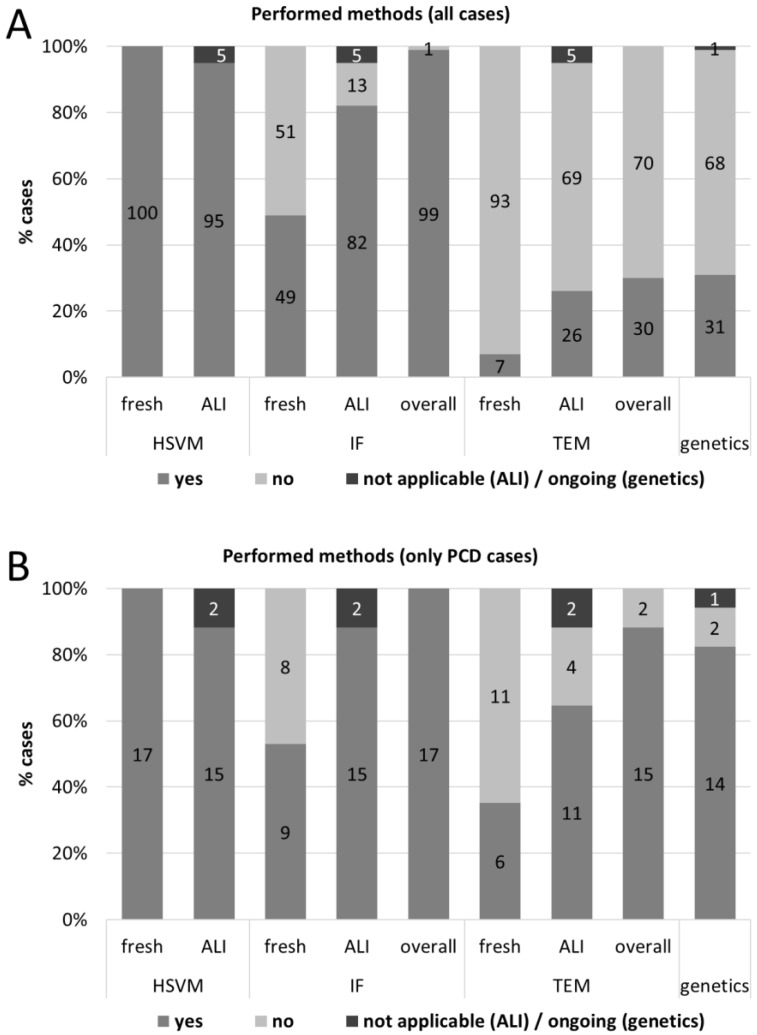
The diagnostic outcome was usually the result of several tests; see Figure 2 for an overview over all patients (A) and PCD cases (B). For all patients, we performed HSVM of the fresh samples and tried to obtain re-differentiated ALI cell cultures. HSVM ALI was performed whenever we succeeded in growing and re-differentiating cells (see below for details on cell culture success rate). For the vast majority of cases, we performed IF, preferable from ALI cell cultures (n = 82, n = 49 additional from fresh samples). For one case, we omitted IF since the clinical suspicion was moderate, HSVM showed normal CBP and CBF, and PCD was no longer the most probable diagnosis pursued. In 30 cases with highly suspicious CBP in HSVM and/or high clinical suspicion, we performed TEM. We carried out TEM analysis from seven fresh samples and from 26 ALI samples (for three patients we performed both). Genetics was done in 31 cases with high clinical suspicion and/or suggestive results of previous tests.
Figure 2.
Overview of the performed tests from the first 100 patients (A) of the PCD-UNIBE diagnostic center and of PCD cases only (B). Numbers in the bars indicate number of cases.
3.3. Air-Liquid Interface (ALI) Cell Culture
We aimed to cultivate cells from all patients. The cell culture grew and re-differentiated successfully for 90% from the first brushings. Reasons for failure of cell cultivation were bacterial contamination (n = 1), fungal contamination (n = 2), bad primary material (n = 7, e.g., not sufficient viable cells, no attachment or growing of the cells). In 10 cases, the cell cultivation was not successful at all; additionally, in one case, the quality of the cell culture was insufficient. For four patients, we omitted a re-brushing: one case could be diagnosed with PCD based on a TEM hallmark defect in the fresh sample; one patient had a pathologic mutation in a known PCD-associated gene; two patients refused a re-brushing. For the other seven cases with failure of cell cultivation, we performed a re-brushing and cultured cells again (success rate of 86%; six re-differentiated successfully, one failure due to fungal contamination).
3.4. Results of the Performed Tests: HSVM, IF, TEM, Genetics
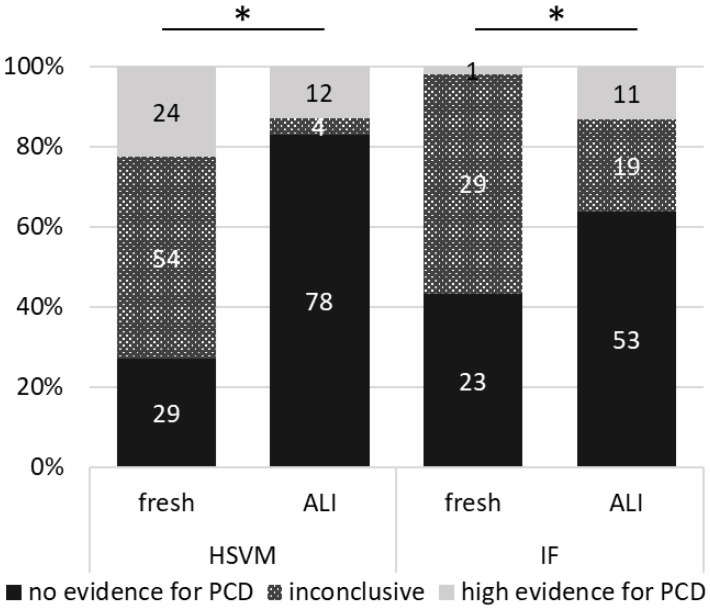
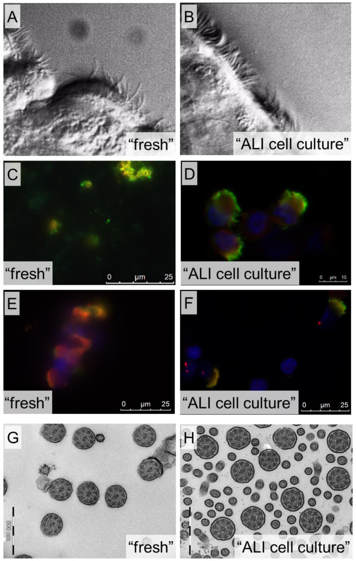
From the 107 fresh HSVM analyses, 29 cases (27%) showed no evidence for PCD (CBP score > 3), 54 cases (50%) were inconclusive, and 24 cases (22%) showed a high evidence for PCD (Figure 3). Results of HSVM done on cell cultures were much clearer: 78 cases (83%) showed no evidence for PCD, four cases (4%) were inconclusive, and 12 cases (13%) showed high evidence for PCD. Interestingly, not all cases were assessed identically from fresh and ALI samples. For 42 cases, the conclusion was similar in both samples: 28 cases were assessed as “no evidence for PCD”, four cases as “inconclusive”, and 10 cases as “high evidence for PCD” based on both, fresh and ALI samples. Forty-one cases were assessed as “inconclusive” based on fresh samples and as “no evidence for PCD” based on the ALI sample. Moreover, importantly, one case was assessed as “no evidence for PCD” based on fresh and as “high evidence for PCD” based on ALI sample. For one case, the fresh sample was “inconclusive”, but the ALI sample showed a “high evidence for PCD”. In addition, nine cases showed “high evidence for PCD” in fresh and “no evidence for PCD” in the ALI samples. The samples of which the cell culture did not successfully re-differentiate were assessed as follows: 1× “no evidence for PCD”, 7× “inconclusive”, 5× “high evidence for PCD”. Figure 4A,B show the typical difference in sample quality for the assessment of the ciliary motility via HSVM.
Figure 3.
Differences in the assessments of ciliary motility via HSVM and presence of structural ciliary proteins via IF stainings in fresh and ALI samples. For HSVM fresh, we analyzed 107 samples in total, as re-brushings were performed for seven patients. Two of the samples could not be analyzed at all since no viable cells were found. We had a usable cell culture for 94 patients. For five patients we had no ALI and for one patient, the cell culture was not stable and lost all cilia before reaching maturity (at 28 days), and could not be analyzed. * Statistically significant difference between HSVM fresh versus ALI for “no evidence for PCD” (p < 0.0001), “inconclusive” (p < 0.0001) and “high evidence for PCD” (p = 0.04) and IF fresh versus ALI for “inconclusive” (p = 0.03). Differences between IF fresh versus ALI were not significant for “no evidence for PCD” (p = 0.18) and “high evidence for PCD” (p = 0.38). Data were compared using the Wilcoxon matched-pairs signed rank test (GraphPad Prism Version 9.0.2).
Figure 4.
Examples of quality differences between fresh and ALI cell culture samples for HSVM (A,B), IF (C–F), and TEM (G,H). In C–F, the green staining shows the tubulin of the ciliary axoneme, the red staining shows the GAS8 (C,D) or DNAH5 (E,F) protein, and the blue staining the cell nuclei.
The assessments of IF show similar characteristics, when comparing fresh vs. ALI, as the ones of HSVM (Figure 3): based on fresh samples statistically significant more inconclusive results (29 cases (55%) versus 19 cases (23%), p = 0.03) and fewer results suggesting “no evidence for PCD” (23 cases (43%) versus 53 cases (64%), p = 0.18) compared to the ALI samples. Furthermore, IF of the ALI samples enabled us more often to obtain a “high evidence for PCD” (one case (2%) versus 11 cases (13%), p = 0.38). Figure 4C–F give two examples of possible differences in the quality of fresh and ALI samples.
We performed TEM analysis for 30 cases (n = 4 only fresh, n = 3 fresh and ALI, n = 23 only ALI). There was “no evidence for PCD” in 22 cases (72%). Six cases were positive for PCD: four cases (14%) showed a class 1 ODA and IDA defect, one (3%) a class 1 microtubular disorganization and IDAs defect, and one (3%) a class 2 ODA defect. Two cases (7%) remained inconclusive due to bad quality of cell cultures. The production of TEM sections and images from ALI cell cultures was far more efficient than from freshly brushed samples. Abundant ciliated cells from large epithelial stripes and the possibility of aligning the cell culture prior to sectioning provided many more axonemal views, which are additionally optimally transected (see also Figure 4G,H). Furthermore, the effort needed for the TEM assessment of cell cultures was roughly one third of the one needed for the sparse, randomly oriented fresh brushing material.
Genetic analysis was carried out in 31 cases, with one analysis still ongoing. In 17 cases (55%), no abnormal variants were found. In nine cases (29%), two pathologic or likely pathologic variants in the same gene were found (7× DNAH11, 1× DNAH5, 1× HYDIN), finally leading to the diagnosis of PCD. Five cases (16%) showed genetic abnormalities, but did not lead to a (direct) PCD diagnosis: (1) in one case, a homozygous mutation of unknown significance in the DNAH11 gene was found (PCD was confirmed due to missing DNAH11 in IF and pathologic HSVM). (2) In another case, only a single likely pathogenic variant in the DNAH11 was found (PCD was confirmed due to missing DNAH11 in IF and pathologic HSVM). (3) In one case three variants of unknown significance in the DNAH5 gene were found (PCD was not confirmed in this case, since HSVM and TEM were normal and DNAH5 was present in IF). (4) In another case, we found two DNAH9 variants of unknown significance in-cis (the missing of DNAH9 in IF and a TEM class 2 ODA & IDA defect diagnosed PCD). (5) The last of the unclear cases showed only one pathogenic variant in the HYDIN gene (PCD was not diagnosed due to HSVM being better than expected for HYDIN mutations and normal nNO levels).
3.5. Overall Diagnostics Outcome
There was no evidence for PCD in 80 cases (Table 2). Seventeen patients were diagnosed with PCD (5× “high evidence for PCD”, 12× “PCD positive”). Three cases remained inconclusive: Two patients refused further investigations, and in one case, the diagnostics is still ongoing (The fresh sample and the cell culture showed only a few cilia. The genetic analysis showed no pathogenic or likely pathogenic variants in 43 genes (including MCIDAS, CCNO, FOXJ1). Thus, we performed a re-brushing of which the results were still pending). Among the 17 patients with diagnosed PCD, there are three cases with ongoing analysis: In one case, PCD was diagnosed based on fresh HSVM and TEM, but the genetic analysis is not yet complete. In two cases, PCD was diagnosed (1x based on HSVM and TEM, 1x based on IF), but the underlying genetic mutation could not be identified yet. Two of the cases with “no evidence for PCD” were clinically highly suspicious (one with repeatedly reduced nNO), but none of the tests (TEM, HSVM, IF, genetics) showed any evidence of PCD. Further investigations are planned for these patients.
Table 2.
Diagnostic outcome of the first 100 patients referred to PCD-UNIBE.
| Diagnostic Outcome | n (%) |
|---|---|
| no evidence for PCD | 80 (80%) |
| high evidence for PCD (=diagnosis of PCD) | 5 (5%) |
| PCD positive (=diagnosis of PCD) | 12 (12%) |
| Inconclusive a | 3 (3%) |
a Currently inconclusively due to refusal for further investigations in two patients and still ongoing diagnostics in one patient.
3.6. Relevance of the Different Methods for the Diagnosis of PCD
The result of the HSVM confirmed the diagnosis of PCD in 13 cases (13% of all 100 cases investigated with this method). For IF, this was the case in 14 cases (14% of the 99 IF investigated cases), for TEM in five (17% of the 30 TEM investigated cases) and genetics in 10 cases (33% of the 30 genetically investigated cases) (Table 3). This shows that none of the methods used was able to diagnose all 17 PCD cases. Thus, a comprehensive approach is essential for an accurate diagnosis of PCD.
Table 3.
Overview of the different methods and whether the outcomes led to the diagnosis of PCD.
| n | HSVM | IF | TEM | Genetics | Remark |
|---|---|---|---|---|---|
| 8 | + | + | - | + | all DNAH11 mutations |
| 2 | + | + | + | (not done) | no genetics performed |
| 2 | - | + | + | - | no mutations found |
| 1 | + | + | - | - | one pathologic variant in DNAH11 |
| 1 | + | - | + | - | genetics still ongoing |
| 1 | + | - | (not done) | + | HYDIN mutation |
| 1 | - | + | - | - | No mutation found |
| 1 | - | - | (not done) | + | DNAH5 mutation, cell culture not successful |
| Total: 17 | 13 | 14 | 5 | 10 |
n = number of patients, written in bold = the sole method leading to the diagnosis of PCD. + = the outcome of this method was leading to the diagnosis of PCD. - = the outcome of this method was neither “high evidence for PCD” nor “PCD positive” and, thus, was NOT leading to the diagnosis of PCD.
3.7. Costs
The costs for the different investigations vary a lot (Table 4) and are highly specific for different countries and healthcare systems. Our standard diagnostics including brushing, cell culture, HSVM fresh, and ALI and IF ALI costs EUR 1229. This is slightly more compared to the costs of a set without cell culture including three brushings, three HSVM fresh, and IF fresh, which adds up to EUR 1165. However, as soon as TEM needs to be performed, the costs for the cell culture are overcompensated: A full set with only fresh material (3× brushings, 3× HSVM fresh, IF fresh, TEM fresh) would cost EUR 3165, while a full set including cell culture (1x brushing, HSVM fresh and ALI, IF ALI, TEM ALI) would be EUR 2025. These reduced costs are in addition to the advantages of higher quality of the results (for details see above), less clinical visits and less burden for the patients due to fewer brushings. Our costs are higher compared to the numbers published earlier by Shoemark et al.: USD 187 (EUR 159 (exchange rate of EUR 0.85 per one dollar was used) and USD 1452 (EUR 1231). The higher costs in our setting are mostly related to higher salaries and higher prices for consumables in Switzerland.
Table 4.
Overview of the costs for the different methods. These costs only include running costs, such as consumables, fee for using microscopes and working hours, but no initial set up costs (e.g., purchase of equipment) and neither costs on the side from clinics or patients for the visits (of which more are needed if only fresh material is used).
| Investigation/Method | Consumables and Fee for Equipment Use | Working Hours | Total |
|---|---|---|---|
| nasal brushing | EUR 6 | EUR 70 | EUR 76 |
| cell culture | EUR 250 | EUR 260 | EUR 510 |
| HSVM fresh | EUR 24 | EUR 180 | EUR 204 |
| HSVM ALI | EUR 24 | EUR 140 | EUR 164 |
| IF fresh 1 | EUR 155 | EUR 170 | EUR 325 |
| IF ALI 1 | EUR 155 | EUR 120 | EUR 275 |
| TEM fresh | EUR 500 | EUR 1500 | EUR 2000 |
| TEM ALI | EUR 250 | EUR 750 | EUR 1000 |
| Reporting/meetings/etc. | EUR 0 | EUR 181 | EUR 181 |
| Genetics 2 | EUR 200 | EUR 2800 | EUR 3000 |
| all standard methods 3 | EUR 459 | EUR 770 | EUR 1229 |
| all methods 4 | EUR 909 | EUR 4501 | EUR 5410 |
Remark about costs: PCD-UNIBE is based in Switzerland where salaries and prices for consumables are usually higher than in other European countries. Original costs were calculated in Swiss francs. For the conversion to Euros, an exchange rate of 1.1 CHF per one EUR was used. 1 These costs apply for the standard panel including DNAH5, GAS8, and RSPH9. Costs are higher if additional proteins are stained. 2 Average costs, costs vary depending on affected gene. 3 Standard methods include nasal brushing, cell culture, HSVM fresh, HSVM ALI, and IF ALI. 4 All methods include nasal brushing, cell culture, HSVM fresh and ALI, IF ALI, TEM ALI, and genetics.
4. Discussion
4.1. Summary
Among the first 100 patients referred to our newly founded PCD-UNIBE diagnostic center, we diagnosed PCD in 17 cases and found no evidence for PCD in 80 patients. Three cases remain inconclusive due to patients’ refusal for further investigations (n = 2) or ongoing diagnostics (n = 1). We performed HSVM of the fresh samples for all patients and IF for 99% of the patients. HSVM was repeated for all patients with a successful ALI cell culture (90% after the initial brushing, 5% after a re-brushing). TEM was performed for 30 and genetics for 31 patients. The use of the cell culture avoided a re-brushing in 90% of the cases and was crucial for most of the cases as it reduced the ratio of inconclusive findings for HSVM and IF significantly. HSVM confirmed diagnosis of PCD in 13, IF in 14, TEM in five, and genetics in 10 cases, respectively. None of the methods used was able to diagnose all 17 PCD cases.
4.2. Diagnostic Impact of Methods/Procedures in Our Setting
By setting up our new comprehensive PCD diagnostic center in 2018, we decided to include ALI cell culture as standard procedure for all patients. Herein, we report on the importance of this as well as of the other included methods. Every single method included in our algorithm was essential for certain cases to diagnose PCD. IF and genetics were the sole diagnostic method in one case each (in the case of genetics, the cell culture did not grow successfully, HSVM and IF of the fresh sample were inconclusive and we omitted a re-brushing since the genetic testing confirmed PCD). For all other cases, at least a combination of two of the four methods were needed to confirm the diagnosis of PCD. Nevertheless, since none of the methods was able to diagnose all cases, we can neither omit one of them nor choose a single method to be the best. Furthermore, we would like to emphasize the importance of ALI cell cultures for our diagnostic procedure. Its value has been described before [25,26,27,28], but we would like to add important aspects. The use of cell culture avoided a re-brushing in 90% of all cases. This is of particular importance for pediatric patients, in whom additional uncomfortable examinations should be avoided. Further results from HSVM, IF, and TEM using cell cultures are much more precise: The ratio of inconclusive assessments was significantly lower for HSVM and IF after cell culture compared to fresh samples. HSVM of cell cultures has already been shown to robustly represent original characteristics while eliminating secondary effects and overcoming low cell yields of the fresh samples [15,26,28,29]. Secondary or artifactual dyskinesia often seen in fresh samples is one of the main critical points raised when HSVM is used as a diagnostic method [17]. Even highly standardized and gentle brushings mostly provide merely tiny cell conglomerates or single cells that are ripped out of their natural environment. This may lead to additional analysis artefacts. Therefore, we principally avoid HSVM conclusions from single cells (as also recognized by international HSVM specialists). We also found a clear improvement of the sampling quality for IF and TEM as the amount of available cell material influences the validity of the diagnosis. While TEM from fresh material often requires time-consuming search of sparsely scattered cells in numerous resin blocks, ALI-derived epithelial stripes provide large lawns of trimmed cilia. This increases the chance of getting optimally oriented orthogonal transects in one or two sections from only two blocks. TEM assessment can thus usually be based on several hundreds of cilia cross sections obtained at half of the effort and costs needed for fresh samples. The same was true for IF: slides from cell culture contain more nicely stained epithelial cells reducing considerably the time needed to perform a valid analysis. An additional advantage of routinely performed cell cultivation is that leftover cells not used for diagnostics in the first round can be cryostored and used as backup cells (if the primary cell culture should not re-differentiate successfully or if further tests are needed (e.g., RNA sequencing from differentiated cells because of unclear genetics results)) and/or for research purposes (if patients consent), as recognized earlier [26].
4.3. Limitations and Strengths
Our study presents a realistic scenario using real world patients’ data. We principally highlight the practical aspects of our workflow, as such descriptions are often lacking in other studies. These could be particularly helpful for diagnostic centers. Our diagnostic algorithm includes an integral set of several methods including cell culture as preparatory step. Thus, we can evaluate the advantages and disadvantages of the different morpho-functional approaches HSVM, IF, TEM, and the potential improvement by the use of cell cultures. By our intention to present a real live report, patient data was exclusively obtained during our routine diagnostic workflow. A limitation of the study is that, according to our diagnostic workflow, TEM and genetics are not performed if previous results clearly showed no evidence for PCD and the clinical suspicion is low. In order to comply with state regulations to reduce healthcare costs, the expensive TEM analytics and genetic panel diagnostics were only performed upon high suspicion based on clinics and other test results. Additionally, genetics need a special approval by the health insurance. Therefore, we did not have results of all methods for all patients. Three cases remained inconclusive due to missing analytic data (2× refusal of further investigation, 1x ongoing analysis), but are consciously part of the first 100 patients referred to PCD-UNIBE. Such situations are an authentic part of the daily work in our PCD diagnostic center.
4.4. Comparison with the Literature
In our study population, prevalence of PCD diagnosis (17%) was rather high compared to a previous study with a prevalence of approximately 10% [22]. There are two reasons for this high diagnostic yield: (i) when we started the comprehensive approach for PCD diagnostic in 2018, we worked up several previously unclear, but clinically highly suspicious cases. The proportion of patients diagnosed with PCD among them was higher than among the routinely referred ones. This may falsely indicate that our comprehensive workflow enables a superior detection rate. (ii) Among the first 100 patients were two families with four and three members, respectively, also contributing to the high prevalence. The family with four members was affected by mutations in the DNAH11 gene. Interestingly, we found many DNAH11 cases (8 out of 10 genetically solved cases), also among the cases we work-up again. Additionally, one of the diagnosed, but genetically unsolved cases have one pathologic variant in the DNAH11 gene (but so far, no second one). The high proportion of DNAH11 mutations, especially in worked up cases, probably reflects the former diagnostic approach with focus on TEM (TEM hallmark defect is still officially needed to reclaim support from the Swiss disability insurance) and HSVM of fresh samples. The fact that cilia of patients with DNAH11 mutations show a normal TEM ultrastructure explains that these cases had not been diagnosed previously. This highlights again that a comprehensive approach based on different methods is essential.
4.5. Clinical and Practical Relevance
Patients with suspicion of PCD have the right to know whether they suffer from PCD or not. Since every method on its own misses a considerable part of PCD cases (e.g., approximately 30% for TEM and genetics if tested individually [17]), all currently available methods are needed. In our study, every method missed at least four PCD cases (24% of all 17 PCD cases). Even though current therapy is similar for patients with a clinically high suspicion of PCD and those with confirmed PCD, it is still important to precisely know the underlying causes of clinical symptoms [30]. A clear diagnosis may be of importance for parents who are considering having more children. In addition, since some PCD mutations are associated with infertility, a clear diagnosis with known underlying genetic defect is important for the future family planning. Last, having a clear diagnosis of PCD and its specific genetic mutation is important with regard to therapeutic regimens. Recently, first evidence-based data for therapies, specifically for PCD (e.g., with macrolide antibiotics [31]), became available, and initiatives for molecular treatments or gene therapies for PCD are ongoing (e.g., CLEAN-PCD study, ClinicalTrials.gov Identifier: NCT02871778).
5. Conclusions
Diagnosis of PCD is challenging and every method included in our diagnostic algorithm contributed to final diagnoses. In our approach, the use of ALI cell cultures for HSVM, IF, and TEM was particularly crucial. However, none of the methods was able to diagnose all 17 PCD cases singlehandedly, highlighting that a comprehensive approach is essential for an accurate PCD diagnostics.
Acknowledgments
We thank all patients for allowing us to evaluate their diagnostic data. Study authors participate in the BEAT-PCD clinical research collaboration, supported by the European Respiratory Society. Our center participates at the European Reference Network ERN Lung PCD core as a supporting member. The current Swiss PCD Research Group includes (alphabetical order): Sylvain Blanchon (Lausanne), Jean-Louis Blouin (University Hospitals of Geneva, Switzerland), Marina Bullo (Inselspital Bern), Carmen Casaulta (Inselspital Bern), Myrofora Goutaki (Institute of Social and Preventive Medicine, University of Bern, Inselspital Bern, Switzerland), Nicolas Gürtler (Pediatric Otorhinolaryngology, Department of Otorhinolaryngology, University Hospital of Basel, Switzerland), Beat Haenni (Anatomy Bern), Andreas Hector (Division of Respiratory Medicine, University Children’s Hospital Zurich, Switzerland), Michael Hitzler (Division of Paediatric Pulmonology, Children’s Hospital Lucerne, 6000 Lucerne, Switzerland), Andreas Jung (Zürich), Lilian Junker (Pneumology, Hospital Thun, Switzerland), Elisabeth Kieninger (Inselspital Bern), Claudia E. Kuehni (Institute of Social and Preventive Medicine, Inselspital Bern), Yin Ting Lam (Institute of Social and Preventive Medicine, University of Bern, Switzerland), Philipp Latzin (Inselspital Bern), Dagmar Lin (Department of Pulmonary Medicine, Inselspital, Bern University Hospital, University of Bern, Bern, Switzerland), Marco Lurà (Division of Paediatric Pulmonology, Children’s Hospital Lucerne, Switzerland), Loretta Müller (Inselspital Bern), Eva Pedersen Institute of Social and Preventive Medicine, University of Bern, Switzerland), Nicolas Regamey (Lucerne), Isabelle Rochat (Pediatric Pulmonology and Cystic Fibrosis Unit, Service of Pediatrics, Department Woman–Mother–Child, Lausanne University Hospital, University of Lausanne), Daniel Schilter (Quartier Bleu, Medical Practice for Pneumology at the Hospital Lindenhof, Bern, Switzerland), Iris Schmid (Quartier Bleu, Medical Practice for Pneumology at the Hospital Lindenhof, Bern, Switzerland), Bernhard Schwizer (Quartier Bleu, Medical Practice for Pneumology at the Hospital Lindenhof, Bern, Switzerland), Andrea Stokes (Inselspital Bern), Daniel Tachsel (University Children’s Hospital Basel (UKBB), Switzerland), Stefan A. Tschanz (Anatomy Bern), Johannes Wildhaber (Department of Paediatrics, Fribourg Hospital HFR, Faculty of Science and Medicine, University of Fribourg, Switzerland).
Supplementary Materials
The following are available online at https://www.mdpi.com/article/10.3390/diagnostics11091540/s1, Excel templates for the analysis of the HSVM videos and the TEM images, and one fresh and one ALI HSVM video.
Appendix A. Supplementary Methods’ Description
Appendix A.1. Nasal Brushing: Procedure and Processing
After the patients clean their nose, nasal epithelial cells (NECs) are obtained by nasal brushings using one pre-wetted (cold 0.9% NaCl in a 15 mL tube) interdental brush (IDB-G50 3 mm, Top Caredent, Zurich, Switzerland; elongated by attaching a 200 μL pipette tip with parafilm; Figure A1) for each nostril, as previously described [32]. Briefly, the brush is inserted into and removed out of the inferior turbinate of the nasal cavity a couple of times, while rotating and slightly pressing against the lateral edges of the nasal cavity. Brushes with obtained cells are immediately stored in a 15 mL tube with RPMI-1640 media with HEPES (Sigma-Aldrich, Merck, Darmstadt, Germany, cat#R7388) (using PneumaCult Expansion Plus (PC-Ex-Pl) media (STEMCELL Technologies, Vancouver, Canada, cat#05040) with additional 20 mM HEPES Buffer (Sigma-Aldrich, cat#H0887) for brushes shipped from other hospitals (storage up to 4–5 h)).
Under a biosafety cabinet, the elongated brushes are washed with the medium in the tube by pipetting the medium slowly over the brush (using 1 mL of media). After this, all cells are remove by gently pushing and pulling the brush through an adapted 200 μL tip (cut of approximately 5 mm of the tip to have a larger hole), while keeping the tip submerged in the medium. This procedure is repeated with all brushes. After centrifugation (room temperature (RT), 5 min, 300 rcf; this setting is used throughout this protocol, unless stated otherwise), the cell pellet is resuspended in 1.6 mL PC-Ex-Pl medium. This 1.6 mL is then used for various diagnostic tests: (i) 60 μL for HSVM; (ii) 400 μL is used for the preparation of eight IF slides (50 μL/slide; Cellspin Cytoslides uncoated, one circle, Tharmac, Wiesbaden, Germany, 310-100, VWR 720-1808, air dried and stored at −20 °C); (iii) 100 μL is fixed for TEM if HSVM shows large conglomerates of epithelial cells with cilia; and (iv) 1 mL (or more, just the remaining) is used for cell culture.
Figure A1.
Interdental brush elongated by attaching a 200-μL pipette tip with parafilm.
Appendix A.2. Cell Culture
Appendix A.2.1. Preparation of Media
For the culturing of the primary NECs, we use the Stemcell PneumaCult media. Additionally, for the preparation of the ready-to-use media, 50 μL and 200 μL aliquots of Hydrocortisone (Stemcell #07926, stored at −20 °C), 1 mL and 100 μL aliquots of Primocin (InvivoGen, Toulouse, France, cat. ant-pm-2, stored at −20 °C) as well as Heparin (Stemcell #07980, stored at 2–8 °C) were needed.
The PC-Ex-Pl medium is used for the submerged periods (passage 0 and passage 1 in plates or flasks, and p2 on the inserts until confluent). This medium kit (Stemcell, #05040) comes with a “Basal Medium” (#05041, 2–8 °C) and a “Supplement” (#05042, −20 °C). Upon receiving a new kit, the basal medium is aliquoted into 10 × 49 mL portions (in 50 mL conical tubes (Greiner, Monroe, NC, USA, cat.no. 277261) and stored at 4 °C. The supplement is thawed and then gently, but very well mixed, aliquoted into 10 × 1000 μL portions and frozen at −20 °C. As needed, the complete medium is prepared: 1 mL of the PC-Ex-Pl supplement is added to the previously prepared 49 mL PC-Ex-Pl basal medium, along with 50 μL of hydrocortisone and 100 μL of primocin. This complete PC-Ex-Pl medium can be stored at 4 °C for up to 4 weeks.
Later, when the cells are confluent and exposed to the air-liquid interface (ALI), the cell culture is nourished through the PneumaCult ALI Maintenance (PC-ALI) medium. The PC-ALI medium kit (Stemcell, #05001) contains a basal medium (Stemcell, #05002), a supplement (Stemcell #05003) and a maintenance supplement (Stemcell, #05006). For the preparation of the complete PC-ALI base medium, first the PC-ALI Supplement needs to be thawed, in either cold water or overnight at 4 °C. When thawed, it can be mixed gently through inversion of the vial. Then, the whole 50 mL of the supplement is added to the 450 mL of basal medium, along with 1 mL of Primocin. Then, this complete base medium is aliquoted to 40 mL in 50 mL Greiner tubes and frozen at −20 °C. Then, when needed, the complete, ready-to-use PC-ALI medium can be prepared. After the 40 mL complete basal medium thaws, 400 μL of PC-ALI maintenance supplement (comes in 1 mL vials; we usually remove the 400 μL we need for the preparation of the complete PC-ALI medium, transfer 400 μL to a 1.5 mL Eppendorf tube and freeze this, and the original tube with the remaining 200 μL again at −20 °C for later use) along with 200 μL of hydrocortisone and 80 μL of heparin are mixed. To avoid the risk of fungal contaminations, 400 μL of amphotericin (Sigma-Aldrich, #A2942) can be added. This complete PC-ALI medium can be used immediately or stored at 4 °C for up to 2 weeks.
Appendix A.2.2. Preparation of Well Plates
Before the brushing arrives at the lab, the 12-well plates should be coated. The two central wells (b2, b3 of a Falcon 12-well plate, REF353043) are coated with 500 μL diluted PureCol (PureCol Collagen, cat.no. 5005B, Advanced Biomatrix, Carlsbad, CA, USA; diluted 1:10 with sterile water) and incubated for at least 30 min at 37 °C (or overnight at RT). After this, the excess PureCol is removed and the wells are washed with 500 μL PBS (without Ca2+ and Mg2+) (Sigma-Aldrich, #D8537). The PBS from one well is aspirated (to add the cells suspension); the PBS of the second well is left in (until the next day, when the supernatant from the first well will be transferred to the second well after the Accutase treatment).
Appendix A.2.3. Procedure of Cell Cultivation
The cells, after being removed from the brushes, are resuspended in 1.6 mL PC-Ex-Pl medium (as described above), and 1 mL is then used to be seeded in one coated well of a 12-well plate (called passage 0). The day after the brushing, medium from the previously seeded well is transferred to a 15 mL tube, centrifuged (300 rcf, 5 min, RT; all subsequent centrifugation steps are performed with the same settings unless otherwise stated), resuspended in 1 mL Accutase (Sigma-Aldrich, #A6964-100 mL), and kept for 30 min at 37 °C. Subsequently, the cell suspension is centrifuged and again resuspended in 1 mL PC-Ex-Pl and added to a second coated and rinsed well in the same 12-well plate; One mL of fresh PC-Ex-Pl is added to this first well with the already adhered cells. We usually change media on Monday mornings, and Wednesday and Friday afternoons. Ideally, the media of passage 0 cells is changed every day or at least every second day. However, we do not change media over the weekends. The cells are cultured until a maximum of 60–80% confluence is reached (usually after 3–4 days). If cells do not reach 60–80% confluence after one week, depending on the growth process, they are still passaged, in the hope that the provision of a new stimulus might improve growth. Cells are lifted using Accutase (8–10 min at 37 °C, 1 mL for a well of a 12-well plate, flushed off with the Accutase at least 5 times). The cells in the Accutase are transferred to a 15 mL tube, containing 10 mL RPMI, and centrifuged. After aspiration of the supernatant, the cell pellet is resuspended in 1 mL PC-Ex-Pl medium and seeded in a pre-coated (incubation with 2 mL diluted PureCol for 30 min at 37 °C, see preparation of well plates) T25 tissue culture flask (blue vented cap, Falcon, Corning, NY, USA; #353108) (with 5 mL of media in total) until again confluence of a maximum of 60–80% is reached (usually after 5–7 days; rather use of the cells at 50% confluence than risking reaching >80%, especially over the weekend). Then, the cells are lifted and collected again with Accutase (see above) and seeded in uncoated tissue culture transwell (Corning Transwell polyester membrane inserts, pore size 0.4 μm, diameter 12 mm, CLS3460-48EA, Sigma-Aldrich) at a density of 100,000 cells/transwell and supplemented with 1 mL in the lower, basolateral chamber, where no cells are, and 0.5 mL PC-Ex-Pl in the upper, apical chamber, where the cells were seeded into. We usually seed out into three inserts. The leftover cells are frozen in vials with roughly one million cells (using 1 mL of CyroStor freezing media, Sigma-Aldrich, #C2874) as backup if the cell culture would not successfully differentiate. Medium is again changed three times/week (see above). One day after the cells reach complete confluence (usually 2–5 days after seeding), they are exposed to the air-liquid interface (ALI) and fed with 700 μL/well PC-ALI (Stemcell, cat#05001, preparations see above) media from the basolateral chamber only.
After seven or more days at ALI, cultures undergo a weekly PBS wash (Dulbecco’s Phosphate Buffered Saline with MgCl2 and CaCl2 (PBS++), Sigma D8537-500 mL) to remove excess mucus. Before the medium is changed, 500 μL of PBS++ is added to the apical chamber of the insert. The plates are then left at RT for 15 min. After this, the apical PBS and the basal medium is removed and 700 μL of fresh PC-ALI medium is added. Normally after about 7–10 days at the ALI, the cell cultures start to produce mucus, and after about 14–21 days, the first moving cilia were seen. Cell cultures used for the experiments are exposed for ≥28 days at the ALI. Cultures that are at ALI for over 4 weeks are transferred to maintenance and the medium is only changed once per week, along with a PBS wash (see above).
Appendix A.2.4. Processing of Mature ALI Cultures
When the culture is allowed to mature at the ALI for 28 days, it is used for different diagnostic tests. We use one insert for the HSVM ALI and the IF slides (half for each method), and one-half insert for the TEM.
For the HSVM and IF analysis, all medium from the basolateral chamber is transferred to the apical one and the cells are scratched off from the whole membrane of this insert using a 1000 μL filter tip. Then, the media with the cell pieces is added to a 15 mL Greiner tube; 60 μL media with small pieces for the HSVM is then transferred to our self-assembled imaging chamber (see under HSVM). The tube with the remaining media is centrifuged. After aspiration of the supernatant, the cell pellet is resuspended in 1 mL Accutase. The tube is placed in the incubator for 30 min. After the incubation, the cells are resuspended thoroughly, but gently, and centrifuged. After aspiration of the supernatant, the cell pellet is resuspended in, usually, 400-μL RPMI medium (RPMI-1640 Medium, with L-glutamine and sodium bicarbonate, Sigma-Aldrich, #R8758) (50 μL per slide, thus 400 μL for 8 slides); 50 μL is then added to each slide (for details, see above under processing of brushing).
For TEM, the cells are fixed in glutaraldehyde (GA, for details of concentrations, see below); 1 mL of GA is added to a 1.5 mL Eppendorf tube. Taking another insert of the mature culture and using a scalpel, one half of the membrane is cut out of the insert holder and placed in the GA tube, which is stored at 4 °C until further processed (the other half is usually fixed in 4% paraformaldehyde (PFA) for potential 3D immunofluorescence stainings).
Appendix A.3. High-Speed Videomicroscopy (HSVM)
Every patient sample is principally assessed twice by HSVM: the freshly brushed material and scratched material from the ALI cell culture. The first analysis is conducted immediately after the cells arrive in the lab (called “HSVM fresh”). The second analysis is performed when the cell culture is successfully re-differentiated after four weeks at the ALI, approximately six weeks after the brushing (called “HSVM ALI”). A 60 μL drop of cell suspension is brought within a round 200 μm thick rubber spacer (CoverWell silicone ring (Grace Bio-Labs, Bend, OR, USA, Ref 645401) on a standard glass slide (microscopy slide (VWR, Dietikon, Switzerland, 631-1553) (Figure A2) and covered tightly with a standard cover slip (22 × 22 mm, DURAN GROUP, Wertheim, Germany, cat.no. 23550327).
Intact three-dimensional cell stripes are obtained by gently scratching off the epithelial tissue from the inserts of the ALI cell cultures (for more details see below). A minimum of 10 videos with side views and usually two top views of the cell groups are recorded at RT (23–25 °C). Each video is entirely processed within our own software (“Cilialyzer”) and assessed by a scoring system developed by S. Tschanz, considering recent publications [18]. The evaluation criteria are (1) ciliary beating pattern (CBP) with a score from 0 to 4, with ”4” indicating a physiological, complete pattern with power and recovery stroke showing appropriate bending for each phase and “0” indicating no pattern at all. (2) Ciliary beating amplitude with a score from 0 to 4, with “4” being a full amplitude, and “0” having no amplitude at all. (3) Intracellular coordination of ciliary beating with a score from 0 to 2, with “2” showing a well-coordinated ciliary motion within a single cell and “0” showing no coordination at all. (4) Intercellular coordination of ciliary beating with a score from 0 to 2, with “2” showing well-coordinated, propagating ciliary motion of cilia over several cells with physiological temporal displacement and cooperation (metachronal wave) and “0” showing no coordination between cells at all. (5) Cilia-associated transport of available particles (cell fragments, erythrocytes etc.) is analyzed with a score of either “1” (transport observed) or “0” (no transport present). (6) Ciliary beat frequency (CBF) is estimated within several regions of interest (ROI): After selecting a ROI, CBF and its standard deviation are computed within the “Cilialyzer” software based on the generation of the grey level Fourier power spectrum. CBF is laboratory and technique-dependent; therefore, it is recommended to establish a standard CBF with healthy control cells. These scores are filled in an Excel template (see Supplementary Material) and the average of each of the six parameters is calculated to obtain a final score. A score >3 for CBP and amplitude and >1.5 for both coordination types and CBF between 7 and 12 Hz are considered as “no evidence for PCD”, while a score <2 for CBP and amplitude and <1 for the two coordination types and a CBF <7 or >12 Hz reflects “high evidence for PCD”. The scores between the just indicated ranges is considered “inconclusive”. Furthermore, prevalent high frequent (>15 Hz) flickering of cilia with low amplitude and absent bending, as well as rotational movements in top-views are considered as “high evidence for PCD”. At PCD-UNIBE, two diagnostic research specialists always assess all of the recordings. Additionally, to each recording specific observations, such as rotation, impaired partial bending, obstructing cell debris or mucus, or cilia length, are noted. These comments are checked for repeat pathologies (e.g., reduced distal bending) throughout the different videos and a sample quality is assessed (e.g., groups of cells, vitality, ciliation, mucus, etc.). The final HSVM verdict is a combination of the scores and the observation aspects in all available samples (usually fresh and ALI cells culture).
Remark about heterogeneity: in regard to the fresh HSVM, we recommend selecting generally vital looking cells with active ciliary movement—considering that frequent artifacts, due to the brushing, can result in secondary dyskinesia, giving the benefit of the doubt that the more active beating represents the more physiological conditions. Conversely, concerning ALI HSVM, secondary dyskinesia effects should have been considerably mitigated: the intact epithelial stripes from the ALI insert should closer represent physiological conditions and any dyskinetic movements should be regarded as a possible pathology.
Figure A2.
Slide with rubber spacer and cover slip.
Appendix A.4. Immunofluorescence (IF)
We prefer to perform immunofluorescence staining with slides derived from the ALI cell cultures, as the quality and number of nicely ciliated cells are usually higher when compared to the fresh slides. However, slides from the fresh sample can be used. We usually first stain for the proteins DNAH5, GAS8, and RSHP9 (our standard panel) and, depending on the findings of those stainings and the HSVM, we perform other stainings. We have established protocols for the following proteins (in addition to the standard panel): DNAH9, DNAH11, DNALI1, dynein axonemal intermediate chain 1 (DNAI1), DNAI1, coiled-coil domain containing 40 (CCDC40), RSPH1, and RSPH4a (see Table S1 for details about the antibodies).
The protocol for DNAH5, DNALI1, GAS8, RSPH9, CCDC40, DNAI1, and DNAI2 is as follows: first, slides are taken from the −20 °C freezer and allowed to defrost at RT. We use, for each patient, one slide per antibody, and for each staining cycle, we include one slide of a healthy control per antibody and an additional slide for a secondary only control for the healthy control and each patient (stained with tubulin and both secondary antibodies). An additional circle is drawn with the Dako Pen (S2002, Dako, Agilent, Santa Clara, CA, USA) around the circle already printed on the slide. Cells are fixed using 200 μL of 4% paraformaldehyde 15 min at RT (fixation buffer: 4% PFA; made with 10 mL 16% PFA (Lucerna Chem, Lucerne, Switzerland, #15710) and 30 mL phosphate-buffered saline (PBS; made from PBS tablets (GIBCO, ThermoFischer, Waltham, MA, USA, #18912-014), two tablets in 1 L Milli-Q water). After one wash with PBS, the cells are permeabilized using 200 μL permeabilization buffer (PBS with 0.2% triton (100 mL PBS, 200 μL Triton-×100 (Sigma Aldrich, #T9284), pipette slowly as it is viscous, allow to fully dissolve) for 10 min at RT. After washing with PBS again, the slides are blocked with 200 μL blocking buffer (PBS with 5% milk powder (10 mL PBS, 0.5 g milk powder (Carl Roth, Karlsruhe, Germany, #T145.2)) for 1 h at RT. Meanwhile, the primary antibodies (details see in Table A1) are prepared in blocking buffer, with the addition of tubulin (details see in Table A1) to each tube. Blocking buffer is removed and slides are dabbed with a paper towel to remove excess blocking buffer. Then, cells are incubated overnight at 4 °C in a wet and closed chamber with the primary antibodies (200 μL each, dilutions for all antibodies, except beta-tubulin, which works with a dilution of 1:1000). The next day, the slides are washed four times with PBS (1× wash down, 1× 3 min, 2× 10 min). Meanwhile, the secondary antibodies are prepared in freshly prepared blocking buffer and kept in dim light (goat anti-mouse 488 in a dilution of 1:2500, goat anti-rabbit 546 in a dilution of 1:1000) and 200 μL is added to the slides, 1.5 h at RT (protected from light). Afterwards, the antibody solutions are removed and the slides are again washed four times with PBS (1× wash down, 1× 3 min, and 2× 10 min). Lastly, the slides are mounted with one drop of Antifade Mounting Medium with DAPI (hardset, ENZO Life Sciences, Farmingdale, NY, USA, #ENZ-53003), covered with a cover glass (Cover glass, 22 × 22 mm, Duran Group, 235503207), and stored at 4 °C protected from light.
Table A1.
Overview of the antibodies for the IF stainings.
| Protein | Manufacturer | Product Number | Dilution |
|---|---|---|---|
| beta-tubulin | Sigma Aldrich | T7451 | 1:1000 |
| CCDC40 | Prestige, Sigma Aldrich | HPA022974 | 1:100 |
| DNAH5 | Prestige, Sigma Aldrich | HPA037470 | 1:100 |
| DNAH9 | Prestige, Sigma Aldrich | HPA052641 | 1:50 |
| DNAH11 | Prestige, Sigma Aldrich | HPA045880 | 1:50 |
| DNAI1 | Prestige, Sigma Aldrich | HPA021649 | 1:100 |
| DNAI2 | Prestige, Sigma Aldrich | HPA050565 | 1:100 |
| DNALI1 | Prestige, Sigma Aldrich | HPA028305 | 1:100 |
| GAS8 | Prestige, Sigma Aldrich | HPA041311 | 1:200 |
| RSPH1 | Prestige, Sigma Aldrich | HPA017382 | 1:100 |
| RSPH4a | Prestige, Sigma Aldrich | HPA031197 | 1:200 |
| RSPH9 | Novus, Bio-Techne | NBP1-86750 | 1:100 |
| goat anti-mouse 488 | Invitrogen | A21121 | 1:2500 |
| goat anti-rabbit 546 | Invitrogen | A11035 | 1:1000 |
For DNAH11 and DNAH9, the staining process differs slightly: Washing steps are always performed using PBS-Triton. After the permeabilization, the cells are washed with 200 μL PBS-Triton for 3 min and 2× 10 min. The blocking buffer is prepared with PBS-Triton and the antibodies are diluted in PBS-Triton. Secondary antibodies are incubated for 30 min only and before mounting the cells, there is one additional washing with PBS (not PBS-Triton). Pictures of at least 10 cells are taken with a Leica DMI4000 B Multipurpose Fluorescence System using the 63× oil objective. Every channel is imaged separately and an overlay of all colors is done.
Appendix A.5. Transmission Electron Microscopy (TEM)
TEM processing is performed as previously described [21]. Briefly, tissue from nasal brushings (only if HSVM shows big pieces of cells with cilia) and or cell culture membranes (see chapter “Processing of mature ALI cultures” for more details) are fixed in 2.5% GA (350 mOsm, buffered with 0.15 M HEPES at pH7.4) and postfixed with 1% OsO4 (EMS, Hatfield, PA, USA) in 0.1 M Na-cacodylate-buffer (Merck, Darmstadt, Germany) at 4 °C for 1 h. After another washing (0.05 M maleic NaOH-buffer three times for 5 min), the tissue is dehydrated with 70%, 80%, and 96% ethanol (Alcosuisse, Rüti b. Büren, Switzerland) for 15 min each at RT. Then, the tissue is immersed in 100% ethanol (Merck, Darmstadt, Germany; three times, 10 min), in acetone (Merck, Darmstadt, Germany; two times, 10 min) and in acetone-Epon (1:1) overnight at RT. The following day, the tissue is embedded in Epon (Sigma-Aldrich, Buchs, Switzerland) and left to harden (60 °C, 5 days). The orientation of tissue from ALI inserts (providing large epithelial stripes) is observable under a light microscope. A small block of epon containing the tissue is subsequently sawed out and realigned, allowing for optimal orientation of semi- and ultra-thin sectioning in order to get numerous orthogonal cilia cross sections.
An ultramicrotome UC6 (Leica Microsystems, Vienna, Austria) is used to produce semi-thin (1 um, for light microscopy, stained 0.5% toluidine blue O (Merck, Darmstadt, Germany) and ultra-thin sections (70–80 nm, for electron microscopy). The ultra-thin sections are mounted on 200 mesh copper grids and stained with a Leica Ultrostainer (Leica Microsystems, Vienna, Austria) using UranyLess (EMS, Hatfield, PA, USA) and lead citrate.
Sections are viewed on a transmission electron microscope (Tecnai Spirit, FEI, Brno, Czech Republic) equipped with a 4 Megapixel digital camera (Veleta, Olympus, Soft Imaging System, Münster, Germany) at 80 kV. Moreover, 30 to 50 images with final magnification of 60,000× are captured providing at least 100–200 orthogonal ciliary transects. One axis tilting of the specimen holder in the range of maximally 20° is sometimes used to increase the delineation of axonemal structures and dynein arms. The ultrastructural analysis is mainly based on the detection of the hallmark signs of the axonemal structure and the dynein motor proteins, according to the international consensus guidelines on TEM in PCD diagnosis [24]. The result of the visual evaluation is transferred to an Excel form, providing scores of PCD probability (this Excel form can be found under Supplementary Material).
Appendix A.6. Genetics—Whole Genome Sequencing at HUG
For genetic analysis, patients’ blood samples (collected in EDTA tubes) are sent to genetic testing centers (Molecular and Genetic Diagnostic Lab, University Hospital Geneva or Human Genetics, Inselspital, Bern University Hospital, Switzerland) to check for genetic variants in all—by the time point of the analysis—known PCD-associated genes by whole exome sequencing (next-generation-sequencing).
Author Contributions
Conceptualization, L.M., S.A.T., C.C. and P.L.; data curation: L.M., S.A.T., A.S., C.E.K. and J.I.; formal analysis: S.T.S., M.N., C.E.K., E.K. and C.C.; funding acquisition: L.M., S.A.T. and P.L.; investigation: L.M., S.T.S., S.A.T., A.S., A.E. and M.B.; methodology: L.M., S.T.S., S.A.T., A.S., A.E. and B.H.; software: M.S. and S.A.T.; project administration: L.M.; supervision: L.M. and P.L.; writing—original draft: L.M., S.T.S., S.A.T., M.N. and B.H.; writing—review and editing: L.M., S.T.S., S.A.T., A.S., A.E., M.N., M.B., C.E.K., S.B., A.J., N.R., B.H., M.S., J.I., E.K., C.C. and P.L. All authors have read and agreed to the published version of the manuscript.
Funding
This project was supported by the Lung League Bern. Sibel T Savas was supported by the German Academic Scholarship Foundation (Studienstiftung des Deutschen Volkes).
Institutional Review Board Statement
The study was conducted according to the guidelines of the Declaration of Helsinki, and approved by the Ethics Committees of the University Children’s Hospital Bern and of the Canton Bern, Switzerland (reference number 2018-02155).
Informed Consent Statement
Informed consent was obtained from all subjects involved in the study.
Data Availability Statement
The data presented in this study are available upon request from the corresponding authors. The data are not publicly available due to privacy issues and data protection.
Conflicts of Interest
P. Latzin reports personal fees from Gilead, Novartis, Polyphor, Roche, Santhera, Schwabe, Vertex, Vifor, and Zambon, outside the submitted work. All other authors have no conflicts of interest to disclose.
Footnotes
Publisher’s Note: MDPI stays neutral with regard to jurisdictional claims in published maps and institutional affiliations.
References
- 1.Lucas J.S., Burgess A., Mitchison H.M., Moya E., Williamson M., Hogg C. Diagnosis and management of primary ciliary dyskinesia. Arch. Dis. Child. 2014;99:850–856. doi: 10.1136/archdischild-2013-304831. [DOI] [PMC free article] [PubMed] [Google Scholar]
- 2.Kuehni C.E., Frischer T., Strippoli M.P., Maurer E., Bush A., Nielsen K.G., Escribano A., Lucas J.S., Yiallouros P., Omran H., et al. Factors influencing age at diagnosis of primary ciliary dyskinesia in European children. Eur. Respir. J. 2010;36:1248–1258. doi: 10.1183/09031936.00001010. [DOI] [PubMed] [Google Scholar]
- 3.Ardura-Garcia C., Goutaki M., Carr S.B., Crowley S., Halbeisen F.S., Nielsen K.G., Pennekamp P., Raidt J., Thouvenin G., Yiallouros P.K., et al. Registries and collaborative studies for primary ciliary dyskinesia in Europe. ERJ Open Res. 2020;6 doi: 10.1183/23120541.00005-2020. [DOI] [PMC free article] [PubMed] [Google Scholar]
- 4.Afzelius B.A. A human syndrome caused by immotile cilia. Science. 1976;193:317–319. doi: 10.1126/science.1084576. [DOI] [PubMed] [Google Scholar]
- 5.Fliegauf M., Benzing T., Omran H. When cilia go bad: Cilia defects and ciliopathies. Nat. Rev. Mol. Cell Biol. 2007;8:880–893. doi: 10.1038/nrm2278. [DOI] [PubMed] [Google Scholar]
- 6.Leigh M.W., Ferkol T.W., Davis S.D., Lee H.S., Rosenfeld M., Dell S.D., Sagel S.D., Milla C., Olivier K.N., Sullivan K.M., et al. Clinical Features and Associated Likelihood of Primary Ciliary Dyskinesia in Children and Adolescents. Ann. Am. Thorac. Soc. 2016;13:1305–1313. doi: 10.1513/AnnalsATS.201511-748OC. [DOI] [PMC free article] [PubMed] [Google Scholar]
- 7.Holzmann D., Felix H. Neonatal respiratory distress syndrome—A sign of primary ciliary dyskinesia? Eur. J. Pediatr. 2000;159:857–860. doi: 10.1007/PL00008354. [DOI] [PubMed] [Google Scholar]
- 8.Noone P.G., Leigh M.W., Sannuti A., Minnix S.L., Carson J.L., Hazucha M., Zariwala M.A., Knowles M.R. Primary ciliary dyskinesia: Diagnostic and phenotypic features. Am. J. Respir. Crit. Care Med. 2004;169:459–467. doi: 10.1164/rccm.200303-365OC. [DOI] [PubMed] [Google Scholar]
- 9.Lucas J.S., Davis S.D., Omran H., Shoemark A. Primary ciliary dyskinesia in the genomics age. Lancet. Respir. Med. 2020;8:202–216. doi: 10.1016/S2213-2600(19)30374-1. [DOI] [PubMed] [Google Scholar]
- 10.Goutaki M., Meier A.B., Halbeisen F.S., Lucas J.S., Dell S.D., Maurer E., Casaulta C., Jurca M., Spycher B.D., Kuehni C.E. Clinical manifestations in primary ciliary dyskinesia: Systematic review and meta-analysis. Eur. Respir. J. 2016;48:1081–1095. doi: 10.1183/13993003.00736-2016. [DOI] [PubMed] [Google Scholar]
- 11.Kartagener M., Stucki P. Bronchiectasis with situs inversus. Arch. Pediatr. 1962;79:193–207. [PubMed] [Google Scholar]
- 12.Honore I., Burgel P.R. Primary ciliary dyskinesia in adults. Rev. Mal. Respir. 2016;33:165–189. doi: 10.1016/j.rmr.2015.10.743. [DOI] [PubMed] [Google Scholar]
- 13.Nyilas S., Bauman G., Pusterla O., Sommer G., Singer F., Stranzinger E., Heyer C., Ramsey K., Schlegtendal A., Benzrath S., et al. Structural and Functional Lung Impairment in Primary Ciliary Dyskinesia. Assessment with Magnetic Resonance Imaging and Multiple Breath Washout in Comparison to Spirometry. Ann. Am. Thorac. Soc. 2018;15:1434–1442. doi: 10.1513/AnnalsATS.201712-967OC. [DOI] [PubMed] [Google Scholar]
- 14.Horani A., Ferkol T.W. Advances in the Genetics of Primary Ciliary Dyskinesia: Clinical Implications. Chest. 2018;154:645–652. doi: 10.1016/j.chest.2018.05.007. [DOI] [PMC free article] [PubMed] [Google Scholar]
- 15.Lucas J.S., Barbato A., Collins S.A., Goutaki M., Behan L., Caudri D., Dell S., Eber E., Escudier E., Hirst R.A., et al. European Respiratory Society guidelines for the diagnosis of primary ciliary dyskinesia. Eur. Respir. J. 2017;49:1601090. doi: 10.1183/13993003.01090-2016. [DOI] [PMC free article] [PubMed] [Google Scholar]
- 16.Shapiro A.J., Davis S.D., Polineni D., Manion M., Rosenfeld M., Dell S.D., Chilvers M.A., Ferkol T.W., Zariwala M.A., Sagel S.D., et al. Diagnosis of Primary Ciliary Dyskinesia. An Official American Thoracic Society Clinical Practice Guideline. Am. J. Respir. Crit. Care Med. 2018;197:e24–e39. doi: 10.1164/rccm.201805-0819ST. [DOI] [PMC free article] [PubMed] [Google Scholar]
- 17.Shapiro A.J., Ferkol T.W., Manion M., Leigh M.W., Davis S.D., Knowles M.R. High-Speed Videomicroscopy Analysis Presents Limitations in Diagnosis of Primary Ciliary Dyskinesia. Am. J. Respir. Crit. Care Med. 2020;201:122–123. doi: 10.1164/rccm.201907-1366LE. [DOI] [PMC free article] [PubMed] [Google Scholar]
- 18.Kempeneers C., Seaton C., Garcia Espinosa B., Chilvers M.A. Ciliary functional analysis: Beating a path towards standardization. Pediatr. Pulmonol. 2019;54:1627–1638. doi: 10.1002/ppul.24439. [DOI] [PubMed] [Google Scholar]
- 19.Coren M.E., Meeks M., Morrison I., Buchdahl R.M., Bush A. Primary ciliary dyskinesia: Age at diagnosis and symptom history. Acta Paediatr. 2002;91:667–669. doi: 10.1111/j.1651-2227.2002.tb03299.x. [DOI] [PubMed] [Google Scholar]
- 20.Polineni D., Davis S.D., Dell S.D. Treatment recommendations in Primary Ciliary Dyskinesia. Paediatr. Respir. Rev. 2016;18:39–45. doi: 10.1016/j.prrv.2015.10.002. [DOI] [PubMed] [Google Scholar]
- 21.Nussbaumer M.K.E., Tschanz S.A., Savas S.T., Casaulta C., Goutaki M., Blanchon S., Jung A., Regamey N., Kuehni C.E., Latzin P., et al. Diagnosis of Primary Ciliary Dyskinesia: Discrepancy according to different algorithms. ERJ Open. 2021;197:12. doi: 10.1164/rccm.201805-0819ST. [DOI] [PMC free article] [PubMed] [Google Scholar]
- 22.Behan L., Dimitrov B.D., Kuehni C.E., Hogg C., Carroll M., Evans H.J., Goutaki M., Harris A., Packham S., Walker W.T., et al. PICADAR: A diagnostic predictive tool for primary ciliary dyskinesia. Eur. Respir. J. 2016;47:1103–1112. doi: 10.1183/13993003.01551-2015. [DOI] [PMC free article] [PubMed] [Google Scholar]
- 23.Schneiter M. Ph.D. Thesis. University of Bern; Bern, Switzerland: 2020. On the Structure and Function of Mucociliary Epithelia. [Google Scholar]
- 24.Shoemark A., Boon M., Brochhausen C., Bukowy-Bieryllo Z., De Santi M.M., Goggin P., Griffin P., Hegele R.G., Hirst R.A., Leigh M.W., et al. International consensus guideline for reporting transmission electron microscopy results in the diagnosis of primary ciliary dyskinesia (BEAT PCD TEM Criteria) Eur. Respir. J. 2020;55 doi: 10.1183/13993003.00725-2019. [DOI] [PubMed] [Google Scholar]
- 25.Hirst R.A., Rutman A., Williams G., O’Callaghan C. Ciliated air-liquid cultures as an aid to diagnostic testing of primary ciliary dyskinesia. Chest. 2010;138:1441–1447. doi: 10.1378/chest.10-0175. [DOI] [PubMed] [Google Scholar]
- 26.Coles J.L., Thompson J., Horton K.L., Hirst R.A., Griffin P., Williams G.M., Goggin P., Doherty R., Lackie P.M., Harris A., et al. A Revised Protocol for Culture of Airway Epithelial Cells as a Diagnostic Tool for Primary Ciliary Dyskinesia. J. Clin. Med. 2020;9:3753. doi: 10.3390/jcm9113753. [DOI] [PMC free article] [PubMed] [Google Scholar]
- 27.Pifferi M., Bush A., Montemurro F., Pioggia G., Piras M., Tartarisco G., Di Cicco M., Chinellato I., Cangiotti A.M., Boner A.L. Rapid diagnosis of primary ciliary dyskinesia: Cell culture and soft computing analysis. Eur. Respir. J. 2013;41:960–965. doi: 10.1183/09031936.00039412. [DOI] [PubMed] [Google Scholar]
- 28.Hirst R.A., Jackson C.L., Coles J.L., Williams G., Rutman A., Goggin P.M., Adam E.C., Page A., Evans H.J., Lackie P.M., et al. Culture of Primary Ciliary Dyskinesia Epithelial Cells at Air-Liquid Interface Can Alter Ciliary Phenotype but Remains a Robust and Informative Diagnostic Aid. PLoS ONE. 2014;9:e89675. doi: 10.1371/journal.pone.0089675. [DOI] [PMC free article] [PubMed] [Google Scholar]
- 29.Rubbo B., Shoemark A., Jackson C.L., Hirst R., Thompson J., Hayes J., Frost E., Copeland F., Hogg C., O’Callaghan C., et al. Accuracy of High-Speed Video Analysis to Diagnose Primary Ciliary Dyskinesia. Chest. 2019;155:1008–1017. doi: 10.1016/j.chest.2019.01.036. [DOI] [PubMed] [Google Scholar]
- 30.Behan L., Rubbo B., Lucas J.S., Dunn Galvin A. The patient’s experience of primary ciliary dyskinesia: A systematic review. Qual. Life Res. Int. J. Qual. Life Asp. Treat. Care Rehabil. 2017;26:2265–2285. doi: 10.1007/s11136-017-1564-y. [DOI] [PMC free article] [PubMed] [Google Scholar]
- 31.Kobbernagel H.E., Buchvald F.F., Haarman E.G., Casaulta C., Collins S.A., Hogg C., Kuehni C.E., Lucas J.S., Moser C.E., Quittner A.L., et al. Efficacy and safety of azithromycin maintenance therapy in primary ciliary dyskinesia (BESTCILIA): A multicentre, double-blind, randomised, placebo-controlled phase 3 trial. Lancet. Respir. Med. 2020;8:493–505. doi: 10.1016/S2213-2600(20)30058-8. [DOI] [PubMed] [Google Scholar]
- 32.Stokes A.B., Kieninger E., Schogler A., Kopf B.S., Casaulta C., Geiser T., Regamey N., Alves M.P. Comparison of three different brushing techniques to isolate and culture primary nasal epithelial cells from human subjects. Exp. Lung Res. 2014;40:327–332. doi: 10.3109/01902148.2014.925987. [DOI] [PubMed] [Google Scholar]
Associated Data
This section collects any data citations, data availability statements, or supplementary materials included in this article.
Supplementary Materials
Data Availability Statement
The data presented in this study are available upon request from the corresponding authors. The data are not publicly available due to privacy issues and data protection.


