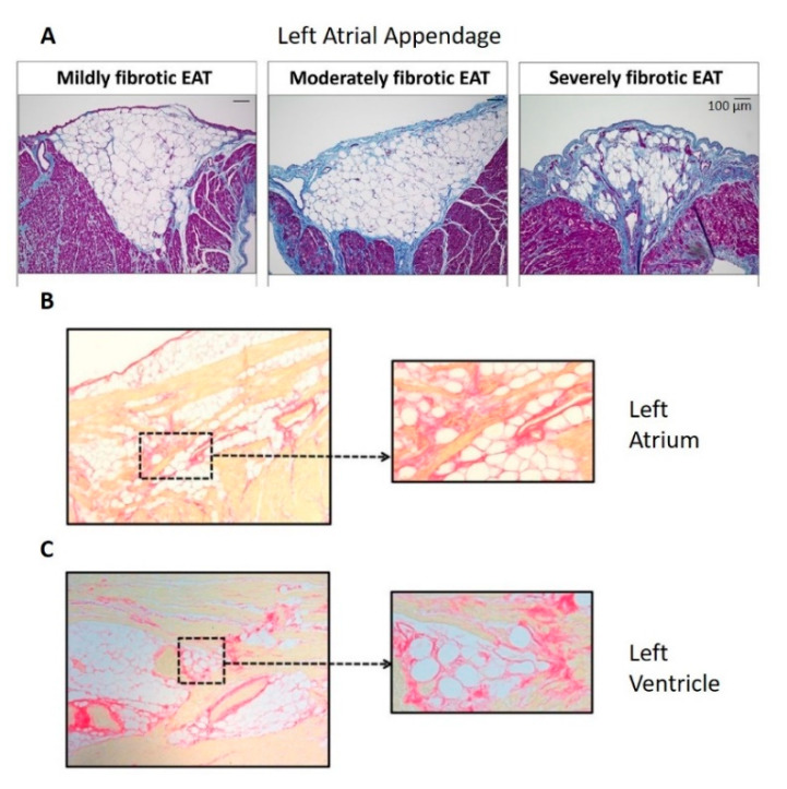
Figure 3.
The extent of fibrosis within the epicardial adipose tissue (EAT) was positively correlated with the extent of myocardial fibrosis (Panel A), within human left atrial appendage (Panel A), left atrium (Panel B) and left ventricle (Panel C). Panel A: Masson’s Trichrome staining in which collagen is blue, adapted from Abe et al. [13]; panels B and C: Picrosirius Red staining in which collagen is red, adapted from Venteclef et al. [93]. No scale was provided for panels B and C in the original paper.

