Abstract
A library of 1,2,3-triazole-incorporated thymol-1,3,4-oxadiazole derivatives (6–18) hasbeen synthesized and tested for anticancer and antimicrobial activities. Compounds 7, 8, 9, 10, and 11 exhibited significant antiproliferative activity. Among these active derivatives, compound 2-(4-((5-((2-isopropyl-5-methylphenoxy)methyl)-1,3,4-oxadiazol-2-ylthio)methyl)-1H-1,2,3-triazol-1-yl)phenol (9) was the best compound against all three tested cell lines, MCF-7 (IC50 1.1 μM), HCT-116 (IC50 2.6 μM), and HepG2 (IC50 1.4 μM). Compound 9 was found to be better than the standard drugs, doxorubicin and 5-fluorouracil. These compounds showed anticancer activity through thymidylate synthase inhibition as they displayed significant TS inhibitory activity with IC50 in the range 1.95–4.24 μM, whereas the standard drug, Pemetrexed, showed IC50 7.26 μM. The antimicrobial results showed that some of the compounds (6, 7, 9, 16, and 17) exhibited good inhibition on Escherichia coli (E. coli) and Staphylococcus aureus (S. aureus). The molecular docking and simulation studies supported the anticancer and antimicrobial data. It can be concluded that the synthesized 1,2,3-triazole tethered thymol-1,3,4-oxadiazole conjugates have both antiproliferative and antimicrobial potential.
Keywords: thymol; 1,2,3-triazole; 1,3,4-oxadiazole; cytotoxicity; antimicrobial; computational studies
1. Introduction
Cancer and infectious diseases are a major health burden on humankind globally [1]. According to the WHO, the mortality rates due to cancer will double in the coming years [2]. Chemotherapy is the mainstay for cancer treatment but suffers lack of selectivity, undesirable side effects, and multidrug resistance [3,4]. On the other hand, due to microbial resistance to the currently available antimicrobial drugs, infectious diseases also pose a serious problem to the medical community [5,6]. Cancer patients who have undergone chemotherapy treatments are more vulnerable to microbial infections due to impaired immunity [7]. Therefore, monotherapy with dual action as an anticancer and antimicrobial might be cost effective and reduce drug administration frequency, side effects, and antimicrobial resistance.
Heterocycles have played an important role in drug discovery. 1,2,3-triazole is a versatile scaffold that exhibits potential pharmacological activities such as anticancer, antimicrobial, anti-inflammatory, antitubercular, antiviral, and anti-HIV activities [8,9,10,11,12,13,14]. Drugs containing 1,2,3-triazole, tazobactam and cefatrizine, are used for antimicrobial and anticancer treatments. Additionally, 1,3,4-oxadiazole derivatives are multifunctional pharmacological pharmacophores which alter the physicochemical and pharmacokinetic properties of a drug. They are found in structures of many marketed drugs such as Raltegravir (antiviral), furamizole (antimicrobial), Zibotentan (anticancer), etc. [15]. It is very well known that natural products, due to their structural diversity and excellent pharmacological potential have provided numerous leads in drug discovery. Thymol, a monoterpenoid, is a major component of Thymus vulgaris L. which possesses anti-inflammatory, antimicrobial, antioxidant, and anticancer potential [16,17,18,19]. Recently, thymol-incorporated 1,2,3-triazole hybrids exerted significant anticancer activity [20].
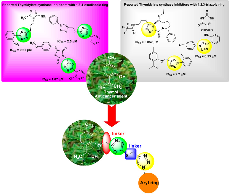
Thymidylate synthase inhibitors are evolving as a captivating target in chemotherapy [21]. These inhibitors induce apoptosis and cause cell cycle arrests by halting deoxythymidine monophosphate, which is responsible for formation of dTTP (a DNA synthesis precursor) [22]. From the literature, it is reported that 1,3,4-oxadiazole and 1,2,3-triazole derivatives exhibit antiproliferative activity by inhibiting thymidylate synthase [23,24,25,26,27,28] (Figure 1). Our research group has been involved in the development of anticancer leads targeting thymidylate synthase. In continuation of our previously reported work [20,23,26,29], we aim to incorporate both these heterocycles with thymol in a single molecule to generate new anticancer leads. Therefore, we report the synthesis of thymol-incorporated 1,3,4-oxadiazole and 1,2,3-triazole hybrids, anticancer and antimicrobial activities, as well as computational studies.
Figure 1.
Rationale for the present work.
2. Results and Discussion
2.1. Chemistry
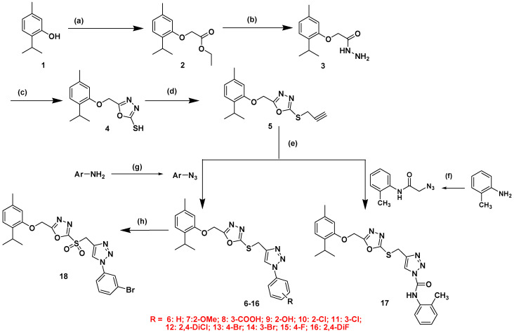
The route for the synthesis of final derivatives 6–18 is shown in Scheme 1. Compounds 6–17 were obtained by the Click chemistry approach using different aromatic azides and S-propargylated 1,3,4-oxadiazole intermediate 5. Intermediate 5 was synthesized from thymol (1) by esterification, followed by treatment with hydrazinium hydroxide in absolute ethanol, which yielded intermediate 3 [30]. This intermediate 3, on treatment with CS2 and KOH, afforded intermediate 4 which, upon propargylation using propargyl bromide in dry acetone, gave the key intermediate 5, which was further used for the synthesis of final compounds 6–17. Compound 18 was synthesized by the oxidation of compound 14 with hydrogen peroxide in dichloromethane. All the final compound structures were confirmed by different analytical techniques.
Scheme 1.
(a) K2CO3, ClCH2COOC2H5, reflux, 10 h; (b) MeOH, N2H4.H2O, reflux, 10 h; (c) i-Abs EtOH, KOH, CS2, stir, 24 h, ii- reflux, 10hrs; (d) acetone, K2CO3, Propargyl bromide; reflux, 5 h; (e) tert-BuOH:H2O (1:1), Na ascorbate, CuSO4·H2O, r.t., 5–11 h; (f) i. Dichloromethane (DCM), TEA, ClCH2COCl, stir, 0 to −5 °C; ii. NaN3, stir, 12 h; (g) HCl, NaNO2, NaN3, CH3COONa, stir, 0 to −5 °C, 1–2 h; (h) DCM, H2O2, stir, 0 to −5 °C, 28 h.
2.2. In Silico Absorption, Distribution, Metabolism and Elimination (ADME)/Pharmacokinetic Studies
In order to save cost and time in the drug development process, in silico studies are now used extensively in drug discovery. The desired pharmacokinetics are very important for a drug so as to avoid the problems of permeability, gastrointestinal absorption, solubility, transportation, and so on [31]. It is very well known that the orally available drug should follow the Lipinski rule of five; for example, it should have a molecular weight less than 500, and the hydrogen bond acceptor/donor should be less than 10 and 5, respectively. Furthermore, the lipophilicity of the drug should be less than five to avoid bioavailability problems. Therefore, all the final thymol hybrids were screened for in silico pharmacokinetics studies.
All the final compounds except 13, 14, and 18 exhibited a molecular weight less than 500, whereas the hydrogen bond acceptor and donor were found to be in an acceptable range for all the compounds, indicating that most of the compounds can be easily transported in the body. The highest % absorption of 73.07 was depicted by compounds 6,10–16 while other compounds showed % absorption in the range 60–70%. All the compounds displayed a desired TPSA in the range 104.16–141.46 and were non-permeable to the brain (Table 1).These data suggested that these compounds possess good pharmacokinetics and drug-likeness properties.
Table 1.
Absorption, distribution, metabolism, and elimination (ADME)/Pharmacokinetics studies of final hybrids 5–18.
| No. | Lipinski Parameters | nROTB e | TPSA f | %ABS g | BBB h | GI ABS i | ||||
|---|---|---|---|---|---|---|---|---|---|---|
| MW a | HBA b | HBD c | iLogP d | Violations | ||||||
| 5 | 302.39 | 4 | 0 | 3.61 | 0 | 6 | 73.45 | 83.65 | Yes | High |
| 6 | 421.52 | 6 | 0 | 4.14 | 0 | 8 | 104.16 | 73.07 | No | High |
| 7 | 451.54 | 7 | 0 | 4.54 | 1 | 9 | 113.39 | 69.88 | No | High |
| 8 | 465.52 | 8 | 1 | 3.93 | 0 | 9 | 141.46 | 60.20 | No | Low |
| 9 | 437.51 | 7 | 1 | 4.08 | 0 | 8 | 124.39 | 66.09 | No | Low |
| 10 | 455.96 | 6 | 0 | 4.22 | 0 | 8 | 104.16 | 73.07 | No | High |
| 11 | 455.96 | 6 | 0 | 4.05 | 0 | 8 | 104.16 | 73.07 | No | High |
| 12 | 490.41 | 6 | 0 | 4.45 | 1 | 8 | 104.16 | 73.07 | No | Low |
| 13 | 500.41 | 6 | 0 | 4.61 | 1 | 8 | 104.16 | 73.07 | No | High |
| 14 | 500.41 | 6 | 0 | 4.19 | 1 | 8 | 107.16 | 73.07 | No | High |
| 15 | 439.51 | 7 | 0 | 4.29 | 0 | 8 | 104.16 | 73.07 | No | High |
| 16 | 457.50 | 8 | 0 | 4.30 | 1 | 8 | 104.16 | 73.07 | No | Low |
| 17 | 492.59 | 7 | 1 | 3.26 | 0 | 11 | 133.26 | 63.02 | No | Low |
| 18 | 532.41 | 8 | 0 | 3.99 | 1 | 8 | 121.38 | 67.13 | No | Low |
a: Molecular weight; b: Hydrogen bond acceptor; c: Hydrogen bond donor; d: Lipophilicity; e: Number of rotatable bonds; f: Topological polar surface area; g: Absorption percentage; calculated by %ABS = 109 − (0.345 × TPSA); h: blood-brain barrier permeability; i: Gastrointestinal absorption.
2.3. Biological Studies
2.3.1. Antiproliferative Activity
The antiproliferative activity of the final derivatives 6–18 was performed on three cancer cell lines (MCF-7, HepG2 and HCT-116) by MTT assay [23]; 5-fluorouracil (5-FU) and doxorubicin were used as standard drugs (positive control). The antiproliferative activity depicted variable anticancer potential (Table 2). Two most promising compounds, 9 and 10 were found to be equipotent to doxorubicin (IC50 1.2 μM) with IC50 1.1 and 1.3 μM, respectively, whereas 17- and 15-times active to 5-florouracil (IC50 18.74 μM) towards MCF-7 cells. Other compounds 7, 8, and 11 were more active than 5-FU towards the same cancer cells with IC50 in the range 2.4 μM–4.9 μM. Compound 17 (IC50 9.39 μM) bearing the amide group was two-fold active to 5-FU, whereas sulfone-incorporated compound 18 was less active with IC50 98.2 μM towards breast cancer cells, MCF-7.
Table 2.
Cytotoxicity (IC50 ± SD, μM) of the final derivatives against MCF-7, HCT-116, and HepG2 cell lines.
| Compound | MCF-7 | HCT-116 | HepG2 |
|---|---|---|---|
| 6 | 22.5 | 36.5 | 41.6 |
| 7 | 2.4 | 3.1 | 1.8 |
| 8 | 3.9 | 5.8 | 5.3 |
| 9 | 1.1 | 2.6 | 1.4 |
| 10 | 1.3 | 3.8 | 2.5 |
| 11 | 4.9 | 16.3 | 18.6 |
| 12 | 59.4 | 47.3 | 40.6 |
| 13 | 65.3 | 78.7 | 69.4 |
| 14 | 41.8 | 32.8 | 23.7 |
| 15 | 23.15 | 31.4 | 49.2 |
| 16 | 34.5 | 56.2 | 58.3 |
| 17 | 9.39 | 17.3 | 39.1 |
| 18 | 98.2 | 118.5 | 95.7 |
| Dox | 1.2 | 2.5 | 1.8 |
| 5-FU | 18.74 | 30.68 | 28.65 |
Values are the mean ± SD of three different experiments. Dox-doxorubicin and 5-FU-5-florouracil (reference drug).
Against HepG2 cells, compounds 7 and 9 were the most potent molecules with IC50 1.8 μM and 1.4 μM, respectively. It was noted that compound 9 was better than doxorubicin (IC50 1.8 μM) in exerting cytotoxicity, whereas 7 was equipotent to doxorubicin. Moreover, both the compounds were 20- and 16-fold active to 5-FU (IC50 28.65 μM). Compounds 8 and 10 displayed better cytotoxicity than 5-FU with IC50 5.3 μM and 2.5 μM, respectively. Other compounds, except 18, showed moderate activity with IC50 in the range 18.6–69.4 μM. Towards HCT-116 cells, compound 9 was the best compound with IC50 2.6 μM and equipotent to doxorubicin.
Structure-activity relationships (SAR) deduced from the activity is as follows: (i) Position of the substituents: Ortho substituted compounds exhibited better activity than meta and para-substituted derivatives. Compound 7 (2-OMe), 9 (2-OH), and 10 (2-Cl) bearing ortho substitution on the phenyl ring were most active followed by compound 8 (3-COOH), 11 (3-Cl), and 13 (4-Br). (ii) Number of the substituents: Increase in the number of substituent decreased the activity: disubstituted derivatives 12 (2,4-diCl) and 16 (2,4-diF) diminished cytotoxicity compared with monosubstituted derivatives 10 and 11 (Cl) and 15 (F). (iii) Incorporation of the sulfone group in the structure displayed weak cytotoxicity.
2.3.2. Thymidylate Synthase Activity
The potent derivatives from cytotoxicity results were selected for the TS enzymatic assay. Compounds 7, 8, 9, 10, and 11 and Pemetrexed (reference drug) were screened for TS inhibition. It was observed all these tested derivatives caused significant inhibition on the TS enzyme. Compound 9 having o-hydroxy was the best compound with IC50 1.95 μM followed by compound 10 with IC50 2.18 μM, whereas Pemetrexed inhibited TS with IC50 7.26 μM. In terms of folds activity, compound 9 was 3.72-fold and 10 was 3.33-fold active to the reference drug, Pemetrexed. Other compounds 7, 8, and 11 were also better than Pemetrexed in TS inhibition with IC50 in the range 3.52–4.24 μM (Table 3). It was interesting to note that the incorporation of 1,3,4-oxadiazole and 1,2,3-triazole under one construct were better in TS inhibition [20]. These derivatives may be investigated further for development of anticancer leads.
Table 3.
Thymidylate synthase (TS) activity of the potent derivatives.
| Compounds | IC50 (µM) |
|---|---|
| 7 | 3.52 ± 1.2 |
| 8 | 3.98 ± 1.1 |
| 9 | 1.95 ± 0.9 |
| 10 | 2.18 ± 1.7 |
| 11 | 4.24 ± 0.8 |
| Pemetrexed | 7.26 ± 1.1 |
IC50 values are the mean ± S.D. of three separate experiments. Pemetrexed (positive control).
2.3.3. Antimicrobial Activity
The antimicrobial activity was performed on E. coli, S. aureus and C. albicans. It was found that compounds 5, 6, 7, 9, 12, 13, 14, 16, and 17 displayed zones of inhibition of 12 mm, 14 mm, 12 mm, 12 mm, 10 mm, 11 mm, 9 mm, 14 mm, and 12 mm, respectively, for E. coli (gram negative), whereas other compounds were not effective for E. coli. Gram-positive (S. aureus) compounds 5, 6, 7, 8,9, 16, and 17 showed zones of inhibition of 9 mm, 10 mm, 12 mm, 10 mm, 12 mm, 15 mm, and 12 mm, respectively. Other samples did not display any zone of inhibition for S. aureus. The antifungal activity against Candida albicans was displayed by compounds 5, 9, and 12 as the formation of inhibition zones was found to be 10 mm, 10 mm, and 12 mm, respectively (Table 4). It was noticed that compounds 5, 6, 7, 9, 16, and 17 had broad-spectrum antimicrobial potential for both gram-negative and positive microbial strains. These data indicated that derivatives 6 and 16 were most potent against E. coli with zones of inhibition of 14 mm each while compound 16 was most active against S. aureus with a zone of inhibition of 15 mm.
Table 4.
Antimicrobial activity of compounds 5–18.
| Compound No. | E. coli | S. aureus | C. albicans |
|---|---|---|---|
| 5 | 12 ± 0.32 | 9 ± 0.30 | 10 ± 0.32 |
| 6 | 14 ± 0.38 | 10 ± 0.40 | --- |
| 7 | 12 ± 0.34 | 12 ± 0.34 | --- |
| 8 | --- | 10 ± 0.40 | --- |
| 9 | 12 ± 0.36 | 12 ± 0.34 | 10 ± 0.032 |
| 10 | --- | --- | --- |
| 11 | --- | --- | --- |
| 12 | 10 ± 0.38 | --- | 12 ± 0.036 |
| 13 | 11 ± 0.34 | --- | --- |
| 14 | 9 ± 0.32 | --- | --- |
| 15 | --- | --- | --- |
| 16 | 14 ± 0.34 | 15±0.38 | --- |
| 17 | 12 ± 0.32 | 12 ± 0.34 | --- |
| 18 | --- | --- | --- |
| Novobiocin | 14 ± 0.64 | 10 ± 0.24 | NT |
| Fluconazole | NT | NT | 16 ± 0.60 |
--- = no zone of inhibition; NT = not tested; E. coli (ATCC 31218); S. aureus (ATCC 29213); C. albicans (ATCC 10231).
2.3.4. Computational Studies
Electronic Properties
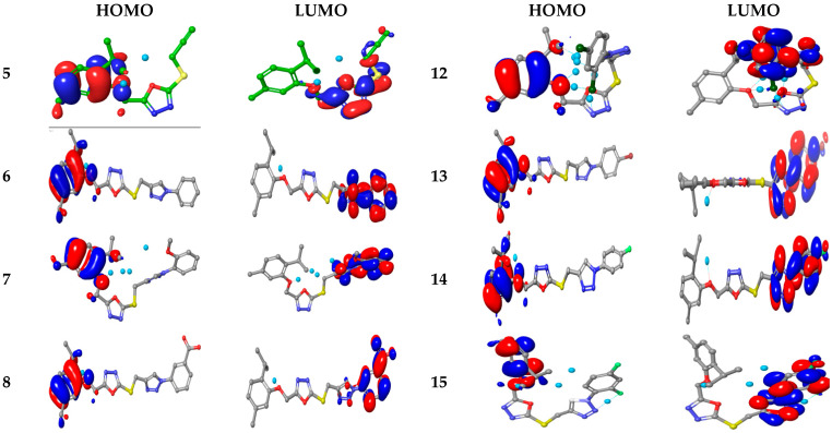
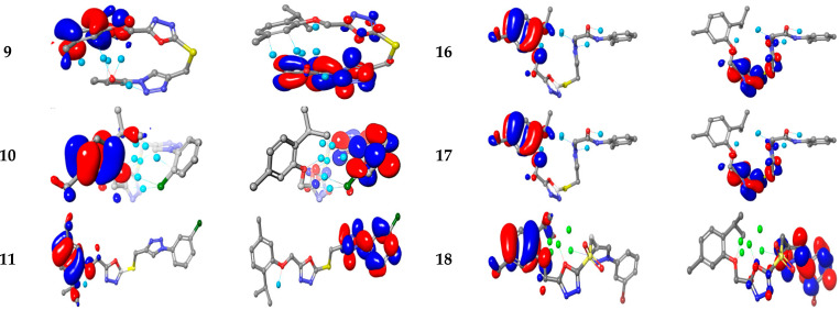
To understand the structural elements of thymol conjugates, frontier molecular orbitals energy are shown in Figure 2. The HOMO/LUMOs and HOMO–1/ LUMO+1 of thymol derivatives were probed at B3LYP/6-311+G(d,p) using the LANL2DZ level [32]. HOMO was observed over the thymol corner for compounds 5–18, whereas the LUMO zone was compressed opposite phenyl corners for 5–15 and18 but in compounds 16 and 17, LUMO overlapped bis-triazole rings. This arrangement revealed that hybrids 5–17 possess intra-molecular charge transfer (ICT) between HOMOs to LUMOs. Compounds 5–18 correlated to the spatial distribution of molecular orbitals, enlightening the most credible locations which are mostly attacked by drugs. The energies of FMOs, for instance HOMO (EHOMO), LUMO (ELUMO), as well as energy gaps (ΔEHOMO/LUMO) between HOMO-LUMO and HOMO-1/LUMO+1 (ΔEHOMO/LUMO+1), are noteworthy parameters to probe molecules (Table 5).
Figure 2.
Ground state charge density of FMOs of thymol derivatives.
Table 5.
The calculated energy for 5–18 derivatives.
| Compd. | E HOMO |
E LUMO |
ΔE HOMO/LUMO |
E HOMO-1 |
E LUMO+1 |
ΔE HOMO-1/LUMO+1 |
IP | η | S | χ | ω |
|---|---|---|---|---|---|---|---|---|---|---|---|
| 5 | −6.026 | −1.802 | −4.224 | −0.150 | −0.062 | −0.0881 | 5.83 | 2.612 | 0.383 | 2.231 | −3.414 |
| 6 | −5.834 | −1.472 | −4.362 | −1.469 | −0.707 | −0.7619 | 5.72 | 2.181 | 0.458 | 3.059 | −3.653 |
| 7 | −5.995 | −1.200 | −4.795 | −1.360 | −1.159 | −0.215 | 1.87 | 2.398 | 0.417 | 2.699 | −3.597 |
| 8 | −5.836 | −1.987 | −3.849 | −1.361 | −0.424 | −0.9361 | 5.71 | 1.924 | 0.520 | 3.976 | −3.912 |
| 9 | −6.104 | −1.227 | −4.877 | −0.963 | −0.435 | −0.5279 | 5.82 | 2.439 | 0.410 | 2.755 | −3.666 |
| 10 | −6.139 | −1.368 | −4.771 | −0.931 | −0.844 | −0.0871 | 6.11 | 2.386 | 0.419 | 2.952 | −3.753 |
| 11 | −5.904 | −1.794 | −4.110 | −1.083 | −0.615 | −0.4680 | 5.69 | 2.055 | 0.487 | 3.604 | −3.849 |
| 12 | −6.181 | −1.588 | −4.593 | −1.197 | −0.990 | −0.2068 | 5.67 | 2.296 | 0.435 | 3.285 | −3.884 |
| 13 | −5.861 | −1.739 | −4.122 | −1.069 | −0.459 | −0.6106 | 5.67 | 2.061 | 0.485 | 3.502 | −3.800 |
| 14 | −5.892 | −1.828 | −4.064 | −1.140 | −0.552 | −0.5875 | 5.90 | 2.032 | 0.492 | 3.666 | −3.860 |
| 15 | −5.849 | −1.516 | −4.332 | −1.026 | −0.422 | −0.6041 | 5.77 | 2.166 | 0.462 | 3.130 | −3.683 |
| 16 | −6.218 | −1.473 | −4.745 | −1.031 | −1.004 | −0.0272 | 6.07 | 2.372 | 0.422 | 3.117 | −3.846 |
| 17 | −5.961 | −1.277 | −4.684 | −0.917 | −0.705 | −0.2122 | 5.92 | 2.342 | 0.427 | 2.796 | −3.619 |
| 18 | −5.989 | −1.821 | −4.168 | −1.625 | −1.059 | −0.5660 | 5.70 | 2.084 | 0.480 | 3.659 | −3.905 |
The chemical potential “IP”, nucleophilicity “χ”, and electrophilicty “ω” were studied using the HOMO–1/LUMO+1 (Table 5).The variations of ΔEHOMO/LUMO for 5–18 hybrids is not clear due to the similar energy values. The promising kinase inhibitors were the organic molecule that donates electrons to the positive-charged amino-acids, and able to accept free electrons from kinase (imine groups). The previous studies displayed a direct relation between the potent antioxidant ability and anticancer drugs [33,34]. The antioxidant potential can be assessed by ionization potential (IP). It is expected that the antioxidant ability for 5–18 might be better due to low IP values. These results suggest that synthesized 5–18 derivatives may have good antioxidant potential.
Molecular Electrostatic and Ionization Potential Maps
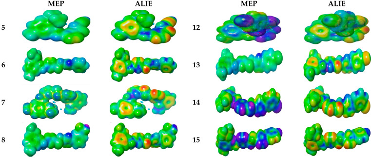
To explore the reactivity of synthesized hybrids, molecular electrostatic potential (MEP) was measured experimentally by diffraction approaches [35]. This property displays the nuclear and electronic distribution over the synthesized hybrids and is indicated by different colors. The high negative-charge region that attacks by electrophile is indicated by red, whereas the blue region is positively charged, which attacks the nucleophile. MEP decreases in the order green > blue > yellow > orange > red, and the red color shows repulsion while the blue color indicates enough attraction. As anticipated, the green color shields aromatic rings of all molecules, whereas the yellow regions cover the triazole ring. These color changes in the molecules help in the identification of electrophilic and nucleophilic sites for the attack by receptors. The green region denotes high nucleophilicity, making the molecules able to recognize the binding site through ionic interaction between the substrate and receptor.
The ALIE (average local ionization energy map) was used to predict favorable molecular sites for electrophiles. The red color surrounds the thymol ring in all 6–18 (Figure 3). The electron density in the yellow color spreads over all 6–18 molecular backbones. The blue color is more localized in the oxadiazole linker. Thereby, these groups are prone to nucleophilic attacks.
Figure 3.
Molecular electrostatic potential surfaces views of 5–18.
2.3.5. Molecular Docking
To know the possible molecular interaction of compounds 5–18 via thymidylate synthase (TS) and DNA gyrase B, the docking experiment was performed. The 3D structure of TS and DNA gyrase enzymes for anticancer and antimicrobial, respectively, were established. 3D loop structure of PDB 6QXG and 4URO complexed with FdUMP and Novobiocin for TS and DNA gyrase, respectively, were used as frameworks for the docking experiment. The 3D models were further checked and verified using Ramachandran plot at PROCHECK [36]. The CASTp Package was used for identifying the active sites for TS and DNA gyrase. TS are reported to bind with Asn 226, His 256, Asp 218, Ser 216, Arg 50, and Arg 215, whereas for DNA gyrase, the binding sites are Arg144, Ser 55, Ala64, Asn65 and Asp89.
Thymidylate Synthase (6QXG)
Compounds 5–18 were successfully docked over active sites for TS. The generated docked poses were energy-minimized by the OPLS force field, and the lowest-binding free-energy ΔG with minimum RMSD between the pose before and after refinement were selected. Finally, the highest ΔG scoring function was employed to assess the binding energy of the compounds. To further validate the binding energy of 5–18, the inhibition constant (Ki) was calculated. The lower the Ki, better is the efficiency of the molecule, and it should be in the range of 0.1–1.0 μM value. Ligand-Efficiency (LE) and Fit-Quality (FQ) are the bioactivity factors which should be ≥0.3 and ≥0.8, respectively [37]. These parameters were compared with reference inhibitors and are shown in Table 6.
Table 6.
Docking energy in (kcal/mol) and bioactivity analysis parameters against 6QXG.
| Cpd. | ΔE | RMSD | Eplace | Econf | Eele | LE | Ki | LEscale | FQ |
|---|---|---|---|---|---|---|---|---|---|
| FdUMP | −7.90 | 0.94 | −430.17 | −15.64 | −14.74 | −8.39 | 2.47 | 3.30 | 5.09 |
| 5FU | −5.28 | 1.60 | −433.91 | −21.32 | −17.47 | −3.30 | 2.41 | −5.28 | −5.28 |
| 5 | −6.42 | 1.26 | 2.48 | −14.45 | −9.31 | −5.08 | 1.98 | 3.05 | 2.03 |
| 6 | −7.36 | 1.64 | 41.66 | −17.27 | −10.34 | −4.48 | 1.54 | 2.82 | 1.67 |
| 7 | −7.50 | 1.10 | 39.34 | −17.66 | −8.68 | −6.81 | 2.16 | 3.17 | 3.64 |
| 8 | −7.59 | 1.92 | 19.80 | −7.35 | −7.87 | −3.95 | 1.73 | 2.68 | 1.27 |
| 9 | −7.46 | 1.63 | 32.10 | −8.59 | −9.55 | −4.56 | 1.94 | 2.82 | 1.74 |
| 10 | −7.14 | 1.01 | 20.09 | −14.91 | −7.02 | −7.04 | 1.56 | 3.24 | 3.80 |
| 11 | −7.04 | 1.53 | 36.13 | −14.00 | −7.68 | −4.61 | 0.16 | 2.88 | 1.73 |
| 12 | −6.87 | 1.73 | 35.84 | −18.02 | −9.06 | −3.97 | 1.81 | 2.77 | 1.20 |
| 13 | −7.39 | 1.40 | 48.40 | −17.34 | −12.24 | −5.27 | 1.43 | 2.96 | 2.32 |
| 14 | −7.50 | 1.33 | 40.76 | −12.81 | −8.17 | −5.63 | 1.49 | 3.00 | 2.63 |
| 15 | −7.04 | 1.46 | 43.98 | −23.03 | −10.03 | −4.82 | 0.94 | 2.92 | 1.90 |
| 16 | −7.17 | 1.65 | 32.20 | −10.86 | −9.15 | −4.36 | 1.88 | 2.81 | 1.54 |
| 17 | −8.16 | 1.60 | −4.51 | −17.16 | −8.75 | −5.10 | 2.03 | 2.84 | 2.26 |
| 18 | −7.45 | 1.56 | 131.24 | −15.92 | −10.20 | −4.78 | 1.77 | 2.86 | 1.91 |
ΔE, final free-binding energy; Econf, binding energy for the ligand-conformer; Eplace, binding energy of the ligand-receptor; E.ele, ligand-receptor Electrostatic interaction; RMSD, the root mean square deviation of the pose of the docking pose compared to the co-crystal ligand position, LE; Ligand efficiency; Ki-consequent inhibition constant (μM).
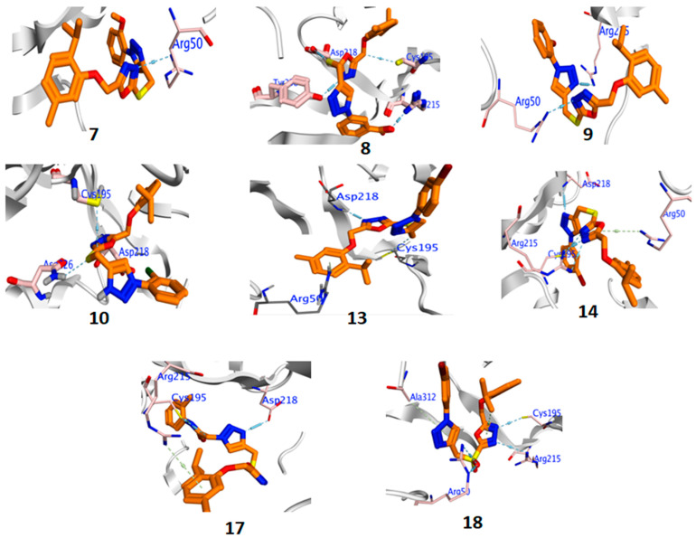
Compound 17 showed a higher glide score than both the reference drugs, fluorodeoxyuridylate (FDUMP) and 5-fluorouracil (FU). Interestingly, all compounds 5–18 showed higher BE than FU. RMSDs were found to be <2 degree supporting docking-process accuracy. The BEs for these derivatives were found to be in the order 17 > FD > 8 > 14 > 18 > 13 > 6 > 16 > 10 > 11 > 12 > 5 > FU. All the compounds 5–18 interacted in a similar manner, like FU. These compounds were more potent than the reference drugs, as indicated by lower inhibition constant values (Ki = 0.16–2.16 μM for compounds 5–18; 2.47 and 2.41 μM for reference drugs). All compounds displayed H-bond formation with important amino acid residues of TS (Figure 4). From analysis of the protein-ligand fingerprint PLIF histogram, it was found that residue Arg215 interacted with 50% of those tested by H-bond interactions and Arg50, Glu87, and ASP218 interacted with the remaining 30% of tested compounds (Figure S30, Supplementary Material).
Figure 4.
Binding interaction of active compounds against TS (6QXG).
DNA Gyrase B (4URO)
Bacterial DNA-gyrase is an attractive antibacterial target, which cleaves the DNA double-helix [38]. The analysis of the co-crystallized DNA-gyrase-novobiocin complex is responsible for restricting the ATPase binding site (Arg144, Ser 55, Ala64, Asn65, Asp89) situated on the cell-wall of organisms. The peptidoglycan layer situated in the cell wall of bacteria inhibits the microorganism via antibacterial agents. From Table 7, compounds 5, 6, 7, 9, 16, and 17 exhibited the highest BE compared with the reference drug, Novobiocin. Other compounds showed lower binding scores with considerable RMSD < 2. These compounds exhibited Ki in the range 1.86–1.99 μM.
Table 7.
Docking energy in (kcal/mol) and bioactivity analysis parameters against 4URO.
| Cpd. | ΔE | RMSD | Eplace | Econf | Eele | LE | Ki | LEscale | FQ |
|---|---|---|---|---|---|---|---|---|---|
| Nov. | −8.41 | 1.16 | 10.98 | −9.99 | −7.80 | −7.24 | 1.95 | 3.56 | 8.26 |
| 5 | −9.18 | 1.97 | 48.83 | −10.04 | −8.81 | −4.67 | 1.86 | 3.12 | 4.12 |
| 6 | −9.57 | 1.11 | 57.74 | −5.82 | −2.67 | −8.65 | 1.82 | 2.66 | 2.01 |
| 7 | −8.95 | 1.58 | 28.83 | −12.31 | −12.87 | −5.66 | 1.89 | 3.16 | 5.49 |
| 8 | −8.26 | 1.19 | 32.72 | −5.94 | −6.46 | −6.93 | 1.97 | 2.85 | 2.81 |
| 9 | −9.26 | 1.94 | 31.38 | −9.99 | −9.03 | −4.77 | 1.86 | 3.10 | 3.83 |
| 10 | −6.09 | 1.79 | 31.82 | −43.28 | −2.51 | −1.17 | 3.34 | 2.67 | 2.10 |
| 11 | −8.49 | 1.38 | 60.48 | −6.87 | −1.93 | −6.14 | 1.94 | 2.74 | 1.57 |
| 12 | −8.21 | 1.96 | 48.99 | −6.93 | −6.42 | −4.19 | 1.98 | 2.97 | 3.17 |
| 13 | −8.26 | 1.87 | 41.56 | −9.63 | −7.28 | −4.42 | 1.97 | 2.66 | 1.52 |
| 14 | −6.09 | 0.82 | 41.09 | −41.70 | −2.56 | −2.56 | 1.96 | 2.70 | 1.72 |
| 15 | −8.08 | 1.23 | 30.40 | −8.24 | −6.28 | −6.58 | 1.99 | 3.43 | 0.86 |
| 16 | −9.62 | 1.26 | 14.00 | −5.65 | −9.83 | −7.61 | 1.82 | 3.07 | 3.51 |
| 17 | −8.68 | 1.48 | 129.68 | −7.59 | −10.45 | −5.85 | 1.92 | 3.05 | 4.56 |
| 18 | −8.26 | 0.70 | 44.95 | −22.82 | −12.90 | −11.83 | 1.97 | 2.91 | 2.95 |
ΔE, final free-binding energy; Econf, binding energy for the ligand-conformer; Eplace, binding energy of the ligand-receptor; E.ele, ligand-receptor Electrostatic interaction; RMSD, the root mean square deviation of the pose of the docking pose compared to the co-crystal ligand position, LE; Ligand efficiency; Ki-consequent inhibition constant (μM), Nov., Novobiocin (positive control).
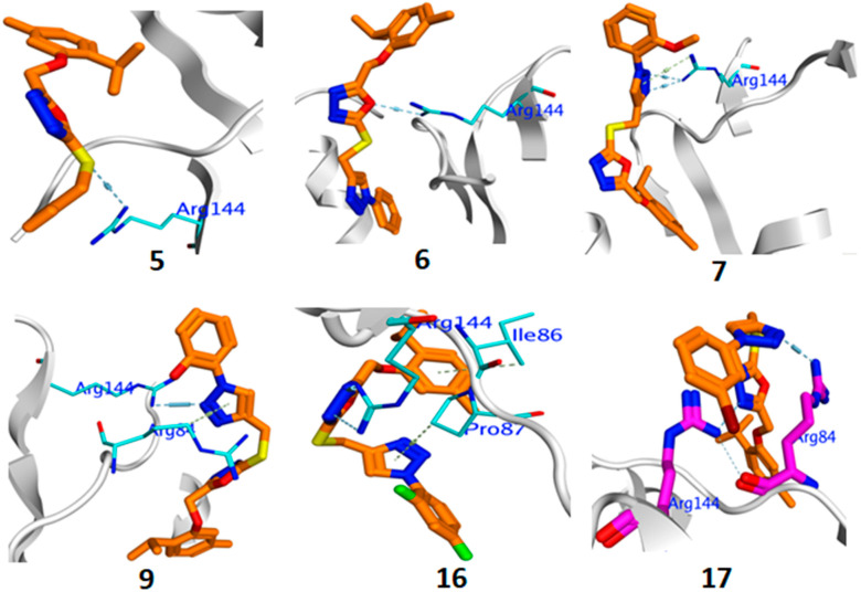
All the compounds formed H-bonds with vital amino acid residues Glu58, Pro87, Ile86, and Arg144 (Figure 5). PLIF analysis showed the Arg144 interacted through H-bond formation with 43% of the tested compounds (Figure S31, Supplementary Material). The inhibition potency of 6–18 against bacterial growth may be explained by the attacking of the peptidoglycan-naked cell-wall of bacteria. Thus, the antimicrobial-mechanism for 5–18 hybrids is due to the high permeability of the microorganism membrane.
Figure 5.
Binding interaction of active compounds against DNA gyrase (4URO).
3. Methods and Materials
3.1. Chemistry
The reagents and solvents used for the synthesis were purchased from Sigma Aldrich (St. Louis, MI, USA) and Loba India Pvt. Ltd. (Mumbai, India) and used directly. NMR analysis was performed on Bruker spectrometer (850 MHz) using CDCl3 as solvent. IR spectra were recorded on Thermoscientific using ATR technique and melting points were recorded using Stuart SMP 40 machine and were uncorrected. LCQ Fleet-LCF10605 Thermoscientific Mass spectrometer was used to measure the molecular weight of the synthesized compounds and elemental analysis was performed using Elementar Analyzer. Compounds 2, 3, and 4 were prepared as previously published [30].
Preparation of 2-((2-isopropyl-5-methylphenoxy)methyl)-5-(prop-2-ynylthio)-1,3,4-oxadiazole (5)
Compound 4 (0.06 mol) was charged into a 250 mL round-bottom flask, and then anhydrous acetone (175 mL) and potassium carbonate (0.066 mol) were added. The reaction mass was stirred for 2 hrs at 50–60 °C and then cooled to room temperature. To the reaction mixture, propargyl bromide (0.066 mol) was added and the reaction was stirred at 40–50 °C for 4 hrs. After completion of the reaction, the reaction mixture was filtered, and filtrate was concentrated to 30 mL and poured into 100 mL water. The product was isolated by extracting with dichloromethane (2 × 100 mL). To the DCM layer, cyclohexane was added under hot conditions to achieve a solid mass, and the solid product was filtered and washed with cyclohexane (20 mL), and then dried.
Yield: 80%, M.p. 58–60 °C, IR (ATR) υmax: 3252 (terminal alkyne C–H), 2985, 2120 (carbob-carbon triple-bond stretching), 1613, 1594, 1507, 1476, 1392, 1383, 1340, 1287 (C=N, C–N), 1176, 1162, 1110, 1096, 1054, 1027 (C–O), 993, 972, 956, 847, 818, 701, 666. 1H NMR (CDCl3, 850 MHz): δ 1.21 (d, J = 7.64 Hz, 6H, –CH(CH3)2, 2.20 (s, 1H, terminal alkyne H), 2.35 (s, 3H, Aromatic methyl), 3.28-3.31 (m, 1H, –C–H (CH3)2, 4.04 (s, 2H, S–CH2–), 5.26 (s, 2H, –O–CH2–), 6.80 (s, 1H), 6.85 (s, 1H), 7.14 (d, J = 8.5 Hz, 1H); 13C NMR (CDCl3, 213 MHz): δ 21.29, 22.83, 26.49, 30.96, 60.14, 73.09, 112.90, 122.96, 126.45, 134.74, 136.66, 154.63, 164.18, 164.34. ESI MS: 302 (M+ + H). C16H18N2O2S (Calculated): C = 63.55; H = 6.00; N = 9.26; O = 10.58; S = 10.60; observed: C = 63.52; H = 6.02; N = 9.24; O = 10.62; S = 10.58.
Synthesis of Final Compounds (6–16)
Compound 5 (0.001 mol) was dissolved in tert-BuOH:water (1:1; 20 mL) in a 250 mL round-bottom flask, and then sodium ascorbate and copper sulphate pentahydrate were added followed by the addition of aromatic azides (0.0011 mole). The reaction mixture was stirred at 20–30 °C for 3–8 h. After completion of the reaction, water (75 mL) and ethylacetate (75 mL) were added to the reaction mixture and stirred for 1 h. The organic layer was separated, washed with water, and dried over anhydrous sodium sulphate. Ethylacetate was evaporated and the compounds were crystallized with a mixture of ethylacetate-cyclohexane or Ethylacetate-hexane.
4-((5-((2-isopropyl-5-methylphenoxy)methyl)-1,3,4-oxadiazol-2-ylthio)methyl)-1-phenyl-1H-1,2,3-triazole (6)
Yield = 68%, M.p. 88–90 °C, IR (ATR) υmax: 3086 (C–H aromatic), 2962 (C–H), 1613, 1593, 1502, 1474, 1413, 1345, 1286, 1254, 1157, 1093, 1053, 1032, 809, 754, 686; 1H NMR (850 MHz, CDCl3): δ 1.18 (d, J = 8.5 Hz, 6H, –CH–(CH3)2), 2.33 (s, 3H, Aromatic methyl), 3.27-3.29 (m, 1H, –CH–(CH3)2), 4.69 (s, 2H, S–CH2–), 5.22 (s, 2H, –O–CH2–), 6.79 (s, 1H), 6.84 (d, J = 8.5 Hz, 1H), 7.13 (d, J = 8.5 Hz, 1H), 7.46 (t, J = 8.5 Hz, 1H), 7.54 (t, J = 8.5 Hz, 2H), 7.73 (d, J = 8.5 Hz, 2H), 8.46 (s, 1H, triazole ring proton); 13C NMR (CDCl3, 213 MHz): δ 21.30, 22.84, 26.43, 30.96, 60.19, 112.93, 120.76, 122.99, 126.45, 128.95, 129.85, 134.79, 136.62, 154.61. ESI MS: 422 (M+ + H). C22H23N5O2S(Calculated): C = 62.69; H = 5.50; N = 16.61; O = 7.59; S = 7.61; observed: C = 62.52; H = 5.54; N = 16.64; O = 7.62; S = 7.58.
4-((5-((2-isopropyl-5-methylphenoxy)methyl)-1,3,4-oxadiazol-2-ylthio)methyl)-1-(2-methoxyphenyl)-1H-1,2,3-triazole (7)
Yield = 65%, M.p. 81-83 °C, IR (ATR) υmax: 2960, 1614, 1586, 1558, 1540, 1505, 1473, 1417, 1391, 1360, 1289, 1255, 1242 (C=N, C–N), 1159, 1095, 1045, 1027, 813, 786, 751, 741, 724, 674; 1H NMR (850 MHz, CDCl3): δ 1.19 (d, J = 8.5 Hz, 6H, –CH–(CH3)2), 2.33 (s, 3H, Aromatic methyl), 3.27-3.30 (m, 1H, –CH–(CH3)2), 3.88 (s, 3H, O–CH3), 4.73 (s, 2H, S–CH2–), 5.23 (s, 2H, –O–CH2–), 6.80 (s, 1H), 6.84 (d, J = 8.5 Hz, 1H), 7.10-7.14 (m, 3H), 7.44 (t, J = 8.5 Hz, 1H), 7.81 (s, 1H), 8.50 (s, 1H, triazole ring proton); 13C NMR (CDCl3, 213 MHz): δ 21.31, 22.84, 26.43, 30.96, 55.94, 60.20, 112.29, 112.93, 121.24, 122.96, 126.44, 130.28, 134.79, 136.61, 154.64. ESI MS: 452 (M+ + H). C23H25N5O2S (Calculated): C = 61.18; H = 5.58; N = 15.51; O = 10.63; S = 7.10; observed: C = 61.14; H = 5.60; N = 15.49; O = 10.65; S = 7.09.
3-(4-((5-((2-isopropyl-5-methylphenoxy)methyl)-1,3,4-oxadiazol-2-ylthio)methyl)- 1H-1,2,3-triazol-1-yl)benzoic acid (8)
Yield = 73%, M.p. 166-168 °C, IR (ATR) υmax: 3120 (COO–H), 2995 (C–H), 1700 (C=O), 1614, 1591, 1493, 1473, 1411, 1252, 1161, 1116, 1095, 1051, 1000, 952, 902, 858, 811, 792, 768, 691. 1H NMR (850 MHz, CDCl3): δ 1.18 (d, J = 8.5 Hz, 6H, –CH–(CH3)2), 2.33 (s, 3H, Aromatic methyl), 3.27–3.30 (m, 1H, –CH–(CH3)2), 4.74 (s, 2H, S–CH2–), 5.21 (s, 2H, –O–CH2–), 6.80 (s, 1H), 6.84 (d, J = 8.5 Hz, 1H), 7.13 (d, J = 8.5 Hz, 1H), 7.68 (t, J = 8.5 Hz, 1H), 8.10 (s, 1H), 8.21 (d, J = 8.5 Hz, 1H), 8.48 (s, 1H, triazole ring proton); 13C NMR (CDCl3, 213 MHz): δ 21.32, 22.84, 26.43, 30.96, 60.17, 112.93, 123.01, 126.45, 130.38, 134.79, 136.64, 154.58, 168.76. ESI MS: 464 (M+ + H). C23H23N5O4S (Calculated): C = 59.34; H = 4.98; N = 15.04; O = 13.75; S = 6.89; observed: C = 59.24; H = 4.99; N = 15.02; O = 13.78; S = 6.86.
2-(4-((5-((2-isopropyl-5-methylphenoxy)methyl)-1,3,4-oxadiazol-2-ylthio)methyl)- 1H-1,2,3-triazol-1-yl)phenol (9)
Yield = 70%, M.p. 86–88 °C, IR (ATR) υmax: 3000 (brd peak, O–H), 2952 (C–H), 1599, 1506, 1472, 1424, 1405, 1290, 1261, 1167, 117, 1096, 1063, 1042 (C–O), 989, 943, 806, 792, 752, 718, 642; 1H NMR (850 MHz, CDCl3): δ 1.19 (d, J = 8.5 Hz, 6H, –CH–(CH3)2), 2.34 (s, 3H, Aromatic methyl), 3.27-3.30 (m, 1H, –CH–(CH3)2), 4.71 (s, 2H, S–CH2–), 5.22 (s, 2H, –O–CH2–), 6.80 (s, 1H), 6.84 (d, J = 8.5 Hz, 1H), 7.13 (d, J = 8.5 Hz, 1H), 7.24 (d, J = 8.5 Hz, 1H), 7.33 (d, J = 7.5 Hz, 1H), 7.42 (d, J = 8.5 Hz, 1H), 9.67 (s, 1H, phenolic proton); 13C NMR (CDCl3, 213 MHz): δ 21.32, 22.84, 26.44, 30.96, 60.19, 112.93, 119.55, 120.52, 123.02, 126.47, 129.93, 143.79, 136.64, 149.18, 154.59. ESI MS: 438 (M+ + H). C22H23N5O3S (Calculated): C = 60.39; H = 5.30; N = 16.01; O = 10.97; S = 7.33; observed: C = 60.35; H = 5.32; N = 15.98; O = 10.99; S = 7.32.
4-((5-((2-isopropyl-5-methylphenoxy)methyl)-1,3,4-oxadiazol-2-ylthio)methyl)-1-(2-chlorophenyl)-1H-1,2,3-triazole (10)
Yield = 75%, M.p. 90–92 °C, IR (ATR) υmax: 2960, 1594, 1505, 1485, 1437, 1287, 1253, 1157, 1116, 1095, 1042, 812, 784, 679.1H NMR (850 MHz, CDCl3): δ 1.17–2.20 (m, 6H, –CH–(CH3)2), 2.34 (s, 3H, Aromatic methyl), 3.29 (brd, s, 1H, –CH–(CH3)2), 4.64 (s, 2H, S–CH2–), 5.25 (s, 2H, –O–CH2–), 6.81–6.85 (m, 2H), 7.14 (d, J = 8.5 Hz, 1H), 7.46–7.82 (m, 4H); 13C NMR (CDCl3, 213 MHz): δ 21.32, 22.85, 26.44, 30.96, 60.23, 112.95, 123.02, 122.99, 126.47, 128.97, 134.79, 143.80, 136.63, 155.50. ESI MS: 456 (M+ + H), 458 (M+ + H+2). C22H22ClN5O2S (Calculated): C = 57.95; H = 4.86; N = 15.36; O = 7.02; S = 7.03; observed: C = 57.90; H = 4.88; N = 15.34; O = 7.04; S = 7.01.
4-((5-((2-isopropyl-5-methylphenoxy)methyl)-1,3,4-oxadiazol-2-ylthio)methyl)-1-(3-chlorophenyl)-1H-1,2,3-triazole (11)
Yield = 68%, M.p. 92–94 °C, IR (ATR) υmax: 2962 (C–H), 1614, 1592, 1495, 1475, 1416, 1380, 1344, 1285, 1251, 1158, 1112, 1098, 1074, 807, 752; 1H NMR (850 MHz, CDCl3): δ 1.18 (d, J = 8.5 Hz, 6H, –CH–(CH3)2), 2.33 (s, 3H, Aromatic methyl), 3.26-3.29 (m, 1H, –CH–(CH3)2), 4.69 (s, 2H, S–CH2–), 5.23 (s, 2H, –O–CH2–), 6.78 (s, 1H), 6.83 (d, J = 8.5 Hz, 1H), 7.13 (d, J = 8.5 Hz, 1H), 7.45–7.48 (m, 2H), 7.59–7.62 (m, 2H), 8.18 (s, 1H); 13C NMR (CDCl3, 213 MHz): δ 21.29, 22.83, 26.44, 26.82, 30.96, 60.15, 112.90, 122.96, 125.51, 126.44, 127.76, 127.94, 128.85, 130.82, 130.90, 134.71, 134.76, 136.62, 146.12,154.60. ESI MS: 456 (M+ + H), 458 (M+ + H+2).C22H22ClN5O2S (Calculated): C = 57.95; H = 4.86; N = 15.36; O = 7.02; S = 7.03; observed: C = 57.91; H = 4.88; N = 15.33; O = 7.04; S = 7.02.
4-((5-((2-isopropyl-5-methylphenoxy)methyl)-1,3,4-oxadiazol-2-ylthio)methyl)-1-(2,4-dichlorophenyl)-1H-1,2,3-triazole (12)
Yield = 78%, M.p. 84–86 °C, IR (ATR) υmax: 2961 (C–H), 1614, 1594, 1505, 1494, 1251, 1169, 1154, 1099, 999, 813, 778, 743, 730, 682. 1H NMR (850 MHz, CDCl3): δ 1.19 (d, J = 8.5 Hz, 6H, –CH–(CH3)2), 2.33 (s, 3H, Ar-CH3), 3.26–3.30 (m, 1H, –CH–(CH3)2), 4.71 (s, 2H, S–CH2–), 5.22 (s, 2H, –O–CH2–), 6.79–6.83 (m, 2H), 7.13 (d, J = 8.5 Hz, 1H), 7.44(d, J = 8.5 Hz, 1H), 7.57–7.62 (m, 2H), 8.45 (s, 1H); 13C NMR (CDCl3, 213 MHz): δ 21.33, 22.84, 26.45, 30.96, 60.16, 112.90, 122.99, 126.45, 128.33, 128.44, 129.51, 130.68, 134.75, 136.49, 136.63, 154.58, ESI MS: 490 (M+ + H), 492 (M+ + H+2).C22H21Cl2N5O2S (Calculated): C = 53.88; H = 4.32; N = 14.28; O = 6.52; S = 6.54; observed: C = 53.82; H = 4.34; N = 14.26; O = 6.53; S = 6.51.
4-((5-((2-isopropyl-5-methylphenoxy)methyl)-1,3,4-oxadiazol-2-ylthio)methyl)-1-(4-bromophenyl)-1H-1,2,3-triazole (13)
Yield = 80%, M.p. 114–116 °C, IR (ATR) υmax: 2963 (C–H), 1615, 1592, 1497, 1476, 1414, 1379, 1285, 1254, 1190, 1153, 1091, 1075, 1045, 986, 817, 809; 1H NMR (850 MHz, CDCl3): δ 1.19 (d, J = 8.5 Hz, 6H, –CH–(CH3)2), 2.34 (s, 3H, Ar-CH3), 3.27–3.29 (m, 1H, –CH–-(CH3)2), 4.72 (s, 2H, S–CH2–), 5.20 (s, 2H, –O–CH2–), 6.77–6.85 (m, 2H), 7.13 (d, J = 8.5 Hz, 1H), 7.66 (s, 3H), 8.5 (s, 1H); 13C NMR (CDCl3, 213 MHz): δ 21.35, 22.85, 26.45, 30.96, 60.22, 112.93, 122.65, 123.03, 126.47, 128.97, 133.18, 134.78, 136.65, 154.59.ESI MS: 500 (M+ + H), 502 (M+ + H+2).C22H22BrN5O2S (Calculated): C = 52.80; H = 4.43; N = 14.00; O = 6.39; S = 6.41; observed: C = 52.74; H = 4.45; N = 14.02; O = 6.41; S = 6.39.
4-((5-((2-isopropyl-5-methylphenoxy)methyl)-1,3,4-oxadiazol-2-ylthio)methyl)-1-(3-bromophenyl)-1H-1,2,3-triazole (14)
Yield = 75%, M.p. 72–74 °C, IR (ATR) υmax: 3091C-H, aromatic), 2958 (C–H), 1614, 1588, 1506, 1493, 1473, 1435, 1252, 1160, 1095, 1051, 1041, 952, 942, 859, 812, 792, 768, 673; 1H NMR (850 MHz, CDCl3): δ 1.19 (d, J = 8.5 Hz, 6H, –CH–(CH3)2), 2.34 (s, 3H, Aromatic methyl), 3.27–3.30 (m, 1H, –CH–(CH3)2), 4.70 (s, 2H, S–CH2–), 5.20 (s, 2H, –O–CH2–), 6.80 (s, 1H), 6.84 (d, J = 8.5 Hz, 1H), 7.13 (d, J = 8.5 Hz, 1H), 7.41–7.42 (m, 2H), 7.60 (d, J = 8.5 Hz, 1H), 7.70 (d, J = 8.5 Hz, 1H), 7.97 (s, 1H), 8.58 (s, 1H); 13C NMR (CDCl3, 213 MHz): δ 21.34, 22.85, 26.44, 30.96, 60.19, 112.93, 123.02, 123.60, 124.01, 126.47, 131.28, 131.96, 134.79, 136.64, 154.60. ESI MS: 500 (M+ + H), 502 (M+ + H+2). C22H22BrN5O2S (Calculated): C = 52.80; H = 4.43; N = 14.00; O = 6.39; S = 6.41; observed: C = 52.77; H = 4.45; N = 14.02; O = 6.41; S = 6.39.
4-((5-((2-isopropyl-5-methylphenoxy)methyl)-1,3,4-oxadiazol-2-ylthio)methyl)-1-(4-florophenyl)-1H-1,2,3-triazole (15)
Yield = 75%, M.p. 118–120 °C, IR (ATR) υmax: 3090, 2958, 2868, 1614, 1588, 1506, 1492, 1473, 1348, 1288, 1252, 1160, 1095, 1051, 1000, 952, 942, 859, 812, 792, 768, 749, 673, 591,1H NMR (850 MHz, CDCl3): δ 1.19 (d, J = 8.5 Hz, 6H, –CH–(CH3)2), 2.33 (s, 3H, Aromatic methyl), 3.27–3.29 (m, 1H, –CH–(CH3)2), 4.67 (s, 2H, S–CH2–), 5.23 (s, 2H, –O–CH2–), 6.80–6.84 (m, 2H), 7.13 (d, J = 8.5 Hz, 1H), 7.22–7.24 (m, 2H), 7.71 (brd, s, 2H), 8.31 (s, 1H); 13C NMR (CDCl3, 213 MHz): δ 21.30, 22.84, 26.44, 30.96, 60.19, 112.93, 116.80, 122.80, 123.01, 126.46, 134.78, 136.64, 154.59, 161.95, 163.12.ESI MS: 440 (M+ + H).C22H22FN5O2S (Calculated): C = 60.12; H = 5.05; N = 15.93; O = 7.28; S = 7.30; observed: C = 60.08; H = 5.07; N = 15.90; O = 7.30; S = 7.31.
4-((5-((2-isopropyl-5-methylphenoxy)methyl)-1,3,4-oxadiazol-2-ylthio)methyl)-1-(2,4-diflorophenyl)-1H-1,2,3-triazole (16)
Yield = 75%, M.p. 130–132 °C, IR (ATR) υmax: 3096 (C–H, aromatic), 2965 (C–H), 1615, 1519, 1506, 1473, 1455, 1284, 1254,1157, 1109, 1093, 1055, 1045, 958, 849, 810, 689, 609. 1H NMR (850 MHz, CDCl3): δ 1.19 (d, J = 8.5 Hz, 6H, –CH–-(CH3)2), 2.33 (s, 3H, Aromatic methyl), 3.29 (brd, s, 1H, –CH–(CH3)2), 4.72 (s, 2H, S–CH2–), 5.22 (s, 2H, –O–CH2–), 6.80–6.84 (m, 2H), 7.08–7.14 (m, 3H), 7.93 (s, 1H), 8.60 (s, 1H); 13C NMR (CDCl3, 213 MHz): δ 21.34, 22.84, 26.45, 30.97, 60.21, 105.55, 112.92, 122.99, 126.45, 134.77, 136.64, 154.61, 161.98, 163.21. ESI MS: 458 (M+ + H). C22H21F2N5O2S (Calculated): C = 57.76; H = 4.63; N = 15.31; O = 6.99; S = 7.01; observed: C = 57.71; H = 4.65; N = 15.28; O = 7.03; S = 7.00.
Preparation of 4-((5-((2-isopropyl-5-methylphenoxy)methyl)-1,3,4-oxadiazol-2-ylthio)methyl)-N-o-tolyl-1H-1,2,3-triazole-1-carboxamide (17)
Compound 17 was prepared according to same methods as used for compounds 6–16. Yield = 60%, M.p. 124–126 °C, IR (ATR) υmax: 3131 (N–H), 3091 (C–H, aromatic), 2958, 1653, 1614, 1588, 1507, 1493, 1435, 1411, 1349, 1288, 1252, 1160, 1095, 1051, 1039, 993, 963, 859, 812, 792, 767, 673. 1H NMR (850 MHz, CDCl3): δ 1.19 (d, J = 8.5 Hz, 6H, –CH–(CH3)2), 2.34 (m, 6H, Aromatic methyl), 3.29 (brd, s, 1H, –CH–(CH3)2), 4.72 (s, 2H, S–CH2–), 5.24 (s, 2H, –O-CH2–), 6.80-6.82 (m, 2H), 7.10–7.14 (m, 4H), 7.93 (s, 1H), 8.60 (s, 1H); ESI MS: 479 (M+ + H). C25H28N6O3S (Calculated): C = 60.96; H = 5.73; N = 17.06; O = 9.74; S = 6.51; observed: C = 60.92; H = 5.76; N = 17.04; O = 9.76; S = 6.49.
Preparation of 4-((5-((2-isopropyl-5-methylphenoxy)methyl)-1,3,4-oxadiazol-2-ylsulfonyl)methyl)-1-(3-bromophenyl)-1H-1,2,3-triazole (18)
Compound 14 (0.0005 mole) was added into a 100 mL round-bottom flask followed by the addition of dichloromethane (100 mL) and hydrogen peroxide (20 mL, 30%). The reaction mass was stirred for 28 hrs. After completion of the reaction, the reaction mixture was added into 100 mL water and extracted with DCM (50 mL). The DCM layer was concentrated and product was crystallized with cyclohexane and DCM.
Yield = 64%, M.p.98–100 °C, IR (ATR) υmax: 2962, 1614, 1591, 1533, 1498, 1476, 1456, 1413, 1379, 1287, 1253, 1155, 1113, 1093, 1046, 986, 810, 779, 752, 688, 593. 1H NMR (850 MHz, CDCl3): δ 1.19 (d, J = 8.5 Hz, 6H, –CH–(CH3)2), 2.34 (s, 3H, Aromatic methyl), 3.26-3.30 (m, 1H, –CH–(CH3)2), 4.96 (s, 2H, S–CH2–), 5.20 (s, 2H, –O–CH2–), 6.80 (s, 1H), 6.82 (d, J = 8.5 Hz, 1H), 7.12 (d, J = 8.5 Hz, 1H), 7.41-7.42 (m, 2H), 7.60 (d, J = 8.5 Hz, 1H), 7.68 (d, J = 8.5 Hz, 1H), 7.97 (s, 1H), 8.58 (s, 1H); ESI MS: 533 (M+ + H), 535 (M+ + H+2). C22H22BrN5O4S (Calculated): C = 49.63; H = 4.16; N = 13.15; O = 12.02; S = 6.02; observed: C = 49.60; H = 4.17; N = 13.13; O = 12.05; S = 6.01.
3.2. Biological Activities
3.2.1. Antiproliferative Activity
The antiproliferative activity of the final compounds 6–18 were performed by MTT method as previously published [26]. The compounds were screened on breast (MCF-7), liver (HepG2), and colorectal (HCT-116) cancer cells, obtained from American Type culture Collection (ATCC); 0.1% of DMSO was used as a vehicle control [26].
3.2.2. Thymidylate Synthase Activity
The activity was carried out as previously published [26].
3.2.3. Antimicrobial Activity
The antimicrobial activity of the final compounds was performed by reported methods [39].The bacterial strains, E. coli (Gram negative), S. aureus (Gram positive), and fungi, Candida albicans, were used for the study. Luria broth Agar and Sabouraud Dextrose Agar were prepared into a petri dish using 25 mL of autoclaved media; 100 μL of diluted broth of freshly prepared E. coli, S. aureus, and C. albicans were spread on Luria Broth Agar and Sabouraud Agar plates, respectively. Then, 7 wells of 50 μL capacity were punched on each plate. Samples 5–18 (10 μL of 10 mg/mL standard prepared in 50% DMSO) were placed in each plate’s well in different plate sets. All the plates were incubated for overnight at 37 °C in an incubator shaker. The inhibition zone was calculated in triplicate. The results are shown in Table 4.
3.2.4. Statistical Analysis
Data are presented as mean standard deviation unless otherwise indicated. Significance of the statistical analysis was acceptable to a level of p < 0.05.
3.3. Computational Details and Molecular Docking
The computational studies were carried out by Jaguar package for DFT calculations. The docking studies were performed on thymidylate synthase (PDB 6QXG) and DNA gyrase (PDB 4URO) for anticancer and antimicrobial studies, respectively. The simulation and docking studies were performed according to our previous published work [20].
4. Conclusions
A library of synthesized 1,2,3-triazole-incorporated thymol-1,3,4-oxadiazole derivatives (6–18) was tested for anticancer and antimicrobial activities. Compounds 7, 8, 9, 10, and 11 exhibited significant antiproliferative activity. Among these active derivatives, compound 2-(4-((5-((2-isopropyl-5-methylphenoxy)methyl)-1,3,4-oxadiazol-2-ylthio)methyl)-1H-1,2,3-triazol-1-yl)phenol (9)was the best compound against all three tested cell lines, MCF-7 (IC50 1.1 μM), HCT-116 (IC50 2.6 μM), and HepG2 (IC50 1.4 μM). Compound 9 was found to be better than the standard drugs, doxorubicin and 5-fluorouracil. These compounds showed anticancer activity through thymidylate synthase inhibition, as they displayed significant TS inhibitory activity with IC50 in the range 1.95–4.24 μM, whereas the standard drug, Pemetrexed, showed IC50 7.26 μM. The antimicrobial results showed that some of the compounds (6, 7, 9, 16, and 17) exhibited good inhibition on E. coli and S. aureus. The molecular docking and simulation studies supported the anticancer and antimicrobial data. It can be concluded that the synthesized 1,2,3-triazole tethered thymol-1,3,4-oxadiazole conjugates have both antiproliferative and antimicrobial potential.
Acknowledgments
Authors acknowledges Albaha University for the necessary facilities for this work.
Supplementary Materials
The following are available online at https://www.mdpi.com/article/10.3390/ph14090866/s1, Figures S1–S9: Proton (1H) NMR of final derivatives; Figures S10–S18: Carbon (13C) NMR of final derivatives; Figures S19–S29: Mass spectra of final derivatives; Figure S30: PLIF histogram with thymidylate synthase; Figure S31: PLIF histogram with DNA gyrase B.8.5H.
Author Contributions
Conceptualization, A.S.A.A. and M.M.A., methodology, S.N., N.M.A., A.A.A.A. and A.A., software, A.A.E., A.M.M. and M.A.A.; validation, S.Y.M.A. and A.M.M.; formal analysis, A.A.E.; investigation, N.M.A., M.A.A. and A.S.A.A.; data curation, A.A.; writing—original draft preparation, S.N. and S.Y.M.A.; writing—review and editing, A.A.A.A. and M.M.A.; supervision, M.M.A.; funding acquisition, A.S.A.A. All authors have read and agreed to the published version of the manuscript.
Funding
This research was funded by Taif University Researchers Supporting Project, grant number TURSP-2020/44.
Institutional Review Board Statement
Not applicable.
Informed Consent Statement
Not applicable.
Data Availability Statement
Data is contained within the article and supplementary material.
Conflicts of Interest
The authors declare no conflict of interest.
Footnotes
Publisher’s Note: MDPI stays neutral with regard to jurisdictional claims in published maps and institutional affiliations.
References
- 1.Kamal A., Bharathi E.J., Reddy J.S., Ramaiah M.J., Dastagiri D., Reddy M.K., Viswanath A., Reddy T.L., Shaik T.B., Pushpavalli S.N.C.V.L., et al. Synthesis and biological evaluation of 3,5-diaryl isoxazoline/isoxazole linked 2,3-dihydroquinazolinone hybrids as anticancer agents. Eur. J. Med. Chem. 2011;46:691. doi: 10.1016/j.ejmech.2010.12.004. [DOI] [PubMed] [Google Scholar]
- 2.Parkin D.M. Global cancer statistics in the year 2000. Lancet Oncol. 2001;2:533–543. doi: 10.1016/S1470-2045(01)00486-7. [DOI] [PubMed] [Google Scholar]
- 3.Hassan M.F., Rostom S.A.F., Badr M.H., Ismail A.E., Almohammadi A.M. Synthesis of some 1,4,6-trisubstituted-2-oxo-1,2-dihydropyridine-3-carbonitriles and their biological evaluation as cytotoxic and antimicrobial agents. Arch. Pharm. Chem. Life Sci. 2015;348:824–834. doi: 10.1002/ardp.201500175. [DOI] [PubMed] [Google Scholar]
- 4.Hanahan D., Weinberg R.A. Hallmarks of cancer, the next generation. Cell. 2011;144:646–674. doi: 10.1016/j.cell.2011.02.013. [DOI] [PubMed] [Google Scholar]
- 5.Aslam B., Wang W., Arshad M.I., Khurshid M.S., Muzammil M.H., Rasool M.A., Nisar R.F., Alvi M.A., Aslam M.U., Qamar M.K.F., et al. Antibiotic resistance: A rundown of a global crisis. Infect. Drug Resist. 2018;11:1645–1658. doi: 10.2147/IDR.S173867. [DOI] [PMC free article] [PubMed] [Google Scholar]
- 6.Sekyere J.O., Asante J. Emerging mechanisms of antimicrobial resistance in bacteria and fungi: Advances in the era of genomics. Future Microbiol. 2018;13:241–262. doi: 10.2217/fmb-2017-0172. [DOI] [PubMed] [Google Scholar]
- 7.Rostom S.A.F., Badr M.H., Abd-El Razik H.A., Ashour H.M.A., Abdel-Wahab A.E. Synthesis of Some Pyrazolines and Pyrimidines Derived from Polymethoxy Chalcones as Anticancer and Antimicrobial Agents. Arch. Pharm. Chem. Life Sci. 2011;344:572–587. doi: 10.1002/ardp.201100077. [DOI] [PubMed] [Google Scholar]
- 8.Venkata S.R.G., Narkhede U.C., Jadhav V.D., Naidu C.G., Addada R.R., Pulya S., Ghosh B. Quinoline Consists of 1H-1,2,3-Triazole Hybrids: Design, Synthesis and Anticancer Evaluation. Chem. Select. 2019;4:14184–14190. [Google Scholar]
- 9.Ashour H.F., Abou-zeid L.A., El-Sayed M.A.A., Selim K.A. 1,2,3-Triazole-Chalcone hybrids: Synthesis, in vitro cytotoxic activity and mechanistic investigation of apoptosis induction in multiple myeloma RPMI-8226. Eur. J. Med. Chem. 2020;189:112062. doi: 10.1016/j.ejmech.2020.112062. [DOI] [PubMed] [Google Scholar]
- 10.Lal K., Yadav P., Kumar A., Kumar A., Paul A.K. Design, synthesis, characterization, antimicrobial evaluation and molecular modeling studies of some dehydroacetic acid-chalcone-1,2,3-triazole hybrids. Bioorg. Chem. 2018;77:236–244. doi: 10.1016/j.bioorg.2018.01.016. [DOI] [PubMed] [Google Scholar]
- 11.Shaikh M.H., Subhedar D.D., Nawale L., Sarkar D., Khan F.A.K., Sangshetti J.N., Shingate B.B. 1,2,3-Triazole derivatives as antitubercular agents: Synthesis, biological evaluation and molecular docking study. Med. Chem. Commun. 2015;6:1104–1116. doi: 10.1039/C5MD00057B. [DOI] [Google Scholar]
- 12.Kim T.W., Yong Y., Shin S.Y., Jung H., Park K.H., Lee Y.H., Lim Y., Jung K.Y. Synthesis and biological evaluation of phenyl-1H-1, 2, 3-triazole derivatives as anti-inflammatory agents. Bioorg. Chem. 2015;59:1–11. doi: 10.1016/j.bioorg.2015.01.003. [DOI] [PubMed] [Google Scholar]
- 13.Brik A., Alexandratos J., Lin Y.C., Elder J.H., Olson A.J., Wlodawer A., Goodsell D.S., Wong C.H. 1,2,3-triazole as a peptide surrogate in the rapid synthesis of HIV-1 protease inhibitors. Chem. Biol. Chem. 2005;6:1167–1169. doi: 10.1002/cbic.200500101. [DOI] [PubMed] [Google Scholar]
- 14.Gonzaga D.T.G., Souza T.M.L., Andrade V.M.M., Ferreira V.F., De C da Silva F. Identification of 1-aryl-1H-1,2,3-triazoles as potential new antiretroviral agents. Med. Chem. 2018;14:242–248. doi: 10.2174/1573406413666170906121318. [DOI] [PubMed] [Google Scholar]
- 15.Siddiqui N., Rana A., Khan S.A., Haque S.E., Alam M.S., Ahsan W., Ahmed S. Synthesis of 8-substituted-4-(2/4substitutedphenyl)-2H-[1,3,5]triazino[2,1-b] [1,3]benzo thiazole-2-thiones and their anticonvulsant, antinociceptive, and toxicity evaluation in mice. J. Enz. Inhib. Med. Chem. 2009;24:1344–1350. doi: 10.3109/14756360902888176. [DOI] [PubMed] [Google Scholar]
- 16.Javan A.J., Javan M.J. Electronic structure of some thymol derivatives correlated with the radical scavenging activity: Theoretical study. Food Chem. 2014;165:451–459. doi: 10.1016/j.foodchem.2014.05.073. [DOI] [PubMed] [Google Scholar]
- 17.Nesterkina M., Kravchenko I. Synthesis and pharmacological properties of novel esters based on monoterpenoids and glycine. Pharmaceuticals. 2017;10:47. doi: 10.3390/ph10020047. [DOI] [PMC free article] [PubMed] [Google Scholar]
- 18.Cui Z., Li X., Nishida Y. Synthesis and Bioactivity of Novel Carvacrol and Thymol Derivatives Containing 5-Phenyl-2-furan. Lett. Drug Des. Discov. 2014;11:877–885. doi: 10.2174/1570180811666140220005252. [DOI] [Google Scholar]
- 19.Diab-Assaf M., Semaan J., El-Sabban M., Al-Jouni S.K., Azar R., Kamal M.A., Harakeh S. Inhibition of Proliferation and Induction of Apoptosis by Thymoquinone via Modulation of TGF Family, p53, p21 and Bcl-2α in Leukemic Cells. Anticancer Agents Med. Chem. 2018;18:210–215. doi: 10.2174/1871520617666170912133054. [DOI] [PubMed] [Google Scholar]
- 20.Alam M.M., Malebari A.M., Nazreen S., Neamatallah T., Almalki A.S.A., Elhenawy A.A., Obaid R.J., Alsherif M.A. Design, synthesis and molecular docking studies of thymol based 1,2,3-triazole hybrids as thymidylate synthase inhibitors and apoptosis inducers against breast cancer cells. Bioorg. Med. Chem. 2021;38:116136. doi: 10.1016/j.bmc.2021.116136. [DOI] [PubMed] [Google Scholar]
- 21.Rahman L., Voeller D., Rahman M., Lipkowitz S., Allegra C., Barrett J.C., Kaye F.J., Zajac-Kaye M. Thymidylate synthase as an oncogene: A novel role for an essential DNA synthesis enzyme. Cancer Cell. 2004;5:341–351. doi: 10.1016/S1535-6108(04)00080-7. [DOI] [PubMed] [Google Scholar]
- 22.Catalano A., Luciani R., Carocci A., Cortesi D., Pozzi C., Borsari C., Ferrari S., Mangani S. X-ray crystal structures of Enterococcus faecalis thymidylate synthase with folate binding site inhibitors. Eur. J. Med. Chem. 2016;123:649–664. doi: 10.1016/j.ejmech.2016.07.066. [DOI] [PubMed] [Google Scholar]
- 23.Alam M.M., Almalki A.S.A., Neamatallah T., Ali N.M., Malebari A.M., Nazreen S. Synthesis of New 1, 3, 4-Oxadiazole-Incorporated 1, 2, 3-Triazole Moieties as Potential Anticancer Agents Targeting Thymidylate Synthase and Their Docking Studies. Pharmaceuticals. 2020;13:390. doi: 10.3390/ph13110390. [DOI] [PMC free article] [PubMed] [Google Scholar]
- 24.Lu G.Q., Li X.Y., Mohamed O.K., Wang D., Meng F.H. Design, synthesis and biological evaluation of novel uracil derivatives bearing 1, 2, 3-triazole moiety as thymidylate synthase (TS) inhibitors and as potential antitumor drugs. Eur. J. Med. Chem. 2019;171:282–296. doi: 10.1016/j.ejmech.2019.03.047. [DOI] [PubMed] [Google Scholar]
- 25.Baraniak D., Baranowski D., Ruszkowski P., Boryski J. Nucleoside dimers analogues with a 1,2,3-triazole linkage: Conjugation of floxuridine and thymidine provides novel tools for cancer treatment. Part II. Nucleosides Nucleotides Nucleic Acids. 2019;38:807–835. doi: 10.1080/15257770.2019.1610891. [DOI] [PubMed] [Google Scholar]
- 26.Alzhrani Z.M.M., Alam M.M., Neamatallah T., Nazreen S. Design, synthesis andinvitroantiproliferative activity of new thiazolidinedione-1,3,4-oxadiazole hybrids as thymidylate synthase inhibitors. J. Enzyme Inhib. Med. Chem. 2020;35:1116–1123. doi: 10.1080/14756366.2020.1759581. [DOI] [PMC free article] [PubMed] [Google Scholar]
- 27.Li X., Wang D., Lu G., Lu K., Zhang T., Li S., Mohamed K.O., Xue W., Qian X., Meng F. Development of a novel thymidylate synthase (TS) inhibitor capable of up-regulating P53 expression and inhibition angiogenesis in NSCLC. J. Adv. Res. 2020;26:95–110. doi: 10.1016/j.jare.2020.07.008. [DOI] [PMC free article] [PubMed] [Google Scholar]
- 28.Du Q.R., Li D.D., Pi Y.Z., Li J.R., Sun J., Fang F., Zhong W.Q., Gong H.B., Zhu H.L. Novel 1,3,4-oxadiazole thioether derivatives targeting thymidylate synthase as dual anticancer/antimicrobial agents. Bioorg. Med. Chem. 2013;21:2286–2297. doi: 10.1016/j.bmc.2013.02.008. [DOI] [PubMed] [Google Scholar]
- 29.Nazreen S. Design, synthesis, and molecular docking studies of thiazolidinediones as PPAR-γ agonists and thymidylate synthase inhibitors. Arch. Der Pharm. 2021:e2100021. doi: 10.1002/ardp.202100021. [DOI] [PubMed] [Google Scholar]
- 30.Balakrishna K., Chimbalkar R., Prashantha G. Synthesis and biological activities of some 1,2,4-triazole and 1,3,4-oxadiazoles. Indian J. Het. Chem. 1996;6:103–106. [Google Scholar]
- 31.Lipinski C.A., Lombardo F., Dominy B.W., Freener P.J. Experimental and computational approaches to estimate solubility and permeability in drug discovery and development settings. Adv. Drug Del. Rev. 2001;46:3–26. doi: 10.1016/S0169-409X(00)00129-0. [DOI] [PubMed] [Google Scholar]
- 32.Bochevarov A.D., Harder E., Hughes T.F., Greenwood J.R., Braden D.A., Philipp D.M., Rinaldo D., Halls M.D., Zhang J., Friesner R.A. Jaguar: A high-performance quantum chemistry software program with strengths in life and materials sciences. Int. J. Quantum Chem. 2013;113:2110–2142. doi: 10.1002/qua.24481. [DOI] [Google Scholar]
- 33.Youssef K.M., El-Sherbeny M.A., El-Shafie F.S., Farag H.A., Al-Deeb O.A., Awadalla S.A.A. Synthesis of Curcumin Analogues as Potential Antioxidant, Cancer Chemopreventive Agents. Arch. Pharm. 2004;337:42–54. doi: 10.1002/ardp.200300763. [DOI] [PubMed] [Google Scholar]
- 34.El Gaafary M., Syrovets T., Mohamed H.M., Elhenawy A.A., El-Agrody A.M., Amr A.E.-G.E., Ghabbour H.A., Almehizia A.A. Synthesis, Cytotoxic Activity, Crystal Structure, DFT Studies and Molecular Docking of 3-Amino-1-(2, 5-dichlorophenyl)-8-methoxy-1H-benzo [f] chromene-2-carbonitrile. Crystals. 2021;11:184. doi: 10.3390/cryst11020184. [DOI] [Google Scholar]
- 35.Abusaif M.S., Fathy M., Abu-Saied M.A., Elhenawy A.A., Kashyout A.B., Selim M.R., Ammar Y.A. New carbazole-based organic dyes with different acceptors for dye-sensitized solar cells: Synthesis, characterization, dssc fabrications and density functional theory studies. J. Mol. Struct. 2021;1225:129297. doi: 10.1016/j.molstruc.2020.129297. [DOI] [Google Scholar]
- 36.Laskowski R.A., MacArthur M.W., Thornton J.M. International Tables for Crystallography. John Wiley & Sons, Inc.; Hoboken, NJ, USA: 2012. PROCHECK: Validation of protein-structure coordinates. [Google Scholar]
- 37.Hopkins A.L., Keseru G.M., Leeson P.D., Rees D.C., Reynolds D.H. The role of ligand efficiency metrics in drug discovery. Nat. Rev. Drug Discov. 2014;13:105–121. doi: 10.1038/nrd4163. [DOI] [PubMed] [Google Scholar]
- 38.Durcik M., Tammela P.S.M., Barancokova M., Tomasic T., Ilas J., Kikelj D., Zidar N. Synthesis and Evaluation of N-Phenylpyrrolamides as DNA Gyrase B Inhibitors. Chem. Med. Chem. 2018;13:186–198. doi: 10.1002/cmdc.201700549. [DOI] [PubMed] [Google Scholar]
- 39.Alghamdi A.A., Alam M.M., Nazreen S. In silico ADME predictions and in vitro antibacterial evaluation of 2-hydroxy benzothiazole-based 1,3,4-oxadiazole derivatives. Turk. J. Chem. 2020;44:1068–1084. doi: 10.3906/kim-1912-55. [DOI] [PMC free article] [PubMed] [Google Scholar]
Associated Data
This section collects any data citations, data availability statements, or supplementary materials included in this article.
Supplementary Materials
Data Availability Statement
Data is contained within the article and supplementary material.


