Abstract
The luminescent coarse-, micro- and nanocrystalline europium(III) terephthalate tetrahydrate (Eu2bdc3·4H2O) metal-organic frameworks were synthesized by the ultrasound-assisted wet-chemical method. Electron micrographs show that the europium(III) terephthalate microparticles are 7 μm long leaf-like plates. According to the dynamic light scattering technique, the average size of the Eu2bdc3·4H2O nanoparticles is equal to about 8 ± 2 nm. Thereby, the reported Eu2bdc3·4H2O nanoparticles are the smallest nanosized rare-earth-based MOF crystals, to the best of our knowledge. The synthesized materials demonstrate red emission due to the 5D0–7FJ transitions of Eu3+ upon 250 nm excitation into 1ππ* state of the terephthalate ion. Size reduction results in broadened emission bands, an increase in the non-radiative rate constants and a decrease in both the quantum efficiency of the 5D0 level and Eu3+ and the luminescence quantum yields. Cu2+, Cr3+, and Fe3+ ions efficiently and selectively quench the luminescence of nanocrystalline europium(III) terephthalate, which makes it a prospective material for luminescent probes to monitor these ions in waste and drinking water.
Keywords: metal-organic framework, luminescence, rare earth, europium, nanoparticle, luminescent probe
1. Introduction
Rare-earth-based metal-organic frameworks (MOFs) are actively used in various fields of science and technology as luminescent sensors [1,2,3,4,5,6,7,8,9,10,11], LED components [12], luminescent probes for bioimaging [13,14], and luminescent thermometers [15,16]. Small-sized crystals of the rare-earth-based MOFs are especially interesting due to their unique properties. Such materials have a large specific surface area, and as a result, they can effectively adsorb other ions and molecules, which is necessary for the development of sensitive luminescent sensors [17,18,19]. The presence of heavy metals in drinking water can cause numerous disorders and diseases of humans and animals [20,21]. Therefore, one must develop new sensors for such pollutants. MOFs are actively used as luminescent and electrochemical sensors for heavy metal ion detection in drinking and wastewater [1,2,3,6,7,10]. Nanosized luminescent MOFs are able to penetrate the cell membrane and are therefore used in bioimaging as luminescent probes [13,14]. The nano-sized rare-earth-based MOFs can be synthesized by several synthetic routes [13,14,22,23,24,25] such as solvothermal, reverse microemulsion, surfactant-assisted, microwave, and ultrasonic methods. The resulting small-sized particles usually have sizes from 40 to 5000 nm.
In our current study, we report the room-temperature ultrasonic-assisted wet chemical method of the synthesis of the small-sized luminescent Eu2bdc3·4H2O MOFs including 8 nm nanoparticles—the smallest nanosized rare-earth-based MOF crystals, to the best of our knowledge. The luminescent properties of the coarse-, micro- and nanocrystalline europium(III) terephthalate are studied. In addition, the selective luminescence quenching by heavy metal ions is also reported.
2. Materials and Methods
2.1. Reagents
Europium chloride hexahydrate was purchased from Chemcraft (Russia). Benzene-1,4-dicarboxylic (terephthalic, H2bdc) acid (>98%) sodium hydroxide (>99%), polyethylene glycol 6000 (PEG-6000, for synthesis), iron(III) chloride hexahydrate (>99%), iron(II) sulphate heptahydrate (>99%), chromium(III) chloride hexahydrate (>99%), magnesium chloride hexahydrate (>99%), nickel(II) chloride hexahydrate (>99%), lead(II) nitrate (>99%), cobalt(II) chloride hexahydrate (>99%), anhydrous zinc chloride (>98%), cadmium chloride hydrate (>98%), barium chloride dihydrate (>99%), copper(II) chloride dihydrate (>99%), and EDTA disodium salt (0.1M aqueous solution) were purchased from Sigma-Aldrich Pty Ltd. (Germany) and used without additional purification. The 0.2 M solutions of the above-mentioned salts were prepared and standardized by complexometric titration with EDTA. An amount of 0.3 moles of the terephthalic acid and 0.6 moles of the sodium hydroxide were dissolved in the distilled water to obtain 1 L of 0.3 M solution of the disodium terephthalate (Na2bdc).
2.2. Synthesis
The europium(III) terephthalate was obtained by mixing the EuCl3 and Na2bdc solutions. Sample 1 was synthesized by a slow mixing of equal volumes of the 2 mM Na2bdc and 1 mM EuCl3 solutions accompanied by vigorous stirring (Table 1). Sample 2 was synthesized by a slow mixing of the equal volumes of the 2 mM Na2bdc solution and the solution containing 1 mM EuCl3 and 20% PEG-6000, accompanied by ultrasonication (40 kHz, 60 W) and vigorous stirring. The white precipitates of europium(III) terephthalate (Samples 1 and 2) were separated from the reaction mixture by centrifugation (4000× g) and washed with deionized water 5 times. Sample 3 was synthesized by a slow mixing of equal volumes of 1 mM Na2bdc and 0.5 mM EuCl3 accompanied by ultrasonication (40 kHz, 60 W) and vigorous stirring. The obtained clear solution was centrifugated at 7500× g; however, no solid was precipitated. The addition of both polar (methanol and acetone) and non-polar solvents (ethanol–dichloromethane mixture) did not result in the salting-out of any solid. Therefore, we used the solution of Sample 3 in the further experiments. All experiments were performed at the temperature of 25 °C.
Table 1.
Overview of the synthesis of europium(III) terephthalates 1–3.
| Sample | C(EuCl3) | C(Na2bdc) | PEG-6000 | Ultrasonication | Stirring |
|---|---|---|---|---|---|
| 1 | 1 mM | 2 mM | - | - | + |
| 2 | 1 mM | 2 mM | 20% | + | + |
| 3 | 0.5 mM | 1 mM | - | + | + |
2.3. Characterization
The morphologies of the microstructures of the synthesized Samples 1 and 2 were characterized using scanning electron microscopy (SEM) with a Zeiss Merlin electron microscope (Zeiss, Germany) equipped with the energy-dispersive X-ray spectroscopy (EDX) module (Oxford Instruments INCAx-act, UK). X-ray powder diffraction (XRD) measurements were performed on a D2 Phaser (Bruker, USA) X-ray diffractometer using Cu Kα radiation (λ = 1.54056 Å). The particle size distribution of the aqueous solution of Sample 3 was revealed by the dynamic light scattering technique with an SZ-100 Series Nanoparticle Analyzer (Horiba Jobin Yvon, Japan) The luminescence spectra were recorded with a Fluorolog-3 fluorescence spectrometer (Horiba Jobin Yvon, Japan). Lifetime measurements were performed with the same spectrometer using a pulsed Xe lamp (pulse duration 3 µs). The absolute values of the photoluminescence quantum yields were recorded using a Fluorolog 3 Quanta-phi device. All measurements were performed at the temperature of 25 °C.
3. Results and Discussion
3.1. Morphology
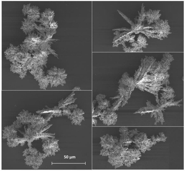
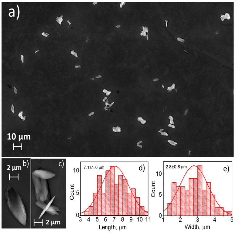
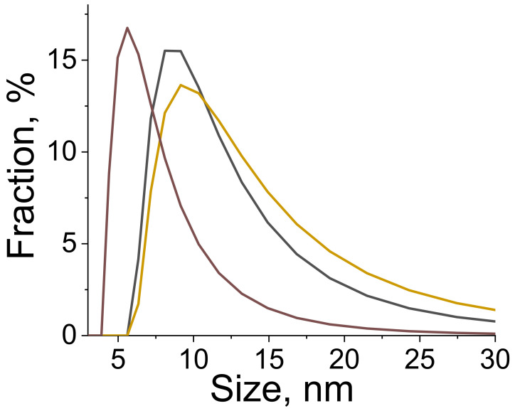
A scanning electron microscope was used to observe the shape and the size of the particles in the synthesized materials. Sample 1, which was synthesized by a slow mixing of equal volumes of sodium terephthalate (2 mM) and europium chloride (1 mM) aqueous solutions, precipitated in the form of a polycrystalline solid with the average particle size of 120 ± 30 µm (Figure 1). The observed species consisted of smaller particles stacked together forming dendrimer-like microparticle assemblies. The addition of the non-ionic surfactant (PEG-6000) to the reaction mixture and ultrasonication without a change in the Eu3+ and bdc2− concentrations (Sample 2) prevented the aggregation of the microparticles and resulted in the formation of individual microparticles (Figure 2a–c). The particles had a leaf-like shape with ratio length:width:height of about 13:5:1. The particles size was obtained from SEM images, the particle size distribution is shown in Figure 2d,e. The average length and width were calculated from these distributions and are equal to 7.1 ± 1.6 and 2.8 ± 0.8 µm, respectively. We found that under ultrasonication the solution remained clear to the eye when the concentration of Eu3+ and bdc2− was decreased twofold (1 mM Na2bdc and 0.5 mM EuCl3) both in the absence and the presence of the surfactant (PEG-6000). We could not precipitate the solid from the reaction mixture using high-speed centrifugation or by salting-out using organic solvents. Therefore, the formation of the nano-sized particles of europium(III) terephthalate was supposed. In order to exclude the contribution of the PEG micelles to the experimental data, in further experiments we carefully studied the aqueous suspension of Sample 3 obtained by a slow mixing of equal volumes of the 1 mM Na2bdc and 0.5 mM EuCl3 accompanied by ultrasonication and vigorous stirring without a PEG-6000 addition. The particle size distribution was revealed by a dynamic light scattering technique, resulting in the average particle size equal to about 8 ± 2 nm (Figure 3). The SEM-EDX study of 3 aggregates formed by drying the reaction mixture on the silicon plate revealed the presence of Eu in the sample but did not determine the particle size due to the insufficient spatial resolution of the used SEM microscope. The direct observation of the species using TEM was also problematic because the high-energy electron radiation (>100 kV) burned out the sample due to the decomposition of an organic linker (terephthalate ion).
Figure 1.
SEM images of Sample 1. The average diameter of polycrystals is 120 ± 30 µm.
Figure 2.
SEM images of Sample 2 (panels a–c). Particle size distribution (length and width) is shown in panels (d,e). The particles have the shape of the elliptic plates with ratio length:width:height of about 13:5:1. The average length and width were found to be equal to 7.1 ± 1.6 and 2.8 ± 0.8 µm, respectively.
Figure 3.
The particle size distribution of the aqueous solution of 3 is revealed by dynamic light scattering as a result of three parallel measurements. The average particle size is equal to about 8 ± 2 nm (spherical approximation).
In our study, we have found that ultrasonication and PEG-6000 addition significantly decreases the particle size and prevents aggregation. The increase in particle size can be achieved via continuous growth of a particle or gradual aggregation of various particles or seed crystals. The contact of particles can reduce the total surface area in the aggregation process resulting in overall energy reduction. Ultrasonication can encourage surface tension between the species caused by the acoustic radiation force on a compressible particle [26]. The effect of PEG addition on the particle size can be explained by the well-known properties of surfactants including polyethylene glycol to be adsorbed on the particles or seed crystals that decrease their surface energy and prevents aggregation [27,28,29]. Surprisingly, we revealed that the twofold decrease in the reagents’ concentration leads to size reduction for several orders. A recent kinetic study of zinc-2-methylimidazole MOF ZIF-8 [30] reported that nucleation and crystal growth rates non-monotonously depend on the concentration of the reagents. During the low concentrations of the metal ions and the organic linker, the 1:1 M:L complex dominates. This state is called the “pre-equilibrium”. Further nucleation is associative and fast because the central atom has several weakly coordinated solvent molecules and can easily react with other 1:1 complexes resulting in the formation of oligomeric secondary building units (SBUs) [31]. Increasing the concentration of the metal ions and the organic linker leads to the domination of 1:2 and 1:3 M:L complexes. The aggregation of 1:2 and 1:3 M:L complexes into SBUs is slower than 1:1 complexes, which results in slower nucleation. Therefore, nucleation is faster than the growth process in solutions containing low concentrations of the metal ions and the organic linker, which explains the formation of the smaller particle size of the MOFs crystallizing at low concentrations.
3.2. Crystal Structure
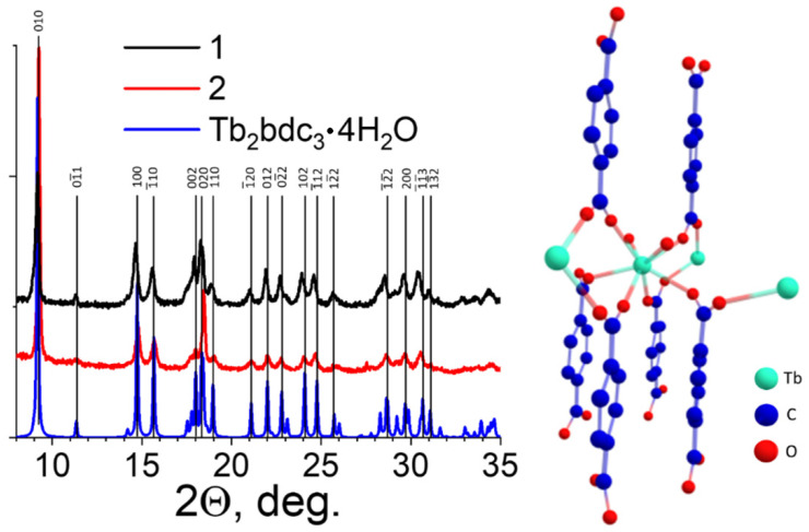
The X-ray powder diffraction (XRD) patterns were measured (Figure 4) for Samples 1 and 2 to discover the crystalline phase of the obtained materials. We could not precipitate Sample 3 from the solution; therefore, the XRD pattern of Sample 3 was not measured. Analysis of XRD patterns demonstrated that synthesized materials 1 and 2 are isostructural with the Tb2bdc3·4H2O [32], the typical crystalline phase of lanthanide terephthalates [22], which indicated that materials 1 and 2 were obtained in a form of Eu2bdc3·4H2O. This structure is a three-dimensional metal-organic framework (MOF), where octacoordinated Eu3+-ions are bound to the two water molecules and six terephthalate ions through the oxygen atoms (Figure 4). XRD peaks of Eu2bdc3·4H2O in Samples 1 and 2 slightly diverge from their counterparts measured for Tb2bdc3·4H2O reported previously [32]. To compare the structures of Eu2bdc3·4H2O and Tb2bdc3·4H2O materials, the refinement of unit cell parameters was performed for the Eu2bdc3·4H2O samples (Table 2). One can observe that the structure of coarse-crystalline Eu2bdc3·4H2O (1) is slightly different from that of Tb2bdc3·4H2O. The ionic radius of the octacoordinated Eu3+ ion (1.066 Å) is slightly larger than that of the octacoordinated Tb3+ ion (1.040 Å) [33], which most likely results in minor differences between Eu2bdc3·4H2O and Tb2bdc3·4H2O structures. The unit cell parameters of microcrystalline Eu2bdc3·4H2O (2) are somewhat different, both from that of coarse-crystalline Eu2bdc3·4H2O (1) and Tb2bdc3·4H2O [32], which is likely caused by the surface defects due to the relatively small particle size of several micrometers.
Figure 4.
The XRD patterns of europium(III) terephthalate powders (1 and 2) and the simulated XRD pattern of Tb2bdc3·4H2O single-crystal structure taken from ref. [32] and the crystal structure of Tb2bdc3·4H2O.
Table 2.
Unit cell parameters for Tb2bdc3·4H2O [32] and Eu2bdc3·4H2O (Samples 1 and 2).
| Sample | a, Å | b, Å | c, Å | α, deg. | β, deg. | γ, deg. | V, Å3 |
|---|---|---|---|---|---|---|---|
| Tb2bdc3·4H2O | 6.14 | 10.07 | 10.10 | 102.25 | 91.12 | 101.52 | 596.63 |
| Eu2bdc3·4H2O (1) | 6.20 | 9.85 | 10.29 | 102.15 | 89.75 | 105.10 | 592.91 |
| Eu2bdc3·4H2O (2) | 6.16 | 9.8 | 10.22 | 101.84 | 90.27 | 104.86 | 582.19 |
3.3. Luminescent Properties
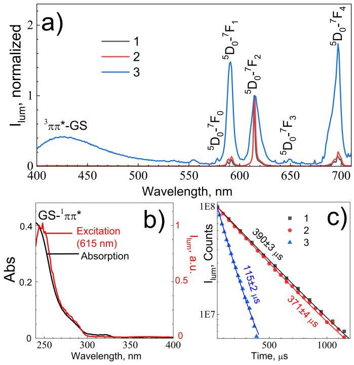
Terephthalate ions are known to intensively absorb ultraviolet light, promoting them into the 1ππ* singlet electronic excited state [32,34,35]. In europium(III) terephthalate, the 1ππ* state efficiently undergoes the 3ππ* triplet electronic excited state by intersystem crossing due to the heavy atom effect [35] followed by an energy transfer to 5D1 level of the Eu3+ ion, due to relatively close energy values of the lowest energy 3ππ* excited state of terephthalate ion [35] (≈20,000 cm−1) and 5D1 level of Eu3+ ion [36] (≈19,000 cm−1). 5D1 level of the Eu3+ ion [36] then undergoes internal conversion followed by emission corresponding to 5D0–7FJ (J = 0–5) transitions. Figure 5a presents the emission spectra of the europium(III) terephthalate series (1–3) upon 250 nm excitation into the 1ππ* singlet electronic excited state of the terephthalate ion. The emission spectra include narrow lines corresponding to the transitions from excited 5D0 to lower 7FJ levels: 5D0–7F0 (578 nm),5D0-–7F1 (590 nm), 5D0–7F2 (615 nm), 5D0–7F3 (649 nm), and 5D0–7F4 (697 nm). The emission spectrum of nanocrystalline 3 also contains spectrally broad band peaking at about 420 nm, which corresponds to the terephthalate phosphorescence [35]. The most prominent transitions in the emission spectra are magnetic dipole 5D0–7F1 and forced electric dipole 5D0–7F2 and 5D0–7F4 transitions. The excitation spectrum (λem = 615 nm) of nanocrystalline 3 resembles its UV–Vis absorption spectrum (Figure 5b) consisting of a 250 nm band as well as a 280 nm shoulder corresponding to the transitions into 1ππ* singlet electronic excited states of the terephthalate ion. One can notice that emission bands corresponding to the f–f transitions of the Eu3+ ion significantly broaden with the particle size reduction. Thus, the 5D0–7F2 band of coarse-crystalline 1, microcrystalline 2, and nanocrystalline 3 have full width at half maximum (fwhm) equal to 48, 66, and 238 cm−1, respectively. The smaller particles have larger surface-to-volume ratio and the number of structural defects, which results in a larger dispersion of energies of electronic levels of Eu3+ ions caused by the larger non-uniformity of the local environment of europium ions [37,38]. The luminescence decay curves of europium(III) terephthalate (Figure 5c) are fitted by single-exponential functions:
| (1) |
where time constant τf corresponds to the observed lifetime of 5D0 level. The observed lifetime of 5D0 level of coarse-crystalline 1, microcrystalline 2, and nanocrystalline 3 were found to be equal to 393 ± 3, 371 ± 4, and 115 ± 2 μs, respectively.
Figure 5.
(a) The emission spectra of europium(III) terephthalate samples of different sized particles (λex = 250 nm) normalized at the 615 nm emission band intensity. Sample numbers are shown in legend; (b) absorption (black line) and excitation (red line, λem = 615 nm) spectra of aqueous solution europium(III) terephthalate nanoparticles 3; (c) 615 nm luminescence decay curves of europium(III) terephthalate samples of different sized particles.
Luminescence decay is affected by the combination of radiative and nonradiative processes. Radiative decay rate is determined by dipole transition strength and local-field correction. Nonradiative processes include multi-phonon relaxation, quenching on impurities (e.g., O-H group of water molecules) and cooperative processes (cross-relaxation, energy migration). Detailed descriptions of these processes were provided in our earlier papers [39,40]. The radiative and nonradiative decay rates of Eu3+-doped phosphors can be calculated from the emission spectrum using 4f–4f intensity theory [41]. Magnetic dipole 5D0–7F1 transition probability A0-1 = AMD,0∙n03 = 14.65∙1.53 = 49 s−1. AMD,0 is the spontaneous emission probability of the magnetic dipole 5D0–7F1, 14.65 s−1, and n0 is the refractive index, 1.5 [34]. Radiative decay rates A0–λ (λ = 2, 4) of the 5D0–7Fλ emission transition can be obtained from this formula:
| (2) |
where I0–λ and ν0–λ are the integral intensity and frequency of the 5D0–7Fλ emission transition. The total radiative decay rate, Ar, could be calculated by summing all the A0–λ radiative decay rates (λ = 1, 2, 4). The total decay rate is reciprocal to the observed lifetime of 5D0 level, shown in Figure 5c, , whereas the nonradiative probability can be calculated as: . Quantum efficiency of 5D0 level is . Decay rates and quantum efficiencies of the 5D0 level of europium(III) terephthalates 1–3 are summarized in Table 3.
Table 3.
Radiative (Ar), nonradiative (Anr) and total (Atotal) decay rates, quantum efficiencies (η) of the 5D0 level of europium(III) and Eu3+ luminescence quantum yields (Φ) upon excitation into 1ππ* singlet electronic excited state of terephthalate ion terephthalates 1–3.
| Sample | Ar (s−1) | Anr (s−1) | Atotal (s−1) | η (%) | Φ (%) |
|---|---|---|---|---|---|
| 1 | 371 | 2193 | 2564 | 14.5 | 10 ± 1 |
| 2 | 290 | 2405 | 2695 | 10.8 | 5 ± 1 |
| 3 | 150 | 8545 | 8695 | 1.7 | 1.5 ± 0.5 |
Analyzing Table 2, one can see the quantum efficiencies of the 5D0 level and Eu3+ luminescence quantum yields decrease in series 1–3 simultaneously with particle size, whereas nonradiative decay rate constants increase upon the size reduction. Smaller particles have larger surface-to-volume ratios, resulting in more efficient quenching of the 5D0 level and Eu3+ by the water molecules in an aqueous solution [42]. Comparing the quantum efficiencies of the 5D0 level and Eu3+ luminescence quantum yields values, one can notice that the η/Φ ratio is equal to 0.5–0.9, which indicates a very efficient energy transfer from initially excited terephthalate chromophore to the 5D0 level of Eu3+ ion.
3.4. Sensing Transition Metal Cations
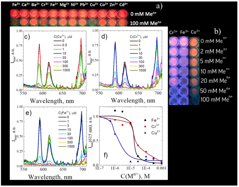
Previous studies demonstrated that the presence of impurities such as ions of transition metals (Fe3+, Cu2+, Pb2+, MnO4−, Cr2O72−) [43], and organic compounds (aromatic, nitroaromatic, carbonyl compounds) can significantly quench the luminescence of the Eu-based metal-organic frameworks [1,2,3,4,5,6,7,8,9] making them prospective for the design of luminescent sensors for various pollutants and explosives. To reveal the selectivity of the europium(III) terephthalate MOF luminescence quenching to the various metal cations, 80 μL of aqueous suspensions of coarse-crystalline 1 (C(Eu3+) = 8 mM) was mixed with the 100 μL of metal salt solutions (C(Mn+) = 100 mM; Mn+ = Fe3+, Ca2+, Ba2+, Cr3+, Fe2+, Mg2+, Ni2+, Pb2+, Cu2+, Co2+, Zn2+, Cd2+) or distilled water. After 30 min, the photographs of these solutions under 254 nm illumination were recorded (Figure 6a,b). It was found that the Eu-based red emission faded only in the presence of Fe3+, Cr3+ and Cu2+ ions (Figure 6a) starting from metal ion concentration 10–50 mM (Figure 6b). The emission spectra of aqueous solutions of nanocrystalline 3 (C(Eu3+) = 5 μM) in the absence and in the presence of various concentrations of Cu2+, Cr3+, and Fe3+ ions (λexc = 250 nm) indicate the quenching of Eu3+ 5D0–7Fλ luminescence by the above-mentioned metal ions (Figure 6c–e). The dependence of the 615 nm emission band intensity on the Cu2+, Cr3+, and Fe3+ concentration is given in Figure 6f. The concentration dependence resembles the step-function, where luminescence intensity sharply falls starting from the certain concentration of metal ion: 1 μM of Cu2+ and 30 μM of Cr3+ or Fe3+. Surprisingly, we revealed that the addition of Fe3+ ions resulted in simultaneous quenching for the Eu3+ 5D0–7Fλ luminescence (591, 615, and 697 nm bands) and the terephthalate phosphorescence (420 nm), whereas the addition of Cu2+ and Cr3+ ions almost failed to reduce the intensity of the terephthalate phosphorescence band at 420 nm (Figures S1–S3, Supplementary Materials). This observation indicates a different quenching mechanism of Eu3+ 5D0–7Fλ luminescence by the above-mentioned metal ions. Most likely, Cu2+, Cr3+, and Fe3+ ions somehow coordinate with the oxygens of terephthalate ligands, but Fe3+ ions quench the 3ππ* triplet electronic excited state of terephthalate ion, whereas Cu2+ and Cr3+ ions quench the 5D0 level of Eu3+. To reveal the complete quenching mechanism, one must study the excited-state dynamics of singlet and triplet electronic states of terephthalate ion, as well as the 5D0 level of Eu3+, depending on the heavy metal ion concentration by time-resolved transient absorption and luminescence spectroscopy methods. We have found that nanocrystalline europium(III) terephthalate MOF 3 demonstrates significantly lower limits of detection on Cu2+, Cr3+, and Fe3+ ions than coarse-crystalline 1 (10–50 mM for coarse-crystalline 1 vs 1–30 μM for nanocrystalline 3, Figure 6b,f). This observation is explained by a larger surface-to-volume ratio of nanoparticles relatively to the bulk material, resulting in a higher luminescence quenching efficiency of the later materials due to a greater number of coordination sites. The sensitivity of our materials to Cu2+, Cr 3+ and Fe3+ ions is comparable with the best reported luminescent MOF-based sensors reported previously (Table 4). Despite the higher sensitivity of electrochemical MOF-based sensors (Table 4), the luminescent sensors can be used for the design of relatively inexpensive express tests on heavy metal ions.
Figure 6.
(a,b) Photographs of aqueous suspension of coarse-crystalline 1 under 254 nm illumination in the absence and presence of various metal ions; emission spectra of aqueous solution of nanocrystalline 3 in the absence and presence of various concentrations of Cu2+ (c), Cr3+ (d), and Fe3+ (e) ions upon 250 nm excitation; (f) Cu2+, Cr3+, and Fe3+ concentration dependence of 615 nm emission intensity of Sample 3.
Table 4.
Limits of detection (LOD) of nanocrystalline europium(III) terephthalate tetrahydrate 3 and previously reported materials for Cu2+, Cr 3+ and Fe3+ ions.
| Sensing Material | Method | Target Contaminant | LOD | Ref. |
|---|---|---|---|---|
| Eu2(bdc)3·4H2O | luminescent | Cu2+ | 1 μM | Current work |
| Tb(BTC)(H2O) | luminescent | Cu2+ | 10 μM | [2] |
| CDs@Eu-DPA MOFs | luminescent | Cu2+ | 26.3 nM | [3,43] |
| [Eu(PDC)1.5(DMF)]·(DMF)0.5(H2O)0.5 | luminescent | Cu2+ | 10 mM | [7,44] |
| Eu2(FMA)2(OX)(H2O)4·4H2O | luminescent | Cu2+ | 100 μM | [7,45] |
| [Eu4(BPT)4(DMF)2(H2O)8] | luminescent | Cu2+ | 10 μM | [7,46] |
| [Tb3(L)2(HCOO)(H2O)5]·DMF·4H2O | luminescent | Cu2+ | 100 μM | [7,47] |
| [Eu(ox)2(H2O)](Me2NH2)(H2O)3 | luminescent | Cu2+ | 10 μM | [10,48] |
| Zr6(O)8(OH2)8(tpdc)4 | luminescent | Cu2+ | 1 μM | [10,49] |
| PCN-222-Pd(II) | luminescent | Cu2+ | 50 nM | [10,50] |
| Me2NH2@MOF-1 | electrochemical | Cu2+ | 10 pM | [3,51] |
| Eu2(bdc)3 nanoparticles | luminescent | Fe3+ | 30 μM | Current work |
| [Me2NH2][In(abtc)]·solvents | luminescent | Fe3+ | 34.5 μM | [52] |
| [LnK(BPDSDC)(DMF)(H2O)]·x(solvent) | luminescent | Fe3+ | 10 μM | [10,53] |
| [Eu(BTPCA)(H2O)]·2DMF·3H2O | luminescent | Fe3+ | 10 μM | [7,54] |
| [Eu(HL)(H2O)2]n·2H2O | luminescent | Fe3+ | 1 μM | [7,55] |
| EuL | luminescent | Fe3+ | 100 μM | [7,56] |
| [H2NMe2]3[Tb(DPA)3] | luminescent | Fe3+ | 10 μM | [7,57] |
| Eu (4′-(4-carboxyphenyl)-2,2′: 6′,2″-terpyridine)3 | luminescent | Fe3+ | 100 μM | [7,58] |
| [H(H2O)8][DyZn4(imdc)4(im)4] | luminescent | Fe3+ | 1 mM | [7,59] |
| Eu3+@Ga2(OH)4(C9O6H4) | luminescent | Fe3+ | 0.28 μM | [7,60] |
| nTbL | luminescent | Fe3+ | 10 μM | [7,61] |
| [Eu(atpt)1.5(phen)(H2O)]n | luminescent | Fe3+ | 500 μM | [7,62] |
| [(CH3)2NH2] ·[Tb(bptc)]·xS | luminescent | Fe3+ | 10 μM | [7,63] |
| Tb-BTB | luminescent | Fe3+ | 10 μM | [7,64] |
| [Eu3(BDC)4.5(H2O)(DMF)2] | luminescent | Fe3+ | 1 μM | [7,65] |
| [Cd(L)(BPDC)]·2H2O | luminescent | Fe3+ | 2 μM | [8,66] |
| [Cd(L)(SDBA)(H2O)]∙0.5H2O | luminescent | Fe3+ | 2 μM | [8,66] |
| [Zn5(hfipbb)4(trz)2(H2O)2] | luminescent | Fe3+ | 10 μM | [8,67] |
| [Eu(Hpzbc)2(NO3)]·H2O | luminescent | Fe3+ | 10 μM | [8,68] |
| [Eu(L)(H2O)2]·NMP·H2O | luminescent | Fe3+ | 100 nM | [8,69] |
| [Tb(L1)1.5(H2O)]·3H2O | luminescent | Fe3+ | 10 μM | [10,70] |
| Bisdiene macrocycle | luminescent | Fe3+ | 0.58 μM | [71] |
| 2-(cyclohexylamino)-3-phenyl-4Hfuro [3,2-c]chromen-4-one | luminescent | Fe3+ | 1.73 μM | [72] |
| [Me2NH2][In(abtc)]·solvents | luminescent | Fe3+ | 34.5 μM | [53] |
| PPCOT/NiFe2O4/C-SWCNT | electrochemical | Fe3+ | 100 pM | [73] |
| Eu2(bdc)3 nanoparticles | luminescent | Cr3+ | 30 μM | Current work |
| Tb(BTC)(H2O) | luminescent | Cr3+ | 10 μM | [2] |
| [TbK(BPDSDC)(DMF)(H2O)2] | 10 μM | [8,74] | ||
| [Eu2L3(DMF)3]·2DMF·5H2O | luminescent | Cr3+ | 75.2 nM | [75] |
| ATNA deriviative | electrochemical | Cr3+ | 130 pM | [76] |
4. Conclusions
In summary, we reported the ultrasound-assisted wet-chemical synthesis and characterization of luminescent coarse-, micro-, and nano-crystalline Eu2bdc3·4H2O MOFs. The particles of coarse-crystalline Eu2bdc3·4H2O, which are synthesized by the mixing of sodium terephthalate and europium chloride aqueous solutions without ultrasound, are dendrimer-like microparticle assemblies with the average particle size of 120 ± 30 µm. The microcrystalline MOFs were prepared by mixing sodium terephthalate and europium chloride aqueous solutions with the addition of PEG-6000 in the presence of ultrasonication. The microparticles have the shape of leaf-like plates and an average size of 7.1 × 2.8 µm. The average size of Eu2bdc3·4H2O nanoparticles, synthesized by the mixing of low-concentration sodium terephthalate and europium chloride aqueous solutions in the presence of ultrasonication, is equal to about 8 ± 2 nm. Thus, the reported Eu2bdc3·4H2O nanoparticles are the smallest nanosized rare-earth-based MOF crystals, to the best of our knowledge. The emission spectra of synthesized materials exhibit narrow lines corresponding to transitions from excited 5D0 to lower 7FJ levels of Eu3+ ion: 5D0–7F0 (578 nm),5D0–7F1 (590 nm), 5D0–7F2 (615 nm), 5D0–7F3 (649 nm), and 5D0–7F4 (697 nm). Size reduction resulted in a broadening of the emission bands. The Eu3+ luminescence quantum yields, upon excitation into 1ππ* singlet electronic excited state of terephthalate ion, were found to be of 10 ± 1%, 5 ± 1% and 1.5 ± 0.5% for coarse-, micro- and nanocrystalline Eu2bdc3·4H2O MOFs, respectively. The nonradiative decay rate of nanocrystalline europium(III) terephthalate was significantly larger that the corresponding values of Eu2bdc3·4H2O MOFs, which resulted from more efficient quenching of the 5D0 level and Eu3+ by the water molecules in aqueous solution due to greater surface-to-volume ratio of nanocrystalline MOF. The Cu2+, Cr3+, and Fe3+ ions efficiently and selectively quench the Eu3+ 5D0−7Fλ luminescence of nanocrystalline Eu2bdc3·4H2O MOFs starting from the relatively low concentrations of metal ion: 1 μM of Cu2+ and 30 μM of Cr3+ or Fe3+. The reported nanocrystalline europium(III) terephthalateis one of the most sensitive luminescent MOF-based sensorfor Cu2+, Cr 3+ and Fe3+ ions (Table 4). Therefore, synthesized nanocrystalline Eu2bdc3·4H2O MOFs can be considered promising luminescent probes for heavy metal ions in waste and drinking water.
Acknowledgments
The measurements were performed at the Research Park of Saint Petersburg State University (“Magnetic Resonance Research Centre”, “SPbU Computing Centre”, “Cryogenic Department”, “Interdisciplinary Resource Centre for Nanotechnology”, “Centre for X-ray Diffraction Studies”, “Chemical Analysis and Materials Research Centre”, “Centre for Optical and Laser Materials Research”, and “Centre for Innovative Technologies of Composite Nanomaterials”). The project was partially performed at the facilities of the Educational Center “Sirius”, Sochi, Russia. The authors acknowledge Elena A. Bessonova, Vladimir Sosnovsky, David Zheglov, Anton Ostrosablin, and Saveliy Zaverukhin for help with the experiments.
Supplementary Materials
The following are available online at https://www.mdpi.com/article/10.3390/nano11092448/s1, Figure S1: (a) Emission spectra of aqueous solution of nanocrystalline 3 in the absence and presence of various concentrations of Cu2+ upon 250 nm excitation; (b) Cu2+ concentration dependence of 420, 591, 615, and 697 nm emission intensities of 3, Figure S2: (a) Emission spectra of aqueous solution of nanocrystalline 3 in the absence and presence of various concentrations of Cr3+ upon 250 nm excitation; (b) Cr3+ concentration dependence of 420, 591, 615, and 697 nm emission intensities of 3, Figure S3: (a) Emission spectra of aqueous solution of nanocrystalline 3 in the absence and presence of various concentrations of Fe3+ upon 250 nm excitation; (b) Fe3+ concentration dependence of 420, 591, 615, and 697 nm emission intensities of 3.
Author Contributions
Conceptualization, S.S.K., E.M.K. and A.S.M.; Methodology, S.S.K. and E.M.K.; Formal Analysis, A.S.M., V.G.N. and I.E.K.; Investigation, S.S.K., V.G.N., I.E.K., E.M.K., A.A.V. and A.A.S.; Resources, N.A.B., M.Y.S. and A.S.M.; Data Curation, A.S.M. and I.E.K., Writing—Original Draft Preparation, A.S.M. and V.G.N.; Writing—Review and Editing, M.N.R., M.S.P., I.I.T., N.A.B. and M.Y.S.; Visualization, V.D.K.; Supervision, A.S.M.; Project Administration, A.S.M.; Funding Acquisition, A.S.M. and M.S.P. All authors have read and agreed to the published version of the manuscript.
Funding
The reported study was funded by the Russian Fund for Basic Research (RFBR), project number 20-33-70025 (A.S.M., N.A.B. and M.Y.S.). The work was partially supported by Ministry of Education and Science of the Russian Federation (project FSRM-2020-0006, M.S.P. and M.N.R.). The visit of A.S.M., V.G.N., A.A.V., A.A.S., I.I.T., N.A.B. and M.Y.S. to the Educational Center “Sirius” was funded by Educational Foundation “Talent and Success”.
Data Availability Statement
Data is contained within this article and corresponding supplementary materials.
Conflicts of Interest
The authors declare no conflict of interest.
Footnotes
Publisher’s Note: MDPI stays neutral with regard to jurisdictional claims in published maps and institutional affiliations.
References
- 1.Zeng X., Zhang Y., Zhang J., Hu H., Wu X., Long Z., Hou X. Facile colorimetric sensing of Pb2+ using bimetallic lanthanide metal-organic frameworks as luminescent probe for field screen analysis of lead-polluted environmental water. Microchem. J. 2017;134:140–145. doi: 10.1016/j.microc.2017.05.011. [DOI] [Google Scholar]
- 2.Cai D., Guo H., Wen L., Liu C. Fabrication of hierarchical architectures of Tb-MOF by a “green coordination modulation method” for the sensing of heavy metal ions. CrystEngComm. 2013;15:6702. doi: 10.1039/c3ce40820e. [DOI] [Google Scholar]
- 3.Fang X., Zong B., Mao S. Metal-Organic Framework-Based Sensors for Environmental Contaminant Sensing. Nano-Micro Lett. 2018;10:64. doi: 10.1007/s40820-018-0218-0. [DOI] [PMC free article] [PubMed] [Google Scholar]
- 4.Feng H.-J., Xu L., Liu B., Jiao H. Europium metal-organic frameworks as recyclable and selective turn-off fluorescent sensors for aniline detection. Dalton Trans. 2016;45:17392–17400. doi: 10.1039/C6DT03358J. [DOI] [PubMed] [Google Scholar]
- 5.Xu H., Liu F., Cui Y., Chen B., Qian G. A luminescent nanoscale metal-organic framework for sensing of nitroaromatic explosives. Chem. Commun. 2011;47:3153. doi: 10.1039/c0cc05166g. [DOI] [PubMed] [Google Scholar]
- 6.Crawford S.E., Ohodnicki P.R., Baltrus J.P. Materials for the photoluminescent sensing of rare earth elements: Challenges and opportunities. J. Mater. Chem. C. 2020;8:7975–8006. doi: 10.1039/D0TC01939A. [DOI] [Google Scholar]
- 7.Mahata P., Mondal S.K., Singha D.K., Majee P. Luminescent rare-earth-based MOFs as optical sensors. Dalton Trans. 2017;46:301–328. doi: 10.1039/C6DT03419E. [DOI] [PubMed] [Google Scholar]
- 8.Zhang Y., Yuan S., Day G., Wang X., Yang X., Zhou H.-C. Luminescent sensors based on metal-organic frameworks. Coord. Chem. Rev. 2018;354:28–45. doi: 10.1016/j.ccr.2017.06.007. [DOI] [Google Scholar]
- 9.Zhang X., Kang X., Cui W., Zhang Q., Zheng Z., Cui X. Floral and lamellar europium(III)-based metal-organic frameworks as high sensitivity luminescence sensors for acetone. New J. Chem. 2019;43:8363–8369. doi: 10.1039/C9NJ00889F. [DOI] [Google Scholar]
- 10.Lustig W.P., Mukherjee S., Rudd N.D., Desai A.V., Li J., Ghosh S.K. Metal-organic frameworks: Functional luminescent and photonic materials for sensing applications. Chem. Soc. Rev. 2017;46:3242–3285. doi: 10.1039/C6CS00930A. [DOI] [PubMed] [Google Scholar]
- 11.Adusumalli V.N.K.B., Runowski M., Lis S. 3,5-dihydroxy benzoic acid-capped caf2:Tb3+ nanocrystals as luminescent probes for the wo42- ion in aqueous solution. ACS Omega. 2020;5:4568–4575. doi: 10.1021/acsomega.9b03956. [DOI] [PMC free article] [PubMed] [Google Scholar]
- 12.Aslandukov A.N., Utochnikova V.V., Goriachiy D.O., Vashchenko A.A., Tsymbarenko D.M., Hoffmann M., Pietraszkiewicz M., Kuzmina N.P. The development of a new approach toward lanthanide-based OLED fabrication: New host materials for Tb-based emitters. Dalton Trans. 2018;47:16350–16357. doi: 10.1039/C8DT02911C. [DOI] [PubMed] [Google Scholar]
- 13.Liu D., Lu K., Poon C., Lin W. Metal-organic Frameworks as Sensory Materials and Imaging Agents. Inorg. Chem. 2014;53:1916–1924. doi: 10.1021/ic402194c. [DOI] [PMC free article] [PubMed] [Google Scholar]
- 14.Younis S.A., Bhardwaj N., Bhardwaj S.K., Kim K.-H., Deep A. Rare earth metal-organic frameworks (RE-MOFs): Synthesis, properties, and biomedical applications. Coord. Chem. Rev. 2021;429:213620. doi: 10.1016/j.ccr.2020.213620. [DOI] [Google Scholar]
- 15.Zhou X., Wang H., Jiang S., Xiang G., Tang X., Luo X., Li L., Zhou X. Multifunctional Luminescent Material Eu(III) and Tb(III) Complexes with Pyridine-3,5-Dicarboxylic Acid Linker: Crystal Structures, Tunable Emission, Energy Transfer, and Temperature Sensing. Inorg. Chem. 2019;58:3780–3788. doi: 10.1021/acs.inorgchem.8b03319. [DOI] [PubMed] [Google Scholar]
- 16.Khudoleeva V., Tcelykh L., Kovalenko A., Kalyakina A., Goloveshkin A., Lepnev L., Utochnikova V. Terbium-europium fluorides surface modified with benzoate and terephthalate anions for temperature sensing: Does sensitivity depend on the ligand? J. Lumin. 2018;201:500–508. doi: 10.1016/j.jlumin.2018.05.002. [DOI] [Google Scholar]
- 17.Dou X., Sun K., Chen H., Jiang Y., Wu L., Mei J., Ding Z., Xie J. Nanoscale metal-organic frameworks as fluorescence sensors for food safety. Antibiotics. 2021;10:358. doi: 10.3390/antibiotics10040358. [DOI] [PMC free article] [PubMed] [Google Scholar]
- 18.Puglisi R., Pellegrino A.L., Fiorenza R., Scirè S., Malandrino G. A facile one-pot approach to the synthesis of gd-eu based metal-organic frameworks and applications to sensing of Fe3+ and Cr2O72− ions. Sensors. 2021;21:1679. doi: 10.3390/s21051679. [DOI] [PMC free article] [PubMed] [Google Scholar]
- 19.Ding S.B., Wang W., Qiu L.G., Yuan Y.P., Peng F.M., Jiang X., Xie A.J., Shen Y.H., Zhu J.F. Surfactant-assisted synthesis of lanthanide metal-organic framework nanorods and their fluorescence sensing of nitroaromatic explosives. Mater. Lett. 2011;65:1385–1387. doi: 10.1016/j.matlet.2011.02.009. [DOI] [Google Scholar]
- 20.Li Q., Liu H., Alattar M., Jiang S., Han J., Ma Y., Jiang C. The preferential accumulation of heavy metals in different tissues following frequent respiratory exposure to PM2.5 in rats. Sci. Rep. 2015;5:16936. doi: 10.1038/srep16936. [DOI] [PMC free article] [PubMed] [Google Scholar]
- 21.Jaishankar M., Tseten T., Anbalagan N., Mathew B.B., Beeregowda K.N. Toxicity, mechanism and health effects of some heavy metals. Interdiscip. Toxicol. 2014;7:60–72. doi: 10.2478/intox-2014-0009. [DOI] [PMC free article] [PubMed] [Google Scholar]
- 22.Daiguebonne C., Kerbellec N., Guillou O., Bünzli J.-C., Gumy F., Catala L., Mallah T., Audebrand N., Gérault Y., Bernot K., et al. Structural and Luminescent Properties of Micro- and Nanosized Particles of Lanthanide Terephthalate Coordination Polymers. Inorg. Chem. 2008;47:3700–3708. doi: 10.1021/ic702325m. [DOI] [PubMed] [Google Scholar]
- 23.Zou H., Wang L., Zeng C., Gao X., Wang Q., Zhong S. Rare-earth coordination polymer micro/nanomaterials: Preparation, properties and applications. Front. Mater. Sci. 2018;12:327–347. doi: 10.1007/s11706-018-0444-x. [DOI] [Google Scholar]
- 24.Escudero A., Becerro A.I., Carrillo-carrión C., Núñez N.O., Zyuzin M.V., Laguna M., González-mancebo D., Ocaña M., Parak W.J. Rare earth based nanostructured materials: Synthesis, functionalization, properties and bioimaging and biosensing applications. Nanophotonics. 2017;6:881–921. doi: 10.1515/nanoph-2017-0007. [DOI] [Google Scholar]
- 25.Wang F., Deng K., Wu G., Liao H., Liao H., Zhang L., Lan S., Zhang J., Song X., Wen L. Facile and Large-Scale Syntheses of Nanocrystal Rare Earth Metal-organic Frameworks at Room Temperature and Their Photoluminescence Properties. J. Inorg. Organomet. Polym. Mater. 2012;22:680–685. doi: 10.1007/s10904-011-9498-2. [DOI] [Google Scholar]
- 26.Yang G., Lin W., Lai H., Tong J., Lei J., Yuan M., Zhang Y., Cui C. Ultrasonics Sonochemistry Understanding the relationship between particle size and ultrasonic treatment during the synthesis of metal nanoparticles. Ultrason. Sonochem. 2021;73:105497. doi: 10.1016/j.ultsonch.2021.105497. [DOI] [PMC free article] [PubMed] [Google Scholar]
- 27.Liu W., Yin R., Xu X., Zhang L., Shi W., Cao X. Structural Engineering of Low-Dimensional Metal-organic Frameworks: Synthesis, Properties, and Applications. Adv. Sci. 2019;6:1802373. doi: 10.1002/advs.201802373. [DOI] [PMC free article] [PubMed] [Google Scholar]
- 28.Taylor K.M.L., Jin A., Lin W. Surfactant-Assisted Synthesis of Nanoscale Gadolinium Metal-Organic Frameworks for Potential Multimodal Imaging. Angew. Chem. Int. Ed. 2008;47:7722–7725. doi: 10.1002/anie.200802911. [DOI] [PubMed] [Google Scholar]
- 29.Pellegrino F., Sordello F., Mino L., Prozzi M., Mansfeld U., Hodoroaba V.-D., Minero C. Polyethylene Glycol as Shape and Size Controller for the Hydrothermal Synthesis of SrTiO3 Cubes and Polyhedra. Nanomaterials. 2020;10:1892. doi: 10.3390/nano10091892. [DOI] [PMC free article] [PubMed] [Google Scholar]
- 30.Yeung H.H.M., Sapnik A.F., Massingberd-Mundy F., Gaultois M.W., Wu Y., Fraser D.A.X., Henke S., Pallach R., Heidenreich N., Magdysyuk O.V., et al. Control of Metal-organic Framework Crystallization by Metastable Intermediate Pre-equilibrium Species. Angew. Chem. Int. Ed. 2019;58:566–571. doi: 10.1002/anie.201810039. [DOI] [PubMed] [Google Scholar]
- 31.Dighe A.V., Nemade R.Y., Singh M.R. Modeling and Simulation of Crystallization of Metal-organic Frameworks. Processes. 2019;7:527. doi: 10.3390/pr7080527. [DOI] [Google Scholar]
- 32.Reineke T.M., Eddaoudi M., Fehr M., Kelley D., Yaghi O.M. From Condensed Lanthanide Coordination Solids to Microporous Frameworks Having Accessible Metal Sites. J. Am. Chem. Soc. 1999;121:1651–1657. doi: 10.1021/ja983577d. [DOI] [Google Scholar]
- 33.Shannon R.D. Revised Effective Ionic Radii and Systematic Studies of Interatomie Distances in in Halides and Chaleogenides. Acta Crystallogr. Sect. A. 1976;A32:751–767. doi: 10.1107/S0567739476001551. [DOI] [Google Scholar]
- 34.Haquin V., Etienne M., Daiguebonne C., Freslon S., Calvez G., Bernot K., Le Pollès L., Ashbrook S.E., Mitchell M.R., Bünzli J.-C., et al. Color and Brightness Tuning in Heteronuclear Lanthanide Terephthalate Coordination Polymers. Eur. J. Inorg. Chem. 2013;2013:3464–3476. doi: 10.1002/ejic.201300381. [DOI] [Google Scholar]
- 35.Utochnikova V.V., Grishko A.Y., Koshelev D.S., Averin A.A., Lepnev L.S., Kuzmina N.P. Lanthanide heterometallic terephthalates: Concentration quenching and the principles of the “multiphotonic emission”. Opt. Mater. (Amst). 2017;74:201–208. doi: 10.1016/j.optmat.2017.02.052. [DOI] [Google Scholar]
- 36.Binnemans K. Interpretation of europium(III) spectra. Coord. Chem. Rev. 2015;295:1–45. doi: 10.1016/j.ccr.2015.02.015. [DOI] [Google Scholar]
- 37.Carrera Jota M.L., García Murillo A., Carrillo Romo F., García Hernández M., Morales Ramírez A.D.J., Velumani S., de la Rosa Cruz E., Kassiba A. Lu2O3:Eu3+ glass ceramic films: Synthesis, structural and spectroscopic studies. Mater. Res. Bull. 2014;51:418–425. doi: 10.1016/j.materresbull.2013.12.029. [DOI] [Google Scholar]
- 38.Liu G., Hong G., Sun D. Synthesis and characterization of SiO2/Gd2O3:Eu core–shell luminescent materials. J. Colloid Interface Sci. 2004;278:133–138. doi: 10.1016/j.jcis.2004.05.013. [DOI] [PubMed] [Google Scholar]
- 39.Kolesnikov I., Povolotskiy A., Mamonova D., Lahderanta E., Manshina A., Mikhailov M. Photoluminescence Properties of Eu3+ Ions in Yttrium Oxide Nanoparticles: Defect vs Normal Sites. RSC Adv. 2016;6:76533–76541. doi: 10.1039/C6RA16814K. [DOI] [Google Scholar]
- 40.Golyeva E.V., Kolesnikov I.E., Lähderanta E., Kurochkin A.V., Mikhailov M.D. Effect of synthesis conditions on structural, morphological and luminescence properties of MgAl2O4:Eu3+ nanopowders. J. Lumin. 2018;194:387–393. doi: 10.1016/j.jlumin.2017.10.068. [DOI] [Google Scholar]
- 41.Kolesnikov I.E., Kolokolov D.S., Kurochkin M.A., Voznesenskiy M.A., Osmolowsky M.G., Lähderanta E., Osmolovskaya O.M. Morphology and doping concentration effect on the luminescence properties of SnO2:Eu3+ nanoparticles. J. Alloys Compd. 2020;822:153640. doi: 10.1016/j.jallcom.2020.153640. [DOI] [Google Scholar]
- 42.Gorbunov A.O., Lindqvist-Reis P., Mereshchenko A.S., Skripkin M.Y. Solvation and complexation of europium(III) ions in triflate and chloride aqueous-organic solutions by TRLF spectroscopy. J. Mol. Liq. 2017;240:25–34. doi: 10.1016/j.molliq.2017.04.136. [DOI] [Google Scholar]
- 43.Hao J., Liu F., Liu N., Zeng M., Song Y., Wang L. Ratiometric fluorescent detection of Cu2+ with carbon dots chelated Eu-based metal-organic frameworks. Sens. Actuators B Chem. 2017;245:641–647. doi: 10.1016/j.snb.2017.02.029. [DOI] [Google Scholar]
- 44.Chen B., Wang L., Xiao Y., Fronczek F.R., Xue M., Cui Y., Qian G. A luminescent metal-organic framework with Lewis basic pyridyl sites for the sensing of metal ions. Angew. Chem. Int. Ed. 2009;48:500–503. doi: 10.1002/anie.200805101. [DOI] [PubMed] [Google Scholar]
- 45.Xiao Y., Cui Y., Zheng Q., Xiang S., Qian G., Chen B. A microporous luminescent metal-organic framework for highly selective and sensitive sensing of Cu2+ in aqueous solution. Chem. Commun. 2010;46:5503–5505. doi: 10.1039/c0cc00148a. [DOI] [PubMed] [Google Scholar]
- 46.Hao Z., Yang G., Song X., Zhu M., Meng X., Zhao S., Song S., Zhang H. A europium(III) based metal-organic framework: Bifunctional properties related to sensing and electronic conductivity. J. Mater. Chem. A. 2014;2:237–244. doi: 10.1039/C3TA13179C. [DOI] [Google Scholar]
- 47.Zhao J., Wang Y.N., Dong W.W., Wu Y.P., Li D.S., Zhang Q.C. A Robust Luminescent Tb(III)-MOF with Lewis Basic Pyridyl Sites for the Highly Sensitive Detection of Metal Ions and Small Molecules. Inorg. Chem. 2016;55:3265–3271. doi: 10.1021/acs.inorgchem.5b02294. [DOI] [PubMed] [Google Scholar]
- 48.Wang X., Qin T., Bao S.S., Zhang Y.C., Shen X., Zheng L.M., Zhu D. Facile synthesis of a water stable 3D Eu-MOF showing high proton conductivity and its application as a sensitive luminescent sensor for Cu2+ ions. J. Mater. Chem. A. 2016;4:16484–16489. doi: 10.1039/C6TA06792A. [DOI] [Google Scholar]
- 49.Carboni M., Lin Z., Abney C.W., Zhang T., Lin W. A metal-organic framework containing unusual eight-connected Zr-oxo secondary building units and orthogonal carboxylic acids for ultra-sensitive metal detection. Chem.-Eur. J. 2014;20:14965–14970. doi: 10.1002/chem.201405194. [DOI] [PubMed] [Google Scholar]
- 50.Chen Y.Z., Jiang H.L. Porphyrinic Metal-Organic Framework Catalyzed Heck-Reaction: Fluorescence “turn-On” Sensing of Cu(II) Ion. Chem. Mater. 2016;28:6698–6704. doi: 10.1021/acs.chemmater.6b03030. [DOI] [Google Scholar]
- 51.Jin J.-C., Wu J., Yang G.-P., Wu Y.-L., Wang Y.-Y. A microporous anionic metal-organic framework for a highly selective and sensitive electrochemical sensor of Cu2+ ions. Chem. Commun. 2016;52:8475–8478. doi: 10.1039/C6CC03063G. [DOI] [PubMed] [Google Scholar]
- 52.Luo Y.H., Xie A.D., Chen W.C., Shen D., Zhang D.E., Tong Z.W., Lee C.S. Multifunctional anionic indium-organic frameworks for organic dye separation, white-light emission and dual-emitting Fe3+ sensing. J. Mater. Chem. C. 2019;7:14897–14903. doi: 10.1039/C9TC05113A. [DOI] [Google Scholar]
- 53.Zhou L.J., Deng W.H., Wang Y.L., Xu G., Yin S.G., Liu Q.Y. Lanthanide-Potassium Biphenyl-3,3′-disulfonyl-4,4′-dicarboxylate Frameworks: Gas Sorption, Proton Conductivity, and Luminescent Sensing of Metal Ions. Inorg. Chem. 2016;55:6271–6277. doi: 10.1021/acs.inorgchem.6b00928. [DOI] [PubMed] [Google Scholar]
- 54.Tang Q., Liu S., Liu Y., Miao J., Li S., Zhang L., Shi Z., Zheng Z. Cation Sensing by a Luminescent Metal-organic Framework with Multiple Lewis Basic Sites. Inorg. Chem. 2013;52:2799–2801. doi: 10.1021/ic400029p. [DOI] [PubMed] [Google Scholar]
- 55.Liang Y.-T., Yang G.-P., Liu B., Yan Y.-T., Xi Z.-P., Wang Y.-Y. Four super water-stable lanthanide–organic frameworks with active uncoordinated carboxylic and pyridyl groups for selective luminescence sensing of Fe3+ Dalton Trans. 2015;44:13325–13330. doi: 10.1039/C5DT01421B. [DOI] [PubMed] [Google Scholar]
- 56.Dang S., Ma E., Sun Z.M., Zhang H. A layer-structured Eu-MOF as a highly selective fluorescent probe for Fe3+ detection through a cation-exchange approach. J. Mater. Chem. 2012;22:16920–16926. doi: 10.1039/c2jm32661b. [DOI] [Google Scholar]
- 57.Weng H., Yan B. Lanthanide coordination polymers for multi-color luminescence and sensing of Fe3+ Inorg. Chem. Commun. 2016;63:11–15. doi: 10.1016/j.inoche.2015.11.013. [DOI] [Google Scholar]
- 58.Zheng M., Tan H., Xie Z., Zhang L., Jing X., Sun Z. Fast response and high sensitivity europium metal organic framework fluorescent probe with chelating terpyridine sites for Fe3+ ACS Appl. Mater. Interfaces. 2013;5:1078–1083. doi: 10.1021/am302862k. [DOI] [PubMed] [Google Scholar]
- 59.Li Y.F., Wang D., Liao Z., Kang Y., Ding W.H., Zheng X.J., Jin L.P. Luminescence tuning of the Dy-Zn metal-organic framework and its application in the detection of Fe(III) ions. J. Mater. Chem. C. 2016;4:4211–4217. doi: 10.1039/C6TC00832A. [DOI] [Google Scholar]
- 60.Xu X.Y., Yan B. Eu(III)-functionalized MIL-124 as fluorescent probe for highly selectively sensing ions and organic small molecules especially for Fe(III) and Fe(II) ACS Appl. Mater. Interfaces. 2015;7:721–729. doi: 10.1021/am5070409. [DOI] [PubMed] [Google Scholar]
- 61.Dang S., Wang T., Yi F., Liu Q., Yang W., Sun Z.M. A Nanoscale Multiresponsive Luminescent Sensor Based on a Terbium(III) Metal-Organic Framework. Chem.-Asian J. 2015;10:1703–1709. doi: 10.1002/asia.201500249. [DOI] [PubMed] [Google Scholar]
- 62.Kang Y., Zheng X.J., Jin L.P. A microscale multi-functional metal-organic framework as a fluorescence chemosensor for Fe(III), Al(III) and 2-hydroxy-1-naphthaldehyde. J. Colloid Interface Sci. 2016;471:1–6. doi: 10.1016/j.jcis.2016.03.008. [DOI] [PubMed] [Google Scholar]
- 63.Zhao X.-L., Tian D., Gao Q., Sun H.-W., Xu J., Bu X.-H. A chiral lanthanide metal-organic framework for selective sensing of Fe( iii ) ions. Dalton Trans. 2016;45:1040–1046. doi: 10.1039/C5DT03283K. [DOI] [PubMed] [Google Scholar]
- 64.Xu H., Hu H.C., Cao C.S., Zhao B. Lanthanide Organic Framework as a Regenerable Luminescent Probe for Fe3+ Inorg. Chem. 2015;54:4585–4587. doi: 10.1021/acs.inorgchem.5b00113. [DOI] [PubMed] [Google Scholar]
- 65.Zhao J.J., Liu P.Y., Dong Z.P., Liu Z.L., Wang Y.Q. Eu(III)-organic framework as a multi-responsive photoluminescence sensor for efficient detection of 1-naphthol, Fe3+ and MnO4− in water. Inorg. Chim. Acta. 2020;511:119843. doi: 10.1016/j.ica.2020.119843. [DOI] [Google Scholar]
- 66.Chen S., Shi Z., Qin L., Jia H., Zheng H. Two new luminescent Cd(II)-metal−organic frameworks as bifunctional chemosensors for detection of cations Fe3+, anions CrO42−, and Cr2O72− in aqueous solution. Cryst. Growth Des. 2017;17:67–72. doi: 10.1021/acs.cgd.6b01197. [DOI] [Google Scholar]
- 67.Hou B.L., Tian D., Liu J., Dong L.Z., Li S.L., Li D.S., Lan Y.Q. A Water-Stable Metal-Organic Framework for Highly Sensitive and Selective Sensing of Fe3+ Ion. Inorg. Chem. 2016;55:10580–10586. doi: 10.1021/acs.inorgchem.6b01809. [DOI] [PubMed] [Google Scholar]
- 68.Li G.-P., Liu G., Li Y.-Z., Hou L., Wang Y.-Y., Zhu Z. Uncommon Pyrazoyl-Carboxyl Bifunctional Ligand-Based Microporous Lanthanide Systems: Sorption and Luminescent Sensing Properties. Inorg. Chem. 2016;55:3952–3959. doi: 10.1021/acs.inorgchem.6b00217. [DOI] [PubMed] [Google Scholar]
- 69.Wen G.X., Wu Y.P., Dong W.W., Zhao J., Li D.S., Zhang J. An Ultrastable Europium(III)-Organic Framework with the Capacity of Discriminating Fe2+/Fe3+ Ions in Various Solutions. Inorg. Chem. 2016;55:10114–10117. doi: 10.1021/acs.inorgchem.6b01876. [DOI] [PubMed] [Google Scholar]
- 70.Cao L.H., Shi F., Zhang W.M., Zang S.Q., Mak T.C.W. Selective Sensing of Fe3+ and Al3+ Ions and Detection of 2,4,6-Trinitrophenol by a Water-Stable Terbium-Based Metal-Organic Framework. Chem. Eur. J. 2015;21:15705–15712. doi: 10.1002/chem.201501162. [DOI] [PubMed] [Google Scholar]
- 71.Qiu L., Zhu C., Chen H., Hu M., He W., Guo Z. A turn-on fluorescent Fe3+ sensor derived from an anthracene-bearing bisdiene macrocycle and its intracellular imaging application. Chem. Commun. 2014;50:4631–4634. doi: 10.1039/c3cc49482a. [DOI] [PubMed] [Google Scholar]
- 72.Sarih N.M., Ciupa A., Moss S., Myers P., Slater A.G., Abdullah Z., Tajuddin H.A., Maher S. Furo[3,2-c]coumarin-derived Fe3+ Selective Fluorescence Sensor: Synthesis, Fluorescence Study and Application to Water Analysis. Sci. Rep. 2020;10:7421. doi: 10.1038/s41598-020-63262-7. [DOI] [PMC free article] [PubMed] [Google Scholar]
- 73.Katowah D.F., Hussein M.A., Alam M.M., Ismail S.H., Osman O.I., Sobahi T.R., Asiri A.M., Ahmed J., Rahman M.M. Designed network of ternary core-shell PPCOT/NiFe2O4/C-SWCNTs nanocomposites. A Selective Fe3+ ionic sensor. J. Alloys Compd. 2020;834:155020. doi: 10.1016/j.jallcom.2020.155020. [DOI] [Google Scholar]
- 74.Wang H.H., Zhou L.J., Wang Y.L., Liu Q.Y. Terbium-biphenyl-3,3′-disulfonyl-4,4′-dicarboxylate framework with sulfonate sites for luminescent sensing of Cr3+ ion. Inorg. Chem. Commun. 2016;73:94–97. doi: 10.1016/j.inoche.2016.10.006. [DOI] [Google Scholar]
- 75.Zhang P.-P., Song B., Li Z., Zhang J.-J., Ni A.-Y., Chen J., Ni J., Liu S., Duan C. A “turn-on” Cr3+ ion probe based on non-luminescent metal-organic framework-new strategy to prepare a recovery probe. J. Mater. Chem. A. 2021;9:13552–13561. doi: 10.1039/D1TA00062D. [DOI] [Google Scholar]
- 76.El-Shishtawy R.M., Rahman M.M., Sheikh T.A., Nadeem Arshad M., Al-Zahrani F.A.M., Asiri A.M. A New Cr3+ Electrochemical Sensor Based on ATNA/Nafion/Glassy Carbon Electrode. Materials. 2020;13:2695. doi: 10.3390/ma13122695. [DOI] [PMC free article] [PubMed] [Google Scholar]
Associated Data
This section collects any data citations, data availability statements, or supplementary materials included in this article.
Supplementary Materials
Data Availability Statement
Data is contained within this article and corresponding supplementary materials.


