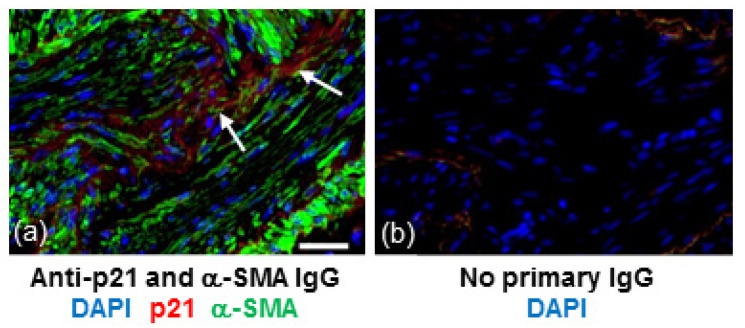
Figure 2.
p21 is detected in areas of α-SMA positive cells within fibrotic regions of IPF lung. (a) Section of lung tissue from a patient with IPF immunostained for p21 (red) and α-SMA (green) and counterstained with DAPI (blue). Arrows show p21 immunoreactivity within clusters of α-SMA positive cells. (b) No IgG control. Scale bar = 25 μm.

