Abstract
Renal hypouricemia is a rare genetic disorder. Hypouricemia can present as renal stones or exercise-induced acute renal failure, but most cases are asymptomatic. Our previous study showed that two recessive variants of SLC22A12 (p.Trp258*, pArg90His) were identified in 90% of the hypouricemia patients from two independent cohorts: the Korean genome and epidemiology study (KoGES) and the Korean Cancer Prevention Study (KCPS-II). In this work, we investigate the genetic causes of hypouricemia in the rest of the 10% of unsolved cases. We found a novel non-synonymous mutation of SLC2A9 (voltage-sensitive uric acid transporter) in the whole-exome sequencing (WES) results. Molecular dynamics prediction suggests that the novel mutation p.Met126Val in SLCA9b (p.Met155Val in SLC2A9a) hinders uric acid transport through a defect of the outward open geometry. Molecular analysis using Xenopus oocytes confirmed that the p.Met126Val mutation significantly reduced uric acid transport but does not affect the SLC2A9 protein expression level. Our results will shed light on a better understanding of SLC2A9-mediated uric acid transport and the development of a uric acid-lowering agent.
Keywords: SLC2A9, hypouricemia
1. Introduction
The homeostasis of serum uric acid (SUA) levels can be achieved by the dynamic processes of production and elimination. Hypouricemia, with a low uric acid level, is defined as an SUA concentration < 2 mg/dL. Hypouricemia is generally asymptomatic in the general population; most hypouricemia patients are identified by chance in regular health examinations [1]. Renal hypouricemia (RUHC) is a rare genetic disorder diagnosed by hypouricemia and increased fractional excretion of uric acid (UA > 10%) [2]. There are two types of RUHC: SLC22A12 mutations cause type 1 RHUC (OMIM: 220150), and SLC2A9 mutations cause type 2 RHUC (OMIM: 612076). Unlike hypouricemia, RHUC is sometimes accompanied by severe complications, such as exercise-induced acute kidney injury (EIAKI) and urolithiasis [3]. In the case of urolithiasis, RUHC is 6–7 times more prevalent than in those with normal levels of SUA [4].
The prevalence of hypouricemia is 0.41% in Koreans [5] which is similar to the prevalence in the Japanese (0.46%) [6]. This may be caused by a founder mutation, the protein-truncating p.Trp258* mutation of SLC22A12 [7]. Population-specific mutations have also been identified in different ethnic groups. Rare missense variants (p.R325W, p.R405C, and p.T467M) of SLC22A12 were reported in European and African American populations [8]. Various ethnic groups, including Israeli-Arab, Iraqi-Jewish, and Roma populations also harbor deleterious mutations of SLC22A12 and SLC2A9 [2,9,10,11,12,13].
In our previous study, approximately 90% of hypouricemia patients showed two variants in SLC22A12, p.W258* and p.Arg90His, in two independent cohorts, the Korean Genome and Epidemiology Study (KoGES, n = 179,318) and the Korean Cancer Prevention Study (KCPS-II, n = 156,701) [14]. However, the seven hypouricemia cases did not present the Asian-specific variants SLC22A12 p.W258* (rs121907892) and p.Arg90His (rs121907896), suggesting that other genes are also involved in the regulation of serum uric acid (SUA) levels.
In this study, we identify a novel variant in SLC2A9 by whole-exome sequencing (WES) of the unsolved cases of hypouricemia. SLC2A9 was initially identified as a glucose transporter that regulates the homeostasis of glucose levels. However, recent studies have demonstrated that SLC2A9 transports uric acid, and that genetic mutations in SLC2A9 have been linked to hyperuricemia and gout [15,16]. Our in silico and in vitro assay suggest that the novel mutation p.Met126Val in SLCA9b significantly reduces urate transport, resulting in hypouricemia.
2. Methods
2.1. Study Participants
This study was approved by the institutional review board of Sungkyunkwan University (IRB# SKKU 2017-12-007) and the Korean Cancer Prevention Study (KCPS-II) cohort from the Severance Hospital, Seoul, Korea (IRB#4-2011-0277) (approval date: Date, February, 2011) [17]. Whole-exome sequencing (WES) was performed in 7 patients with unexplained hypouricemia.
2.2. DNA Preparation and Whole-Exome Sequencing
Genomic DNA was obtained from peripheral blood leukocytes. DNA quality and quantity were assessed by an OD260/280 ratio of 1.8–2.0, 1% agarose gel electrophoresis, and PicoGreen® dsDNA Assay (Invitrogen, Waltham, MA, USA). SureSelect sequencing libraries were prepared (Agilent SureSelect All Exon kit 50 Mb, Santa Clara, CA, USA) and the enriched library was sequenced using the HiSeq 2500 sequencing system (Illumina, San Diego, CA, USA). Image analysis and base calling were performed with the pipeline software using default parameters. Mapping was completed using the human reference genome assembly (GRCh37/hg19), and all variants were called and annotated using the CLC Genomic Workbench (version 9.0.1) software (QIAGEN bioinformatics, Redwood City, CA, USA).
2.3. WES Variant Filtering Analysis
The overall variant-identifying process referred to the standard guidelines of investigating variants for Mendelian disorders from WES data [18,19]. We performed the analysis, assuming an autosomal recessive or X-linked recessive pattern according to the observed inheritance mode in hereditary RHUC [20]. First, based on the prevalence of hypouricemia without other medical conditions, such as hypertension or diabetes mellitus (31/179,318), the Hardy-Weinberg equation was used to calculate the allele frequency threshold to 0.01 and we excluded variants with MAF > 1% in the dbSNP database (version 150), 1000 Genome Projects phase 3 data (2504 individuals), and Genome Aggregation Database (gnomAD, http://gnomad.broadinstitute.org/ accessed date: 27 August 2020) [21]. Second, variants present in the homozygous or hemizygous state in 46 healthy Koreans without hypouricemia were excluded. Third, non-synonymous variants, insertion/deletion (indel), or splice-site variants were selected. In the further analysis, we excluded single heterozygous variants so that homozygous variants and putative compound heterozygous variants finally remained. For males, hemizygous variants in the X chromosome were considered to be retained. The numbers of variants are listed in Table S3.
2.4. Direct Sanger Sequencing
Confirmation of called variants was conducted via direct Sanger sequencing. The DNA sequences spanning the variants were amplified using specific primers (Table S4) and sequenced using an Applied Biosystems genetic analyzer 3500XL (Applied Biosystems, Foster City, CA, USA).
2.5. In Silico Analysis of Novel Missense Variants and Molecular Dynamics
2.5.1. In Silico Prediction
Prior to the analysis, known pathogenic variants of SLC2A9 were screened in the Human Gene Mutation Database (HGMD). For the newly discovered missense SLC2A9 and other candidate variants, we queried whether mutated amino acid residues were highly conserved across the vertebrate orthologs, using the UCSC Genome Browser (https://genome.ucsc.edu/; accessed date: 27 August 2020). Given the functional role of nitrogen excretion in the evolutionary process, we identified amino acid sequences in several mammals (Rhesus macaque, Mus musculus and Canis lupus familiaris) Third, the prediction of the functional effect of missense variants was performed using the latest version of PolyPhen-2 (http://genetics.bwh.harvard.edu/pph2; accessed date: 27 August 2020), SIFT (sorting intolerant from tolerant, http://sift.jcvi.org; accessed date: 27 August 2020), Condel (consensus deleteriousness score of non-synonymous single nucleotide variants, http://bbglab.irbbarcelona.org/fannsdb; accessed date: 27 August 2020), and the mutation taster (http://www.mutationtaster.org; accessed date: 27 August 2020) algorithms [22,23,24,25].
2.5.2. Molecular Dynamics
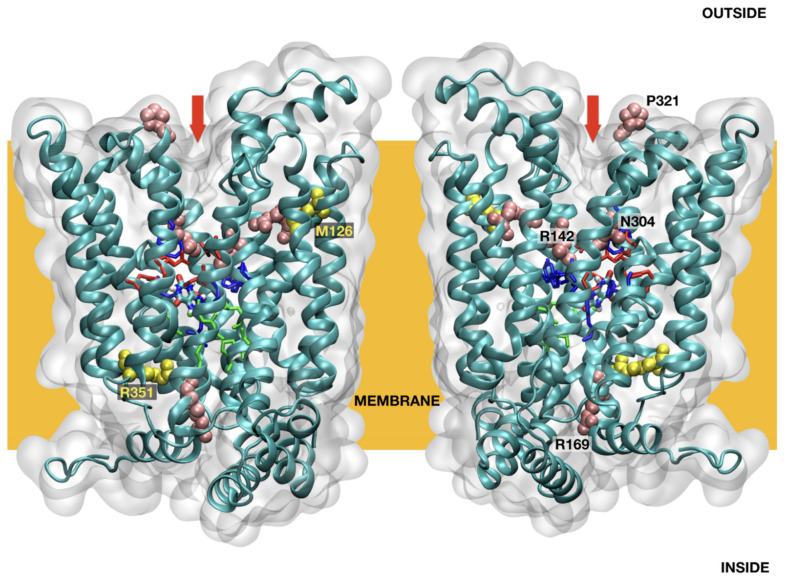
All homology models of SLC2A9 were combined using feedback restrained molecular dynamics [26,27] to form a consensus model (Figure 1). FRMD affords a simple protocol to maximally retain structural features during a molecular dynamics trajectory while minimizing the distortions imposed by an external restraint. All molecular dynamics calculations were performed with NAMD, using an Amberff99SB force field in the NVT ensemble at typical settings (T = 298 K, 2fs integration time, 12A cutoffs), as obtained using QwikMD in VMD with default parameters to prepare the input files [28]. The molecular dynamics results reported are from 125 ns trajectories unless otherwise stated. The overall organization of SLC2A9 is similar to that described by Clemencon [29] (Figure 1). The mutation sites do not cluster in any obvious arrangement or loci, nor do the mutated residues appear to follow a simple distribution pattern (Figure 1). A qualitative evaluation of the mutation effect was made, based on some simple criteria. Mutations affecting the binding site or the entry/exit channel section of the model suggest a direct effect on transport. Mutations resulting in structural changes may affect transport indirectly, by either changing the shape of the transporter or impending its dynamic rearrangement as required for transport (Table 1). The structural effect was evaluated as an increase in the root mean square displacement (RMSD) deviation, computed during 25 ns of molecular dynamics (after 25 ns of equilibration), measured against the conformations obtained during a 25 ns trajectory for the initial sequence.
Figure 1.
SLC2A9b model structure overview.
Table 1.
Demographic characteristics.
| Characteristics | Unexplained Group | SLC2A9 Compound Heterozygote |
|---|---|---|
| n = 6 | n = 1 | |
| Age (years) | 43 ± 12 | 40 |
| BMI † (kg/m2) | 25.1 ± 2.9 | 23.8 |
| Waist circumference, cm | 81 ± 5 | 72 |
| Blood pressure, mmHg | ||
| Systolic | 110 ± 3 | 124 |
| Diastolic | 71 ± 12 | 64 |
| Smoking status | ||
| Never a smoker, no. (%) | 1 (16.67) | 1 (100) |
| Ever a smoker, no. (%) | 5 (83.33) | 0 (0) |
| Alcohol consumption | ||
| Never a drinker, no. (%) | 2 (33.33) | 1 (100) |
| Ever a drinker, no. (%) | 4 (66.67) | 0 (0) |
| Uric acid, mg/dL | 0.78 ± 0.52 | 0.80 |
| Total cholesterol, mg/dL | 214 ± 34 | 174 |
| Triglycerides, mg/dL | 169 ± 69 | 178 |
| Fasting glucose, mg/dL | 86 ± 14 | 92 |
| LDL cholesterol, mg/dL | 116 ± 22 | 90.4 |
| HDL cholesterol, mg/dL | 64 ± 18 | 48 |
| Creatinine, mg/dL | 0.80 ± 0.25 | 0.70 |
Values are mean ± standard deviation (SD) for continuous data. † The body mass index (BMI) was calculated as weight in kilograms divided by height in meters squared.
2.5.3. Molecular/Functional Studies
Generation of SLC2A9b Expression Vectors
The recombinant plasmid SLC2A9b-eGFP/pcDNA-DEST47 [30] was a gift from Wolf Frommer (Addgene plasmid # 18730; http://n2t.net/addgene:18730, accessed date: 27 August 2020; RRID:Addgene_18730). To generate the SLC2A9b-V5 expression vector, the SLC2A9b-V5 DNA fragment was inserted into the pCS107 vector after PCR amplification with appropriate primers as follows, with SLC2A9b forward: 5′-GGCCATCGATAGCC ACCATGAAGCTCAGTAAAAAGGACCGAGGAGAAGATGAAGAAAGTGATTCAGCG-3′; and SLC2A9-V5 reverse: 5′-GCCTGCGGCCGCTTACGTAGAATCGAGACCGAGGAGAGGGTTAGGGATAGGC TTACCAG GCCTTCCATTTATCTTACCATCAG-3′. The amplified DNAs were assembled into the pCS107 vector using T4 DNA Ligase (M0202S, NEB) after ClaI and NotI endonuclease treatment. Then, 10 µL of the ligase reaction mixture was transformed into 120 µL of chemically competent DH5α (18258012; Thermo Fisher Scientific, Waltham, MA, USA) cells and screened on ampicillin-containing LB plates.
Site-Directed Mutagenesis for the Met126Val Mutant
To generate a site-specific point-mutation of SLC2A9b-Met126Val, the QuikChange II Site-Directed Mutagenesis Kit (200524; Agilent Technologies, Santa Clara, CA, USA) was used with appropriate primers, as follows, with Met126Val forward: 5′-GAGCGAGCAGGCCACCAGCAATGCAGCAG-3′; Met126Val reverse: 5′-CTGCTGCATTGCTGGTGGCCTGCTCGCTC-3′. Confirmation of the introduction of the Met126Val mutation into the vector was confirmed by Sanger sequencing. Plasmid DNA was purified for transfection and oocyte experiments using a GenElute endotoxin-free plasmid maxiprep kit (NA0310; Sigma-Aldrich, Burlington, MA, USA).
In Vitro Transcription
All mutants and wild-type cDNAs were linearized by Asp718 (Roche). In vitro transcription was performed with SP6 mMESSAGE mMACHINE Kit (AM1340, Thermo Fisher Scientific, Waltham, MA, USA).
SLC2A9b Expression in X. laevis Oocytes
Stages V–VI oocytes were collected from X. laevis (Nasco, Chicago, IL, USA). Oocytes were injected with 10 ng of either wild-type or mutant RNAs or the equivalent volume of water, and incubated for 48 h in oocyte medium (50% L-15 + glutamine, 40% HEPES/insulin stock, 10% fetal calf serum, 100 mg/mL gentamycin). Animal care and use for this study were performed in accordance with the recommendations of AAALAC for the care and use of laboratory animals in an AAALAC-approved facility. Experimental procedures were specifically approved by the animal care and use committee of the National Cancer Institute-Frederick ASP #18-433, in compliance with AAALAC guidelines.
Western Blot Analysis
Using ten oocytes of the experimental groups, lysates were prepared with ice-cold TNSG buffer (20 mM Tris-HCl pH 7.5, 137 mM NaCl and 1% NP-40). The lysates were separated on a 10% SDS polyacrylamide gel and transferred onto a PVDF membrane (88025, Thermo Fisher Scientific, Waltham, MA, USA). The membranes were incubated overnight with anti-mouse V5-HRP monoclonal antibody (R961-25, Thermo Fisher Scientific, Waltham, MA, USA) (1:2000) after blocking with 10% skim milk. The SLC2A9b signal was revealed by ECL (32106, Thermo Fisher Scientific, Waltham, MA, USA) and exposed on Kodak films.
Uric Acid Uptake Assay
10 ng mRNA of SLC2A9-wild type or mutants were injected into the cytoplasm, at the midline of stage VI Xenopus laevis oocytes defolliculated with collagenase (2.5 mg/mL). Two days after injection, we performed the uric acid uptake assay at room temperature in ND-96 buffer (96 mM NaCl, 2 mM KCl, 1.8 mM CaCl2, 1 mM MgCl2, 5 mM Hepes, pH 7.4) for 120 min. We used 100 mM [14C] of uric acid (ARC 0513A-50 µCi, American Radiolabeled Chemicals, St. Louis MO, USA) to assess urate uptake. Following five washes in ND-96 lacking radiolabel, ten oocytes of experimental groups were collected, lysed in 200 mL of 10% SDS, and subjected to scintillation counting. We harvested ten pools of ten oocytes per experimental point. Statistical data analyses were performed using Prism 8.
Confocal Microscopy
Briefly, the oocytes were fixed in 4% paraformaldehyde in PBS overnight at 4 °C. The oocytes were then embedded in 4% low-melting agarose gel and were sectioned to a thickness of 100 μm with the vibratome (LEICA VT 1200S). The primary antibodies, anti-V5 (1:500, G189, ABM) and the secondary antibodies, anti-mouse Alexa Fluor 594 (A32744, Invitrogen) were incubated at 4 °C overnight. The samples were washed, mounted and imaged using a Zeiss LSM-880 laser-scanning confocal microscope.
3. Results
3.1. Demographics
Baseline patient characteristics are summarized in Table 1. The 7 sequenced participants had unsolved hypouricemia (UA 0.79 ± 0.47 mg/dL; age 42 ± 11 years; BMI 24.9 ± 2.7 kg/m2; total cholesterol level 209 ± 35 mg/dL) that were not due to known causes (e.g., SUA-lowering drugs).
3.1.1. Identification of Novel Variants in SLC2A9b by Whole-Exome Sequencing
WES analysis was performed on 7 subjects identified from the KoGES and KCPS-II cohorts as previously described [14], with an average depth of coverage of 85-fold. We performed variant calling and downstream filtering analyses, assuming an autosomal recessive inheritance or sex-linked hemizygous patterns. One individual (NIH17A8568242) carried compound heterozygous variants (p.Met155Val (c.463A > G, exon 5), p.Arg380Gly (c.1138C > T, exon 10)) of SLC2A9a (long form). The corresponding residues for SLC2A9b (short form) are p.Met126Val and p.Arg351Gly, respectively. p.Arg380Gly was previously reported by HGMD (The Human Gene Mutation Database). The p.Met155Val variant was confirmed by Sanger sequencing (Supplementary Figure S1). The global minor allele frequency of the novel variant is 0.000022 at the gnomAD database (http://gnomad.broadinstitute.org; accessed date: 27 August 2020).
In the remaining six individuals, we found 12 candidate genes for unexplained cases (8 genes for homozygous: ATP8B2, KRTAP5-8, PIK3CB, ASIC3, ADAM8, RBM12, PWWP2B, SULT1A2; 4 genes for hemizygous: ASB12, RLIM, GPR101, PPEF1) The possible disease-causing variants are listed in Supplementary Table S1 (recessive mode). In a systematic review, most of the genes were not found to be involved in biological pathways affecting UA levels. However, the p.Arg78His variant (rs145118752) of ASB12 on chromosome X, which was discovered in two cases, NIH17A8004492 and YID182829, was found in 0.018% of the global population and 0.16% when limited to East Asia only (gnomAD 2020.11, http://gnomad.broadinstitute.org/; accessed date: 27 August 2020). We also found that these two individuals are unrelated (kinship coefficient = 0.001).
3.1.2. In Silico and Molecular Dynamics Prediction of SLC2A9b
The functional prediction for the novel variant of SLC2A9b, p.Met126Val, is predicted as pathogenic by using mutation taster, PolyPhen-2, SIFT and CADD (disease-causing, damaging, deleterious, and 18.24, respectively). The amino acid is highly conserved across vertebrate species down to zebrafish.
3.1.3. Molecular Dynamics Prediction of SLC2A9b and Its Affinity for Uric Acid
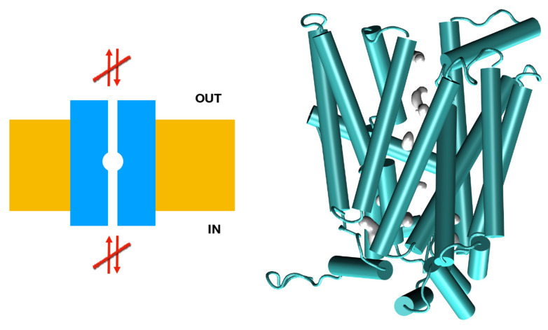
The consequence of the amino acid substitution in SLC2A9b was investigated using a molecular dynamic prediction analysis (Figure 1). Molecular dynamics simulations of the p.Met126Val mutant model suggest the mutation of Met126 to Val126 results in compacting of the helical bundle, resulting in an extremely stable arrangement, with RMSF values for the region surrounding the Val126 residue being 30% lower than those observed in the reference model (Figure 2). This stable arrangement may explain the unexpectedly large effect of this seemingly inconsequential mutation in the transporter’s overall function, stiffening the vestibular areas occluding the binding pocket, and resulting in a less functional rocker structure.
Figure 2.
Mechanistic interpretation of the effects of the M126V mutation of SLC2A9b. The M126V model suggests that this mutation renders the vestibular regions unavailable. Left: schematic representation of the channel (in blue), membrane (yellow), and flow (arrows). Right: a snapshot of the M126V model during an MD trajectory, in cartoon representation in green. Internal space is represented by showing the solvent’s accessible surface in gray.
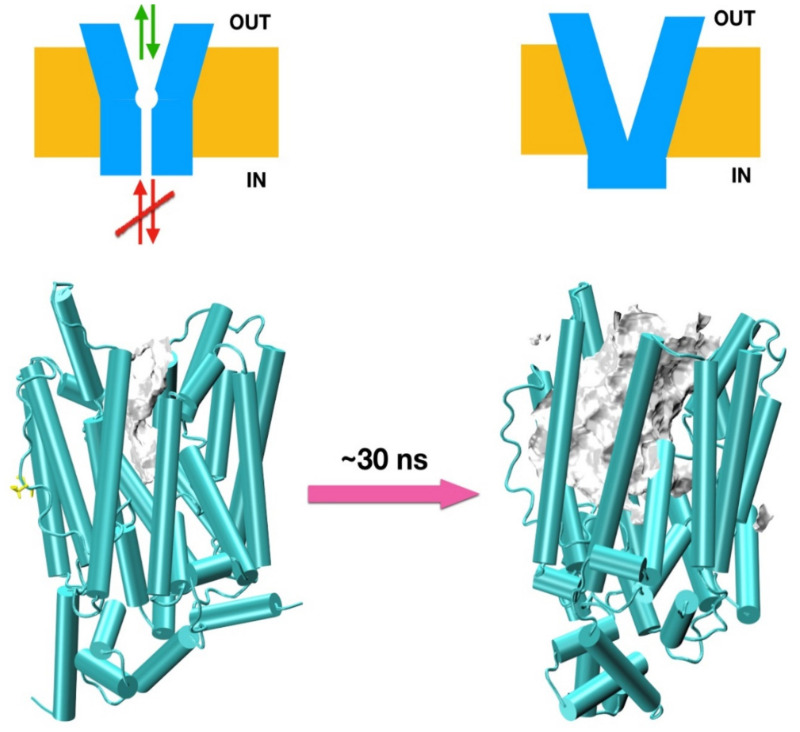
Molecular dynamics simulations of the p.Arg351Trp model suggest the mutation of Arg351 to Trp351 breaks a well-structured chain of 12 charged resides including Lys, Arg, Tyr, and Glu spanning over 20 A (Figure 3) and stabilizing the intracellular domain. This polar structure plays a crucial role in directing anions to the intracellular vestibular area. The p.Arg351Trp mutation affects the inside binding site, decreasing the ∆B1 binding energy of urate to −1.2 Kcal/mol, possibly affecting urate transport. The dislodging of this domain may have an impact on the binding of external effectors as well. Energy barriers for 2 variants are illustrated in Table S2.
Figure 3.
Effects of the R351W mutation of SLC2A9b. Top: schematic representation of the channel (in blue), membrane (yellow), and flow (arrows). Bottom: snapshots of the R351W model during an MD trajectory in cartoon representation in green. Internal space is represented by showing the solvent accessible surface in gray (compared to similar surfaces describing the vestibular areas in Figure 2). The initial model structure is surprisingly stable, but it deforms under molecular dynamics simulations, as seen in the snapshot on the right after ~30 ns of MD trajectory. Notice the very substantial reorganization of the internal helices.
3.2. Molecular Analysis
3.2.1. SLC2A9b-p.Met126Val Expression Analysis in X. laevis Oocytes
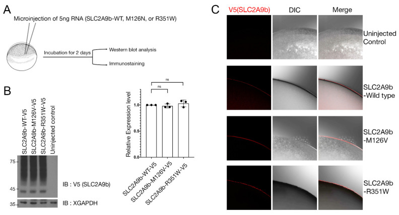
SLC2A9 has two isoforms, a long isoform (SLC2A9a) and a short isoform (SLC2A9b), that differ in their N-terminal region and plasma membrane localization. SLC2A9a localizes to the basolateral side of the plasma membrane, while SLC2A9b traffics to the apical side [31,32]. Previous studies have demonstrated that the basolateral SLC2A9a is involved in uric acid efflux, while the apical SLC2A9b plays a role in uric acid absorption [33,34,35]. To investigate whether the novel exonic mutation, p.Met126Val, affects the molecular function of SLC2A9b, the oocyte expression system was utilized. Xenopus oocytes are known as an excellent tool to study the molecular function of SLC2A9 [29]. We generated the V5 tagged as the SLC2A9b p.Met126Val variant, corresponding to the p.Met155Val of SLC2A9a. One of the well-known exonic variants, p.Arg351Trp, corresponding to p. Arg380Trp of the SLC2A9a, was employed as a positive control [36]. To examine whether the p.Met126Val exonic mutation causes any change in the expression level of SLC2A9b. mRNAs of the SLC2A9b wild-type, M126V, and R351W were generated by in vitro transcription, and then, 5ng of each mRNA were injected into Xenopus oocytes. After 2 days of incubation to allow the translation of injected mRNA, Western blot analysis and immunostaining were performed (Figure 4A). Western blot analysis revealed no difference in the protein expression level of the exonic variants compared to the wild-type SLC2A9b (Figure 4B). Since SLC2A9 is a membrane protein, and the plasma membrane localization influences the SLC2A9 function, we investigated the subcellular localization of the SLC2A9b p.Met126Val variant using immuno-staining. Confocal microscopy analysis revealed that SLC2A9b-WT, SLC2A9b-p.Met126Val-V5 and SLC2A9b-p.Arg351Trp-V5 mutants were localized at the plasma membrane in Xenopus oocytes (Figure 4C).
Figure 4.
Expression of WT and mutant SLC2A9b in Xenopus oocytes. (A) Schematic representation of the experimental procedure. The same amount of wild-type or mutants SLC2A9b RNAs were injected into oocytes. The oocytes were harvested after 2 days and then, Western blot analysis or immunostaining was performed. (B) SLC2A9b wild type and mutants showed similar protein expression levels. The histogram depicts the relative protein expression level (n = 3). Quantification with one-way ANOVA (Dunnett’s multiple comparisons test), p = 0.4363. Data represent the mean ± S.D. of three individual experiments. ns: no statistical differences between the groups. (C) Immunostaining was performed using anti-V5 antibodies. Both WT and mutants SLC2A9b showed plasma membrane localization in Xenopus oocytes. DIC; differential interference contrast image.
Our results suggest that the novel exonic mutation, p.Met126Val, shows similar protein expression level subcellular localization compared to the wild type.
3.2.2. Urate Transport Activity of SLC2A9b-p.Met126Val in Xenopus Oocytes
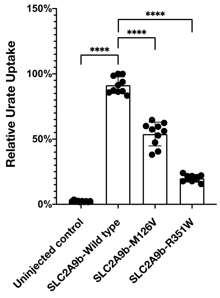
Next, we analyzed urate transport activity using [14C]-uric acid as a substrate in Xenopus oocytes. Our result showed that the well-known exonic mutation, p.Arg351Trp, decreased uric acid transport activity by 70% compared to the wild type. Interestingly, the p.Met126Val mutation also reduced uric acid transport activity by 45% at 100 mM [14C]-uric acid concentration (Figure 5).
Figure 5.
Met126Val mutation reduces uric acid uptake. Uric acid uptake assay was performed as described in the Methods section. M126V mutation in SLC2A9b reduced uric acid uptake by 45% and R351W mutation decreased by 70%. Histogram depicts relative urate uptake level (n = 10). Quantification with one-way ANOVA (Dunnett’s multiple comparisons test), **** p < 0.0001. Data represent the mean ± S.D. of three individual experiments. **** p < 0.0001.
Our result suggests that our novel exonic mutation, p.Met126Val, may also contribute to hypouricemia.
4. Discussion
In this study, we comprehensively evaluated the contribution of SLC2A9 to severe hypouricemia by first identifying variant (p.Met126Val) using WES, followed by molecular dynamics prediction and functional validation.
With regard to SLC2A9b, the p.Met126Val variant was identified in the case of NIH17A8568242 as a compound heterozygote with p.Arg351Trp. Molecular dynamics analysis supported a loss-of-function role considering its RMSD value reflecting structural changes in protein flexibility. Two missense mutations (p.Arg351Trp, rs121908321, and p.Arg169Cys, rs121908322 of SLC2A9b) are well-documented as causal for type 2 RHUC. Our experiments with Xenopus oocytes showed that p.Met155Val for the SLC2A9a (p. Met126Val for SLC2A9b) variant in SLC2A9 causes a defect in uric acid transport. This is consistent with the individual who presented SUA levels that were near 0. SLC2A9 is the most frequently reported gene associated with SUA levels, along with ABCG2, in GWAS studies of hyperuricemia and gout [37]. Intronic SNPs (rs4529048, rs7674711, and rs11936395) of SLC2A9 have been associated with both increased SUA levels and increased risk of gout [38,39]. However, the missense variant (p.Val253Ile, rs16890979) of SLC2A9 has been reported both as a protective SNP for gout and in lower UA levels [40,41]. Moreover, SLC2A9 showed a statistically significant gene–gene interaction, with variants in the intergenic region located 80 kb downstream (WDR1-ZNF518B) [42]. A comprehensive study is needed to evaluate the effect of different transcriptional factors and the variation in regulatory elements on the gene expression of SLC2A9. Recently, large-scale WES using 19,517 participants (15,821 of European ancestry and 3696 of African ancestry) identified variants of SLC22A12 and SLC2A9 that were associated with lower levels of SUA. Identified polymorphisms in uric acid transporter genes associated with lowering UA differ by ethnic group, due to a combination of founder effects, population isolation, and random drift. Collaborative international research with established cohorts, with GWAS and SUA measures, using a multi-ethnic approach is needed to explain the missing heritability of SUA and to further our understanding of the genetic architecture of SUA levels.
The two isoforms of SLC2A9 differ at their N-terminal regions due to binding by the transcriptional factors to different promoters [35]. Both isoforms increase uric acid uptake when overexpressed in HEK293 cells and Xenopus laevis oocytes, with a peak UA uptake as early as 20 min upon uric acid incubation [43]. The overexpression of the SLC2A9 mutant isoforms SLC2A9a-Leu75Arg (SLC2A9b-Leu46Arg) in oocytes resulted in a reduced uric acid uptake when compared to the reference protein [43]. However, SLC2A9b-Leu46Arg showed a greater reduction (80% decrease) in UA uptake than that of SLC2A9a-Leu75Arg (60%) [35]. Our in vitro studies show that the overexpression of the SLC2A9b-reference is sufficient to raise the intracellular concentration of UA when treated for 2 h. Throughout both of our in silico and in vitro studies, we showed that p.Met126Val of SLC2A9b is a causing variant for hypouricemia. One limitation of our approach is that we could not evaluate the change of uric acid binding affinity once it was transported into the cell. What we measured was the final concentration of uric acid at the equilibrium point. The change of binding affinity inside the membrane can be answered in future studies using a patch-clamp and electrophysiological evaluation.
We also identified two males with extreme hypouricemia who carried the X-linked ASB12 variant of unknown significance. Little is known about the functional significance of this gene for uric acid transport. Since it is postulated that the ASB family may be involved in protein degradation via mediating the ubiquitin-proteasome system or signal transduction [44], it may be involved in the trafficking or intracellular degradation of the UA transporter. One limitation of this study is that family members were not available for the analysis of genotype-phenotype segregation through multiple generations. Given that known causative genes for hypouricemia remain unidentified, the genetic inheritance of hypouricemia could be more common than was indicated by our results.
5. Conclusions
We described the clinical and molecular characteristics of hypouricemia, caused by compound heterozygous mutations of SLC2A9. Our clinical and molecular findings may contribute to the understanding of the physiology of renal uric acid. We also proposed candidate genes for hypouricemia from unexplained cases for further study.
Acknowledgments
The bioresources for this study were provided by the National Biobank of Korea, Centers for Disease Control and Prevention, Republic of Korea. We appreciate Young Sup Cho and Hyekyung Son for their academic advice during this project. The content of this publication does not necessarily reflect the views or policies of the Department of Health and Human Services, nor does the mention of trade names, commercial products, or organizations imply endorsement by the U.S. Government.
Supplementary Materials
The following are available online at https://www.mdpi.com/article/10.3390/biomedicines9091172/s1, Supplementary Table S1: Possible variants identified in 6 individuals with hypouricemia by WES; Supplementary Table S2: Predicted functional impacts of amino acid changes of SLC2A9b; Supplementary Table S3: Variant filtering process or unsolved cases; Supplementary Table S4: Primer information for rs369512758 (SLC2A9); Supplementary Figure S1: Sanger confirmation of the novel variant p.M126V of SLC2A9b.
Author Contributions
Conceptualization, S.-K.C., R.C., J.Y.; Validation, S.-K.C., R.C., J.Y.; Investigation, V.A.D., M.T., W.J., S.-H.J.; writing—original draft preparation, S.-K.C.; writing—review and editing, S.-K.C., J.Y.; supervision, I.O.D. and C.A.W. All authors have read and agreed to the published version of the manuscript.
Funding
This research was supported partly by the Intramural Research Program of the NIH, National Cancer Institute, Center for Cancer Research. This project has been funded in part with federal funds from the National Cancer Institute, National Institutes of Health, under contract HHSN26120080001E.
Institutional Review Board Statement
Not applicable.
Informed Consent Statement
Not applicable.
Data Availability Statement
Data presented in this study are available on request from the corresponding author.
Conflicts of Interest
The authors declare no conflict of interest.
Footnotes
Publisher’s Note: MDPI stays neutral with regard to jurisdictional claims in published maps and institutional affiliations.
References
- 1.Cho S.K., Chang Y., Kim I., Ryu S. U-Shaped Association Between Serum Uric Acid Level and Risk of Mortality: A Cohort Study. Arthritis Rheumatol. 2018;70:1122–1132. doi: 10.1002/art.40472. [DOI] [PubMed] [Google Scholar]
- 2.Sebesta I., Stiburkova B., Bartl J., Ichida K., Hosoyamada M., Taylor J., Marinaki A. Diagnostic tests for primary renal hypouricemia. Nucleosides Nucleic Acids. 2011;30:1112–1116. doi: 10.1080/15257770.2011.611483. [DOI] [PubMed] [Google Scholar]
- 3.Cheong H.I., Kang J.H., Lee J.H., Ha I.S., Kim S., Komoda F., Sekine T., Igarashi T., Choi Y. Mutational analysis of idiopathic renal hypouricemia in Korea. Pediatric Nephrol. 2005;20:886–890. doi: 10.1007/s00467-005-1863-3. [DOI] [PubMed] [Google Scholar]
- 4.Ichida K., Hosoyamada M., Hisatome I., Enomoto A., Hikita M., Endou H., Hosoya T. Clinical and molecular analysis of patients with renal hypouricemia in Japan-influence of URAT1 gene on urinary urate excretion. J. Am. Soc. Nephrol. 2004;15:164–173. doi: 10.1097/01.ASN.0000105320.04395.D0. [DOI] [PubMed] [Google Scholar]
- 5.Cho S.K., Kim B., Myung W., Chang Y., Ryu S., Kim H.N., Kim H.L., Kuo P.H., Winkler C.A., Won H.H. Polygenic analysis of the effect of common and low-frequency genetic variants on serum uric acid levels in Korean individuals. Sci. Rep. 2020;10:9179. doi: 10.1038/s41598-020-66064-z. [DOI] [PMC free article] [PubMed] [Google Scholar]
- 6.Kawasoe S., Ide K., Usui T., Kubozono T., Yoshifuku S., Miyahara H., Maenohara S., Ohishi M., Kawakami K. Distribution and Characteristics of Hypouricemia within the Japanese General Population: A Cross-Sectional Study. Medicina. 2019;55:61. doi: 10.3390/medicina55030061. [DOI] [PMC free article] [PubMed] [Google Scholar]
- 7.Enomoto A., Kimura H., Chairoungdua A., Shigeta Y., Jutabha P., Cha S.H., Hosoyamada M., Takeda M., Sekine T., Igarashi T., et al. Molecular identification of a renal urate anion exchanger that regulates blood urate levels. Nature. 2002;417:447–452. doi: 10.1038/nature742. [DOI] [PubMed] [Google Scholar]
- 8.Tin A., Li Y., Brody J.A., Nutile T., Chu A.Y., Huffman J.E., Yang Q., Chen M.H., Robinson-Cohen C., Mace A., et al. Large-scale whole-exome sequencing association studies identify rare functional variants influencing serum urate levels. Nat. Commun. 2018;9:4228. doi: 10.1038/s41467-018-06620-4. [DOI] [PMC free article] [PubMed] [Google Scholar]
- 9.Stiburkova B., Taylor J., Marinaki A.M., Sebesta I. Acute kidney injury in two children caused by renal hypouricaemia type 2. Pediatric Nephrol. 2012;27:1411–1415. doi: 10.1007/s00467-012-2174-0. [DOI] [PubMed] [Google Scholar]
- 10.Dinour D., Bahn A., Ganon L., Ron R., Geifman Holtzman O., Knecht A., Gafter U., Rachamimov R., Sela B., Burckhardt G., et al. URAT1 mutations cause renal hypouricemia type 1 in Iraqi Jews. Nephrol. Dial. Transplant. 2011;26:2175–2181. doi: 10.1093/ndt/gfq722. [DOI] [PubMed] [Google Scholar]
- 11.Tasic V., Hynes A.M., Kitamura K., Cheong H.I., Lozanovski V.J., Gucev Z., Jutabha P., Anzai N., Sayer J.A. Clinical and functional characterization of URAT1 variants. PLoS ONE. 2011;6:e28641. doi: 10.1371/journal.pone.0028641. [DOI] [PMC free article] [PubMed] [Google Scholar]
- 12.Stiburkova B., Sebesta I., Ichida K., Nakamura M., Hulkova H., Krylov V., Kryspinova L., Jahnova H. Novel allelic variants and evidence for a prevalent mutation in URAT1 causing renal hypouricemia: Biochemical, genetics and functional analysis. Eur. J. Hum. Genet. 2013;21:1067–1073. doi: 10.1038/ejhg.2013.3. [DOI] [PMC free article] [PubMed] [Google Scholar]
- 13.Bhasin B., Stiburkova B., De Castro-Pretelt M., Beck N., Bodurtha J.N., Atta M.G. Hereditary renal hypouricemia: A new role for allopurinol? Am. J. Med. 2014;127:e3–e4. doi: 10.1016/j.amjmed.2013.08.025. [DOI] [PubMed] [Google Scholar]
- 14.Cha D.H., Gee H.Y., Cachau R., Choi J.M., Park D., Jee S.H., Ryu S., Kim K.K., Won H.H., Limou S., et al. Contribution of SLC22A12 on hypouricemia and its clinical significance for screening purposes. Sci. Rep. 2019;9:14360. doi: 10.1038/s41598-019-50798-6. [DOI] [PMC free article] [PubMed] [Google Scholar]
- 15.Cho S.K., Winkler C.A., Lee S.J., Chang Y., Ryu S. The Prevalence of Hyperuricemia Sharply Increases from the Late Menopausal Transition Stage in Middle-Aged Women. J. Clin. Med. 2019;8:296. doi: 10.3390/jcm8030296. [DOI] [PMC free article] [PubMed] [Google Scholar]
- 16.Merriman T.R. An update on the genetic architecture of hyperuricemia and gout. Arthritis Res. Ther. 2015;17:98. doi: 10.1186/s13075-015-0609-2. [DOI] [PMC free article] [PubMed] [Google Scholar]
- 17.Cho S.K., Kim S., Chung J., Jee S.H. Discovery of URAT1 SNPs and association between serum uric acid levels and URAT1. BMJ Open. 2015;5:e009360. doi: 10.1136/bmjopen-2015-009360. [DOI] [PMC free article] [PubMed] [Google Scholar]
- 18.MacArthur D.G., Manolio T.A., Dimmock D.P., Rehm H.L., Shendure J., Abecasis G.R., Adams D.R., Altman R.B., Antonarakis S.E., Ashley E.A., et al. Guidelines for investigating causality of sequence variants in human disease. Nature. 2014;508:469–476. doi: 10.1038/nature13127. [DOI] [PMC free article] [PubMed] [Google Scholar]
- 19.Yang Y., Muzny D.M., Reid J.G., Bainbridge M.N., Willis A., Ward P.A., Braxton A., Beuten J., Xia F., Niu Z., et al. Clinical whole-exome sequencing for the diagnosis of mendelian disorders. N. Engl. J. Med. 2013;369:1502–1511. doi: 10.1056/NEJMoa1306555. [DOI] [PMC free article] [PubMed] [Google Scholar]
- 20.Sperling O. Hereditary renal hypouricemia. Mol. Genet. Metab. 2006;89:14–18. doi: 10.1016/j.ymgme.2006.03.015. [DOI] [PubMed] [Google Scholar]
- 21.Kim Y., Han B.G. Cohort Profile: The Korean Genome and Epidemiology Study (KoGES) Consortium. Int. J. Epidemiol. 2017;46:1350. doi: 10.1093/ije/dyx105. [DOI] [PMC free article] [PubMed] [Google Scholar]
- 22.Adzhubei I.A., Schmidt S., Peshkin L., Ramensky V.E., Gerasimova A., Bork P., Kondrashov A.S., Sunyaev S.R. A method and server for predicting damaging missense mutations. Nat. Methods. 2010;7:248–249. doi: 10.1038/nmeth0410-248. [DOI] [PMC free article] [PubMed] [Google Scholar]
- 23.Kumar P., Henikoff S., Ng P.C. Predicting the effects of coding non-synonymous variants on protein function using the SIFT algorithm. Nat. Protoc. 2009;4:1073–1081. doi: 10.1038/nprot.2009.86. [DOI] [PubMed] [Google Scholar]
- 24.Gonzalez-Perez A., Lopez-Bigas N. Improving the assessment of the outcome of nonsynonymous SNVs with a consensus deleteriousness score, Condel. Am. J. Hum. Genet. 2011;88:440–449. doi: 10.1016/j.ajhg.2011.03.004. [DOI] [PMC free article] [PubMed] [Google Scholar]
- 25.Schwarz J.M., Cooper D.N., Schuelke M., Seelow D. MutationTaster2: Mutation prediction for the deep-sequencing age. Nat. Methods. 2014;11:361–362. doi: 10.1038/nmeth.2890. [DOI] [PubMed] [Google Scholar]
- 26.Yokoyama S., Cai Y., Murata M., Tomita T., Yoneda M., Xu L., Pilon A.L., Cachau R.E., Kimura S. A novel pathway of LPS uptake through syndecan-1 leading to pyroptotic cell death. eLife. 2018;7:e37854. doi: 10.7554/eLife.37854. [DOI] [PMC free article] [PubMed] [Google Scholar]
- 27.Cachau R.E., Erickson J.W., Villar H.O. Novel procedure for structure refinement in homology modeling and its application to the human class Mu glutathione S-transferases. Protein Eng. 1994;7:831–839. doi: 10.1093/protein/7.7.831. [DOI] [PubMed] [Google Scholar]
- 28.Phillips J.C., Stone J.E., Vandivort K.L., Armstrong T.G., Wozniak J.M., Wilde M., Schulten K. Petascale Tcl with NAMD, VMD, and Swift/T; Proceedings of the 2014 First Workshop for High Performance Technical Computing in Dynamic Languages; New Orleans, LA, USA. 17 November 2014; pp. 6–17. [Google Scholar]
- 29.Clémençon B., Lüscher B.P., Fine M., Baumann M.U., Surbek D.V., Bonny O., Hediger M.A. Expression, purification, and structural insights for the human uric acid transporter, GLUT9, using the Xenopus laevis oocytes system. PLoS ONE. 2014;9:e108852. doi: 10.1371/journal.pone.0108852. [DOI] [PMC free article] [PubMed] [Google Scholar]
- 30.Takanaga H., Chaudhuri B., Frommer W.B. GLUT1 and GLUT9 as major contributors to glucose influx in HepG2 cells identified by a high sensitivity intramolecular FRET glucose sensor. Biochim. Biophys. Acta. 2008;1778:1091–1099. doi: 10.1016/j.bbamem.2007.11.015. [DOI] [PMC free article] [PubMed] [Google Scholar]
- 31.Augustin R., Carayannopoulos M.O., Dowd L.O., Phay J.E., Moley J.F., Moley K.H. Identification and characterization of human glucose transporter-like protein-9 (GLUT9): Alternative splicing alters trafficking. J. Biol. Chem. 2004;279:16229–16236. doi: 10.1074/jbc.M312226200. [DOI] [PubMed] [Google Scholar]
- 32.Mandal A.K., Mercado A., Foster A., Zandi-Nejad K., Mount D.B. Uricosuric targets of tranilast. Pharm. Res. Perspect. 2017;5:e00291. doi: 10.1002/prp2.291. [DOI] [PMC free article] [PubMed] [Google Scholar]
- 33.Anzai N., Ichida K., Jutabha P., Kimura T., Babu E., Jin C.J., Srivastava S., Kitamura K., Hisatome I., Endou H., et al. Plasma urate level is directly regulated by a voltage-driven urate efflux transporter URATv1 (SLC2A9) in humans. J. Biol. Chem. 2008;283:26834–26838. doi: 10.1074/jbc.C800156200. [DOI] [PubMed] [Google Scholar]
- 34.Matsuo H., Chiba T., Nagamori S., Nakayama A., Domoto H., Phetdee K., Wiriyasermkul P., Kikuchi Y., Oda T., Nishiyama J., et al. Mutations in glucose transporter 9 gene SLC2A9 cause renal hypouricemia. Am. J. Hum. Genet. 2008;83:744–751. doi: 10.1016/j.ajhg.2008.11.001. [DOI] [PMC free article] [PubMed] [Google Scholar]
- 35.Dinour D., Gray N.K., Campbell S., Shu X., Sawyer L., Richardson W., Rechavi G., Amariglio N., Ganon L., Sela B.A., et al. Homozygous SLC2A9 mutations cause severe renal hypouricemia. J. Am. Soc. Nephrol. 2010;21:64–72. doi: 10.1681/ASN.2009040406. [DOI] [PMC free article] [PubMed] [Google Scholar]
- 36.Ruiz A., Gautschi I., Schild L., Bonny O. Human Mutations in SLC2A9 (Glut9) Affect Transport Capacity for Urate. Front. Physiol. 2018;9:476. doi: 10.3389/fphys.2018.00476. [DOI] [PMC free article] [PubMed] [Google Scholar]
- 37.Kottgen A., Albrecht E., Teumer A., Vitart V., Krumsiek J., Hundertmark C., Pistis G., Ruggiero D., O’Seaghdha C.M., Haller T., et al. Genome-wide association analyses identify 18 new loci associated with serum urate concentrations. Nat. Genet. 2013;45:145–154. doi: 10.1038/ng.2500. [DOI] [PMC free article] [PubMed] [Google Scholar]
- 38.Sull J.W., Park E.J., Lee M., Jee S.H. Effects of SLC2A9 variants on uric acid levels in a Korean population. Rheumatol. Int. 2013;33:19–23. doi: 10.1007/s00296-011-2303-2. [DOI] [PubMed] [Google Scholar]
- 39.Vitart V., Rudan I., Hayward C., Gray N.K., Floyd J., Palmer C.N., Knott S.A., Kolcic I., Polasek O., Graessler J., et al. SLC2A9 is a newly identified urate transporter influencing serum urate concentration, urate excretion and gout. Nat. Genet. 2008;40:437–442. doi: 10.1038/ng.106. [DOI] [PubMed] [Google Scholar]
- 40.Meng Q., Yue J., Shang M., Shan Q., Qi J., Mao Z., Li J., Zhang F., Wang B., Zhao T., et al. Correlation of GLUT9 Polymorphisms with Gout Risk. Medicine. 2015;94:e1742. doi: 10.1097/MD.0000000000001742. [DOI] [PMC free article] [PubMed] [Google Scholar]
- 41.Dehghan A., Köttgen A., Yang Q., Hwang S., Kao W.L., Rivadeneira F., Boerwinkle E., Levy D., Hofman A., Astor B.C., et al. Association of three genetic loci with uric acid concentration and risk of gout: A genome-wide association study. Lancet. 2008;372:1953–1961. doi: 10.1016/S0140-6736(08)61343-4. [DOI] [PMC free article] [PubMed] [Google Scholar]
- 42.Wei W.H., Guo Y., Kindt A.S., Merriman T.R., Semple C.A., Wang K., Haley C.S. Abundant local interactions in the 4p16.1 region suggest functional mechanisms underlying SLC2A9 associations with human serum uric acid. Hum. Mol. Genet. 2014;23:5061–5068. doi: 10.1093/hmg/ddu227. [DOI] [PMC free article] [PubMed] [Google Scholar]
- 43.Caulfield M.J., Munroe P.B., O’Neill D., Witkowska K., Charchar F.J., Doblado M., Evans S., Eyheramendy S., Onipinla A., Howard P., et al. SLC2A9 is a high-capacity urate transporter in humans. PLoS Med. 2008;5:e197. doi: 10.1371/journal.pmed.0050197. [DOI] [PMC free article] [PubMed] [Google Scholar]
- 44.Kohroki J., Nishiyama T., Nakamura T., Masuho Y. ASB proteins interact with Cullin5 and Rbx2 to form E3 ubiquitin ligase complexes. FEBS Lett. 2005;579:6796–6802. doi: 10.1016/j.febslet.2005.11.016. [DOI] [PubMed] [Google Scholar]
Associated Data
This section collects any data citations, data availability statements, or supplementary materials included in this article.
Supplementary Materials
Data Availability Statement
Data presented in this study are available on request from the corresponding author.


