. 2021 Sep 14;302(2):460–469. doi: 10.1148/radiol.2021210986

Figure 1:

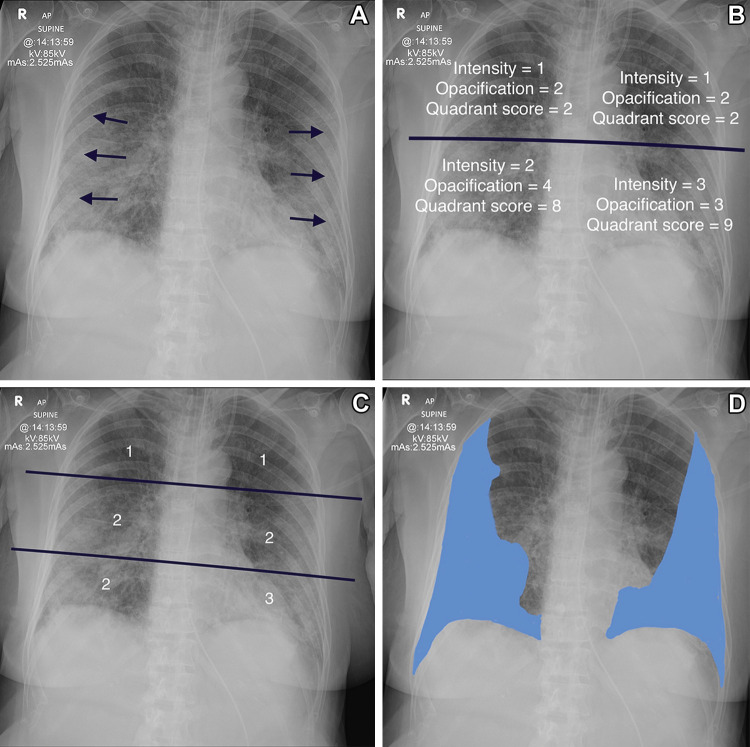
Determination of the various scoring systems. (A) Anteroposterior radiograph in 78-year-old woman shows the classic changes of COVID-19 pneumonitis, which consist of opacification in a peripheral and basal distribution (arrows). (B) Radiograph shows calculation of the radiographic assessment of lung edema (RALE) score. The radiograph is divided into four quadrants. Each quadrant is assigned an intensity score and an opacification score. These are multiplied together for each quadrant, and all four scores are added together. The patient has a RALE score of 21. (C) Radiograph shows calculation of the Brixia score. The lungs are divided into six zones, and the degree of opacification is scored as follows: interstitial opacities, interstitial and alveolar opacities (interstitial predominate), and interstitial and alveolar opacities (alveolar predominate), scored as 1, 2, and 3, respectively. The patient has a Brixia score of 11 (1 + 2 + 2 + 1 + 2 + 3). The highest possible Brixia score is 18. (D) Radiograph shows percentage opacification, a simple visual estimate of the total percentage of lung parenchymal opacification.

Determination of the various scoring systems. (A) Anteroposterior radiograph in 78-year-old woman shows the classic changes of COVID-19 pneumonitis, which consist of opacification in a peripheral and basal distribution (arrows). (B) Radiograph shows calculation of the radiographic assessment of lung edema (RALE) score. The radiograph is divided into four quadrants. Each quadrant is assigned an intensity score and an opacification score. These are multiplied together for each quadrant, and all four scores are added together. The patient has a RALE score of 21. (C) Radiograph shows calculation of the Brixia score. The lungs are divided into six zones, and the degree of opacification is scored as follows: interstitial opacities, interstitial and alveolar opacities (interstitial predominate), and interstitial and alveolar opacities (alveolar predominate), scored as 1, 2, and 3, respectively. The patient has a Brixia score of 11 (1 + 2 + 2 + 1 + 2 + 3). The highest possible Brixia score is 18. (D) Radiograph shows percentage opacification, a simple visual estimate of the total percentage of lung parenchymal opacification.