

Abstract
Wood-inhabiting fungi play crucial roles as decomposers in forest ecosystems and, in this study, two new wood-inhabiting corticioid fungi, Hyphodermapuerense and H.tenuissimumspp. nov., are proposed, based on a combination of morphological features and molecular evidence. Hyphodermapuerense is characterised by effused basidiomata with smooth to floccose hymenial surface, a monomitic hyphal system with clamped generative hyphae and ellipsoid basidiospores. Hyphodermatenuissimum is characterised by resupinate basidiomata with tuberculate to minutely-grandinioid hymenial surface, septate cystidia and cylindrical to allantoid basidiospores. Sequences of ITS and nLSU rRNA markers of the studied samples were generated and phylogenetic analyses were performed with Maximum Likelihood, maximum parsimony and Bayesian Inference methods. These analyses showed that the two new species clustered into Hyphoderma, in which H.puerense grouped with H.moniliforme and H.tenuissimum formed a singleton lineage. In addition, an identification key to Chinese Hyphoderma is provided.
Keywords: Corticioid fungi, diversity, Hyphodermataceae , molecular phylogeny, taxonomy, Yunnan Province
Introduction
Fungi are eukaryotic microorganisms that play fundamental ecological roles as decomposers and mutualists of plants and animals. They drive carbon cycling in forest soils, mediate mineral nutrition of plants and alleviate carbon limitations of other soil organisms (Tedersoo et al. 2014). Fungi form an ecologically important branch of the tree of life, based on their distinct and diverse characters (James et al. 2020).
Hyphoderma Wallr. was typified by H.setigerum (Fr.) Donk (Donk 1957) and the genus is characterised by resupinate to effuse-reflexed basidiomata of ceraceous consistency and a smooth to tuberculate or hydnoid hymenophore. Hyphoderma species are characterised by a monomitic (rarely dimitic) hyphal structure with clamp connections on generative hyphae, presence of cystidia or not, suburniform to subcylindrical to cylindrical basidia and ellipsoid to subglobose, smooth, thin-walled basidiospores (Wallroth 1833; Bernicchia and Gorjón 2010). Currently, about 105 species have been accepted in Hyphoderma worldwide (Donk 1957; Nakasone 2008; Wu et al. 2010; Baltazar et al. 2016; Martín et al. 2018; Guan and Zhao 2021a, 2021b; Ma et al. 2021). Index Fungorum (http://www.indexfungorum.org; accessed on 16 July 2021) and MycoBank (https://www.mycobank.org; accessed on 16 July 2021) register 199 specific and infraspecific names in Hyphoderma.
Hyphoderma has been studied using molecular data, particularly the internal transcribed spacer (ITS) region and the large subunit nuclear ribosomal RNA gene (nLSU). Larsson (2007) showed that H.obtusum J. Erikss. and H.setigerum clustered into the Meruliaceae Rea and formed a sister taxon to Hypochniciumpolonense (Bres.) Å. Strid. Telleria et al. (2012) proposed a new species, Hyphodermamacaronesicum Tellería, M. Dueñas, Beltrán-Tej., Rodr.-Armas & M.P. Martín and then discussed the relationships with the closely-related taxa in Hyphoderma. Research into the Hyphodermasetigerum complex showed that H.pinicola Yurch. & Sheng H. Wu represented a fifth species in this complex (Yurchenko and Wu 2014b). A revised family-level classification of the Polyporales revealed that four Hyphoderma species grouped into the residual polyporoid clade, belonging to Hyphodermataceae in that they grouped with three related genera in Meripilaceae: Meripilus P. Karst., Physisporinus P. Karst. and Rigidoporus Murrill (Justo et al. 2017).
In this study, two undescribed species of corticioid fungi from forest ecosystems were collected in the Yunnan Province, China. We present morphological and molecular phylogenetic evidence that support the recognition of two new species in Hyphoderma, based on the nuclear ribosomal internal transcribed spacer region (ITS1, 5.8S and ITS2) and the nuclear ribosomal nLSU (28S) gene.
Materials and methods
Morphology
The studied specimens are deposited at the Herbarium of Southwest Forestry University (SWFC), Kunming, Yunnan Province, P.R. China. Macromorphological descriptions are based on field notes and photos captured in the field and lab. Colour terminology follows Petersen (Petersen 1996). Micromorphological data were obtained from the dried specimens when observed under a light microscope following Dai (2012). The following abbreviations are used: KOH = 5% potassium hydroxide water solution, CB = Cotton Blue, CB– = acyanophilous, IKI = Melzer’s Reagent, IKI– = both inamyloid and indextrinoid, L = mean spore length (arithmetic average for all spores), W = mean spore width (arithmetic average for all spores), Q = variation in the L/W ratios between the specimens studied and n = a/b (number of spores (a) measured from given number (b) of specimens).
Molecular phylogeny
The CTAB rapid plant genome extraction kit-DN14 (Aidlab Biotechnologies Co., Ltd, Beijing) was used to obtain genomic DNA from the dried specimens following the manufacturer’s instructions (as done in Zhao and Wu 2017). The nuclear ribosomal ITS region was amplified with the primers ITS5 and ITS4 (White et al. 1990). The nuclear ribosomal LSU gene was amplified with the primers LR0R and LR7 (Vilgalys and Hester 1990; Rehner and Samuels 1994). The PCR procedure for ITS was as follows: initial denaturation at 95 °C for 3 min followed by 35 cycles at 94 °C for 40 s, 58 °C for 45 s and 72 °C for 1 min and a final extension of 72 °C for 10 min. The PCR procedure for nLSU was as follows: initial denaturation at 94 °C for 1 min followed by 35 cycles at 94 °C for 30 s, 48 °C for 1 min and 72 °C for 1.5 min and a final extension of 72 °C for 10 min. The PCR products were purified and sequenced at Kunming Tsingke Biological Technology Limited Company, Kunming, Yunnan Province, P.R. China. All newly-generated sequences were deposited in NCBI GenBank (Table 1).
Table 1.
List of species, specimens and GenBank accession numbers of sequences used in this study.
New species is shown in bold; * type material.
The sequences were aligned in MAFFT version 7 (Katoh et al. 2019) using the G-INS-i strategy. The alignment was adjusted manually using AliView version 1.27 (Larsson 2014). Each dataset was aligned separately at first and then the ITS1, 5.8S, ITS2 and nLSU regions were combined with Mesquite version 3.51. The combined dataset was deposited in TreeBASE (submission ID 28564). Climacocystisborealis (Fr.) Kotl. and Pouzar and Diplomitoporuscrustulinus (Bres.) Domański were selected as outgroup (Fig. 1) as inspired by a previous study (Justo et al. 2017).
Figure 1.

Maximum parsimony strict consensus tree illustrating the phylogeny of the two new species and related species in Hyphoderma, based on ITS1+5.8S+ITS2+nLSU sequences. Branches are labelled with maximum likelihood bootstrap values > 70%, parsimony bootstrap values > 50% and Bayesian posterior probabilities > 0.95, respectively.
Maximum parsimony analysis in PAUP* version 4.0a169 (http://phylosolutions.com/paup-test/) was applied to the combined ITS1+5.8S+ITS2+nLSU dataset. All characters were equally weighted and gaps were treated as missing data. Trees were inferred using the heuristic search option with TBR branch swapping and 1,000 random sequence additions. Max-trees were set to 5,000, branches of zero length were collapsed and all parsimonious trees were saved. Clade robustness was assessed using bootstrap (BT) analysis with 1,000 pseudoreplicates (Felsenstein 1985). Descriptive tree statistics – tree length (TL), composite consistency index (CI), composite retention index (RI), composite rescaled consistency index (RC) and composite homoplasy index (HI) – were calculated for each maximum parsimonious tree generated. The combined dataset was also analysed using Maximum Likelihood (ML) in RAxML-HPC2 through the CIPRES Science Gateway (Miller et al. 2012). Branch support (BS) for the ML analysis was determined by 1,000 bootstrap pseudoreplicates.
MrModeltest 2.3 (Nylander 2004) was used to determine the best-fit evolution model for each dataset (ITS1+5.8S+ITS2+nLSU) for Bayesian Inference (BI). BI was calculated with MrBayes version 3.2.7a (Ronquist et al. 2012). Four Markov chains were run for two runs from random starting trees for 3 million generations (Fig. 1). The first 25% of all generations was discarded as burn-in. A majority rule consensus tree was computed from the remaining trees. Branches were considered as significantly supported if they received a maximum likelihood bootstrap support value (BS) of > 70%, a maximum parsimony bootstrap support value (BT) of > 70% or a Bayesian posterior probability (BPP) of > 0.95.
Results
Molecular phylogeny
The ITS1+5.8S+ITS2+nLSU dataset comprised sequences from 86 fungal specimens representing 46 taxa. The dataset had an aligned length of 2,034 characters, of which 1,360 characters were constant, 131 were variable and parsimony-uninformative and 543 (35%) were parsimony-informative. Maximum parsimony analysis yielded 108 equally parsimonious trees (TL = 3,317, CI = 0.3361, HI = 0.6946, RI = 0.7051 and RC = 0.2370). The best model of nucleotide evolution for the ITS1+5.8S+ITS2+nLSU dataset estimated and applied in the Bayesian analysis was found to be GTR+I+G. Bayesian analysis and ML analysis resulted in a similar topology as in the MP analysis. The Bayesian analysis had an average standard deviation of split frequencies = 0.008952 (BI) and the effective sample size (ESS) across the two runs is double the average ESS (avg. ESS) = 1,771. The Bayesian tree is shown here (Fig. 1).
The phylogram inferred from ITS1+5.8S+ITS2+nLSU sequences (Fig. 1) highlights the two undescribed species in Hyphoderma; H.puerense as a sister to H.moniliforme and H.tenuissimum that forms an independent monophyletic lineage (100% parsimony bootstrap support, 100% likelihood bootstrap support and 1.00 BPP).
Taxonomy
Hyphoderma puerense
C.L. Zhao & Q.X. Guan sp. nov.
73F48FA2-9D28-59A2-A6AC-AE1A438B6452
838411
Figure 2.
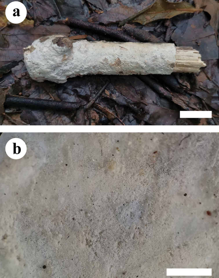
Basidiomata of Hyphodermapuerense (holotype). Scale bars: 2 cm (a); 1 mm (b).
Figure 3.

Microscopic structures of Hyphodermapuerense (holotype) a basidiospores b basidia and basidioles c cystidia d a section of hymenium. Scale bars: 5 µm (a); 10 µm (b–d).
Holotype.
China. Yunnan Province, Puer, Jingdong County, Huilianghe Village, GPS co-ordinates 24°04'45"N, 100°56'32"E, altitude 1246 m a.s.l., on fallen angiosperm branch, leg. C.L. Zhao, 4 January 2019, CLZhao 9476 (SWFC).
Etymology.
puerense (Lat.): referring to the locality (Puer) of the specimens.
Description.
Basidioma annual, resupinate, adnate, byssoid, without odour and taste when fresh, up to 15 cm long, 3 cm wide, 100–260 µm thick. Hymenial surface smooth to floccose, cream when fresh, cream to slightly buff on drying. Margin sterile, thinning out, narrow, cream.
Hyphal system monomitic, generative hyphae with clamps, colourless, thick-walled, frequently branched, interwoven, 2.5–4.5 µm in diameter; IKI-, CB-; tissues unchanged in KOH; subhymenial hyphae densely covered by crystals.
Cystidia tubular, encrusted with small crystals, 25–97 × 5.5–9.5 µm.
Basidia clavate to subcylindrical, slightly constricted in the middle to somewhat sinuous, with 4 sterigmata and a basal clamp, 20–30 × 4.5–6 µm.
Basidiospores ellipsoid, colourless, thin-walled, smooth, IKI-, CB-, (5.5–)6–7.5(–8) × 3–4.5(–5) µm, L = 6.53 µm, W = 3.71 µm, Q = 1.73–1.79 (n = 60/2).
Habitat and ecology.
Climate of the sample collection site is subtropical monsoon climate area, the forest type is evergreen angiosperm forest and samples were collected on fallen angiosperm branches.
Additional specimens examined.
China. Yunnan Province, Puer, Jingdong County, Huilianghe Village, GPS co-ordinates 24°04'45"N, 100°56'32"E, altitude 1246 m a.s.l., on fallen angiosperm branch, leg. C.L. Zhao, 5 January 2019, CLZhao 9583 (SWFC).
Hyphoderma tenuissimum
C.L. Zhao & Q.X. Guan sp. nov.
523885D7-51A5-5383-B36E-DB68F4E670D0
838412
Figure 4.
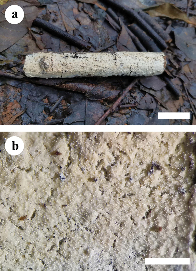
Basidiomata of Hyphodermatenuissimum (holotype). Scale bars: 2 cm (a); 1 mm (b).
Figure 5.

Microscopic structures of Hyphodermatenuissimum (holotype) a basidiospores b basidia and basidioles c cystidia d a section of hymenium. Scale bars: 10 µm (a–d).
Holotype.
China. Yunnan Province, Chuxiong, Zixishan Forestry Park, GPS co-ordinates 25°01'26"N, 101°24'37"E, altitude 2313 m a.s.l., on fallen angiosperm branch, leg. C.L. Zhao, 1 July 2018, CLZhao 7221 (SWFC).
Etymology.
tenuissimum (Lat.): referring to the thin basidiomata.
Description.
Basidioma annual, resupinate, adnate, membranaceous when fresh, hard membranaceous upon drying, up to 20 cm long, 3 cm wide, 30–100 µm thick. Hymenial surface tuberculate to minutely-grandinioid, slightly buff when fresh, buff upon drying, cracking. Margin sterile, slightly buff, 1 mm wide.
Hyphal system monomitic, generative hyphae with clamps, colourless, thick-walled, frequently branched, interwoven, 3–5 µm in diameter, IKI-, CB-; tissues unchanged in KOH.
Cystidia large, cylindrical, with 4–12 clamped septa, with abundant encrustations, 50–220 × 6.5–13 µm.
Basidia clavate to subcylindrical, constricted, somewhat sinuous, with 4 sterigmata and a basal clamp connection, 17–31 × 4.5–8 µm.
Basidiospores cylindrical, colourless, thin-walled, smooth, with oil drops inside, IKI–, CB–, 7–10.5(–11) × 3–4.5(–5) µm, L = 8.75 µm, W = 4.15 µm, Q = 2.02–2.18 (n = 120/4).
Habitat and ecology.
Climate of the sample collection site is subtropical monsoon climate area, the forest type is evergreen angiosperm forest and samples were collected on fallen angiosperm branches.
Additional specimens examined.
China. Yunnan Province, Chuxiong, Zixishan National Forestry Park, GPS co-ordinates 25°01'26"N, 101°24'37"E, altitude 2263 m a.s.l., on fallen angiosperm branch, leg. C.L. Zhao, 1 July 2018, CLZhao 6930, CLZhao 7003 (SWFC); Wenshan, Pingba Town, Wenshan National Nature Reserve, GPS co-ordinates 23°18'19"N, 104°42'47"E, altitude 1976 m a.s.l., on fallen angiosperm branch, leg. C.L. Zhao, 25 July 2019, CLZhao 16210 (SWFC).
Discussion
In the present study, two new species, Hyphodermapuerense and H.tenuissimum are described, based on phylogenetic analyses and morphological characters.
Phylogenetically, the two new taxa were found to belong to Hyphoderma, in which H.puerense forms a sister species to H.moniliforme and H.tenuissimum forms an independent monophyletic lineage (100% BS, 100% BP and 1.00 BPP).
Morphologically, Hyphodermapuerense is similar to H.obtusiforme J. Erikss. & Å. Strid and H.obtusum in having a smooth hymenium, non-septate cylindrical cystidia and ellipsoid basidiospores. However, H.obtusiforme differs from H.puerense by both larger basidia (30–40 × 8–9 µm) and basidiospores (10–14 × 5–7 µm; Eriksson and Ryvarden 1975). Hyphodermaobtusum also differs from H.puerense by larger basidia (30–35 × 6–8 µm) and basidiospores (8–9 × 5–6.5 µm; Eriksson 1958). Hyphodermapuerense is similar to H.roseocremeum (Bres.) Donk in having smooth hymenium and non-septate cylindrical cystidia. However, Hyphodermaroseocremeum differs through the presence of larger basidiospores (8–12 × 3–4 µm; Bernicchia and Gorjón 2010).
Morphologically, Hyphodermatenuissimum is similar to H.floccosum C.L. Zhao & Q.X. Guan, H.mopanshanense, H.nudicephalum Gilb. & M. Blackw., H.pinicola, H.setigerum and H.subsetigerum Sheng H. Wu in having septocystidia and cylindrical basidiospores. However, Hyphodermafloccosum differs from H.tenuissimum by having a floccose hymenial surface and tubular cystidia (Guan and Zhao 2021b); H.mopanshanense is separated from H.tenuissimum by having porulose to pilose hymenial surface and smaller basidia (15–18.5 × 3–4.5 µm; Ma et al. 2021); H.nudicephalum differs from H.tenuissimum in the nature of the septocystidial apex (lacking encrustation; swollen up to 14 µm; Gilbertson and Blackwell 1988); H.pinicola is separated from H.tenuissimum by having basidia with two sterigmata and larger basidiospores (13–16 × 4–4.5 µm; Yurchenko and Wu 2014b); H.setigerum differs by having a combination of thin basidiomata with very long septocystidia (Bernicchia and Gorjón 2010); and H.subsetigerum differs from H.tenuissimum by having narrower basidia (20–30 × 4.5–5.5 µm) and smaller basidiospores (6–8 × 2.8–3.2 µm; Wu 1997).
Nilsson et al. (2003) highlighted the phylogeography of Hyphodermasetigerum (Basidiomycota) in the Northern Hemisphere in a study based on molecular analysis, morphological studies and crossing tests. Nine preliminary taxa were shown to exist inside the H.setigerum complex; in the present study, H.tenuissimum belongs to the H.setigerum complex, based on the morphological character of long septocystidia and phylogenetic evidence. A previous study indicated the importance of vicariance in the evolution of this species complex (Nilsson et al. 2003) and our study shows that the specimens of H.tenuissimum are collected in Zixishan National Forestry Park (GPS co-ordinates 25°01'26"N, 101°24'37"E), Chuxiong, Yunnan Province, China, which is distinct from H.setigerum s. str. (Norway: Oppland and Finland: Pohjois-Häme). The present samples of H.subsetigerum and H.nudicephalum were collected in Yunnan Province, China, but neither of these taxa groups together closely with H.tenuissimum (Fig. 1).
In the current phylogenetic tree, two partially annotated GenBank sequences (KJ668522 and KJ714002) of Hyphoderma sp. (South Korea) cluster closely with four sequences of the new species Hyphodermatenuissimum, although whether they really belong to this species remains to be assessed. It is certainly conceivable that they do, which would mean that Hyphodermatenuissimum has been collected and sequenced at least six times in Asia. Regarding the new taxon H.puerense (Fig. 1), four partially annotated GenBank sequences (KR868735, KR868736, KR868737 and DQ340327) form a reasonably well-supported clade together with our two specimens of H.puerense. We interpret this to mean that all six taxa represent H.puerense. All of the samples in this clade are from Asia, which supports the point of the importance of vicariance in the evolution in this genus.
Key to 30 accepted species of Hyphoderma in China
| 1 | Cystidia absent | 2 |
| – | Cystidia present | 5 |
| 2 | Hymenial surface grandinioid | H. acystidiatum |
| – | Hymenial surface smooth | 3 |
| 3 | Basidiospores > 10.5 µm in length | H. densum |
| – | Basidiospores < 10.5 µm in length | 4 |
| 4 | Hymenophore cracked; basidiospores > 8.5 µm in length | H. fissuratum |
| – | Hymenophore uncracked; basidiospores < 8.5 µm in length | H. sibiricum |
| 5 | Hymenophore smooth | 6 |
| – | Hymenophore tuberculate, porulose, grandinioid or odontoid | 15 |
| 6 | Two types of cystidia present | 7 |
| – | One type of cystidia present | 8 |
| 7 | Moniliform cystidia absent | H. microcystidium |
| – | Moniliform cystidia present | H. sinense |
| 8 | Hymenophore uncracked | 9 |
| – | Hymenophore cracked | 11 |
| 9 | Basidiospores > 11 µm in length | H. definitum |
| – | Basidiospores < 11 µm in length | 10 |
| 10 | Basidiospores > 8.5 µm in length | H. microporoides |
| – | Basidiospores < 8.5 µm in length | H. puerense |
| 11 | Cystidia moniliform | 12 |
| – | Cystidia cylindrical | 13 |
| 12 | Basidiospores > 9 µm in length | H. litschaueri |
| – | Basidiospores < 9 µm in length | H. moniliforme |
| 13 | Basidiospores ellipsoid < 10 μm in length | H. rimulosum |
| – | Basidiospores cylindrical > 10 μm in length | 14 |
| 14 | Basidiospores > 12 µm in length | H. cremeum |
| – | Basidiospores < 12 µm in length | H. subclavatum |
| 15 | Hymenophore odontoid or grandinioid | 16 |
| – | Hymenophore tuberculate, porulose | 19 |
| 16 | Hymenophore odontoid | 17 |
| – | Hymenophore grandinioid | 18 |
| 17 | Basidiospores < 4.5 µm in width | H. transiens |
| – | Basidiospores > 4.5 µm in width | H. formosanum |
| 18 | Basidiospores larger 7–10.5 × 3–4.5 µm | H. tenuissimum |
| – | Basidiospores smaller 6–8 × 2.8–3.2 μm | H. subsetigerum |
| 19 | Cystidia of two types | 20 |
| – | Cystidia of one type | 23 |
| 20 | Septate cystidia absent | 21 |
| – | Septate cystidia present | 22 |
| 21 | Basidiospores < 4 µm in width | H. variolosum |
| – | Basidiospores > 4 µm in width | H. crystallinum |
| 22 | Basidia 2-sterigmata, basidiospores > 13 µm in length | H. pinicola |
| – | Basidia 4-sterigmata, basidiospores < 13 µm in length | H. floccosum |
| 23 | Septate cystidia present | 24 |
| – | Septate cystidia absent | 25 |
| 24 | Hymenophore porulose to pilose, basidia < 5 µm in width | H. mopanshanense |
| – | Hymenophore tuberculate, basidia > 5 µm in width | H. setigerum |
| 25 | Hymenophore porulose | H. obtusiforme |
| – | Hymenophore tuberculate, colliculose | 26 |
| 26 | Cystidia < 30 µm in length | H. cremeoalbum |
| – | Cystidia > 30 µm in length | 27 |
| 27 | Basidia > 30 µm in length | 28 |
| – | Basidia < 30 µm in length | 29 |
| 28 | Hymenophore cracking, cystidia < 10 µm in width | H. medioburiense |
| – | Hymenophore not cracking, cystidia > 10 µm in width | H. clavatum |
| 29 | Hymenophore colliculose | H. nemorale |
| – | Hymenophore tuberculate | H. membranaceum |
Supplementary Material
Acknowledgements
The research was supported by the Yunnan Fundamental Research Project (Grant No. 202001AS070043) and Science Research Foundation of Yunnan Provincial Department of Education Project (Project No. 2021Y275).
Citation
Guan Q-X, Li Y-F, Zhao C-L (2021) Morphological and phylogenetic evidence for recognition of two new species of Hyphoderma (Basidiomycota) from southern China, with a key to all Chinese Hyphoderma. MycoKeys 83: 145–160. https://doi.org/10.3897/mycokeys.83.69909
Funding Statement
Yunnan Fundamental Research Project (Grant No. 202001AS070043) and Science Research Foundation of Yunnan Provincial Department of Education Project (Project No. 2021Y275)
References
- Baltazar JM, Silveira RMB, Rajchenberg M. (2016) Type studies of J. Rick’s corticioid homobasidiomycetes (Agaricomycetes, Basidiomycota) housed in the Herbarium Anchieta (PACA). Phytotaxa 255(2): 101–132. 10.11646/phytotaxa.255.2.1 [DOI] [Google Scholar]
- Bernicchia A, Gorjón SP. (2010) Fungi Europaei 12: Corticiaceae s.l. Alassio: Edizioni Candusso.
- Dai YC. (2012) Polypore diversity in China with an annotated checklist of Chinese polypores. Mycoscience 53(1): 49–80. 10.1007/s10267-011-0134-3 [DOI] [Google Scholar]
- Donk MA. (1957) Notes on resupinate Hymenomycetes IV. Fungus 27: 1–29. [Google Scholar]
- Eriksson J. (1958) Studies in the Heterobasidiomycetes and Homobasidiomycetes – Aphyllophorales of Muddus National Park in North Sweden. Symbolae Botanicae Upsalienses 16(1): 1–172. [Google Scholar]
- Eriksson J, Ryvarden L. (1975) The Corticiaceae of North Europe; Fungiflora: Oslo, Norway, Volume 3, 288–546.
- Felsenstein J. (1985) Confidence intervals on phylogenetics: An approach using bootstrap. Evolution 39: 783–791. 10.1111/j.1558-5646.1985.tb00420.x [DOI] [PubMed] [Google Scholar]
- Floudas D, Hibbett DS. (2015) Revisiting the taxonomy of Phanerochaete (Polyporales, Basidiomycota) using a four gene dataset and extensive ITS sampling. Fungal Biology 119(80): 679–719. 10.1016/j.funbio.2015.04.003 [DOI] [PubMed] [Google Scholar]
- Gilbertson RL, Blackwell M. (1988) Some new or unusual corticioid fungi from the Gulf Coast region. Mycotaxon 33: 375–386. [Google Scholar]
- Guan QX, Zhao CL. (2021a) Taxonomy and phylogeny of the wood-inhabiting fungal genus Hyphoderma with descriptions of three new species from East Asia. Journal of Fungi 7(4): e308. 10.3390/jof7040308 [DOI] [PMC free article] [PubMed]
- Guan QX, Zhao CL. (2021b) Two new corticioid species, Hyphodermasinense and H.floccosum (Hyphodermataceae, Polyporales), from southern China. Mycosystema 40(03): 447–461. 10.13346/j.mycosystema.200382 [DOI] [Google Scholar]
- James TY, Stajich JE, Hittinger CT, Rokas A. (2020) Toward a fully resolved fungal tree of life. Annual Review of Microbiology 74(1): 291–313. 10.1146/annurev-micro-022020-051835 [DOI] [PubMed] [Google Scholar]
- Jang Y, Jang S, Min M, Hong JH, Lee H, Lee H, Lim YW, Kim JJ. (2015) Comparison of the diversity of basidiomycetes from dead wood of the Manchurian fir (Abiesholophylla) as evaluated by fruiting body collection, mycelial isolation, and 454 sequencing. Microbial Ecology 70(3): 634–645. 10.1007/s00248-015-0616-5 [DOI] [PubMed] [Google Scholar]
- Justo A, Miettinen O, Floudas D, Ortiz-Santana B, Sjökvist E, Linder D, Nakasone K, Niemelä T, Larsson K, Ryvarden L, Hibbetta DS. (2017) A revised family-level classification of the Polyporales (Basidiomycota). Fungal Biology 121(9): 798–824. 10.1016/j.funbio.2017.05.010 [DOI] [PubMed] [Google Scholar]
- Katoh K, Rozewicki J, Yamada KD. (2019) MAFFT online service: multiple sequence alignment, interactive sequence choice and visualization. Briefings in Bioinformatics 20(4): 1160–1166. 10.1093/bib/bbx108 [DOI] [PMC free article] [PubMed] [Google Scholar]
- Larsson A. (2014) AliView: a fast and lightweight alignment viewer and editor for large data sets. Bioinformatics 30(22): 3276–3278. 10.1093/bioinformatics/btu531 [DOI] [PMC free article] [PubMed] [Google Scholar]
- Larsson KH. (2007) Re-thinking the classification of corticioid fungi. Mycological Research 111(9): 1040–1063. 10.1016/j.mycres.2007.08.001 [DOI] [PubMed] [Google Scholar]
- Ma X, Huang RX, Zhang Y, Zhao CL. (2021) Hyphodermafissuratum and H.mopanshanense spp. nov. (Polyporales) from southern China. Mycoscience 62(1): 36–41. 10.47371/Mycosci.2020.08.004 [DOI] [PMC free article] [PubMed] [Google Scholar]
- Martín MP, Zhang LF, Fernández-López J, Dueñas M, Rodríguez-Armas JL, Beltrán-Tejera E, Telleria MT. (2018) Hyphodermaparamacaronesicum sp. nov. (Meruliaceae, Polyporales, Basidiomycota), a cryptic lineage to H.macaronesicum. Fungal Systematics and Evolution 2: 57–68. 10.3114/fuse.2018.02.05 [DOI] [PMC free article] [PubMed] [Google Scholar]
- Miller MA, Pfeiffer W, Schwartz T. (2012) The CIPRES Science Gateway: enabling high-impact science for phylogenetics researchers with limited resources. Association for Computing Machinery 39: 1–8. 10.1145/2335755.2335836 [DOI] [Google Scholar]
- Nakasone KK. (2008) Type studies of corticioid Hymenomycetes described by Bresadola. Cryptogamie Mycologie 29(3): 231–257. 10.1002/yea.1629 [DOI] [Google Scholar]
- Nilsson RH, Hallenberg N, Nordén B, Maekawa N, Wu SH. (2003) Phylogeography of Hyphodermasetigerum (Basidiomycota) in the Northern Hemisphere. Mycological Research 107(6): 645–652. 10.1017/S0953756203007925 [DOI] [PubMed] [Google Scholar]
- Nylander JAA. (2004) MrModeltest v.2. Program Distributed by the Author. Evolutionary Biology Centre, Uppsala University, Uppsala.
- Petersen JH. (1996) The Danish Mycological Society’s colour-chart. Foreningen til Svampekundskabens Fremme, Greve.
- Rehner SA, Samuels GJ. (1994) Taxonomy and phylogeny of Gliocladium analysed from nuclear large subunit ribosomal DNA sequences. Mycological Research 98(6): 625–634. 10.1016/S0953-7562(09)80409-7 [DOI] [Google Scholar]
- Ronquist F, Teslenko M, van der Mark P, Ayres DL, Darling A, Hohna S, Larget B, Liu L, Suchard MA, Huelsenbeck JP. (2012) MrBayes 3.2: efficient Bayesian phylogenetic inference and model choice across a large model space. Systematic Biology 61(3): 539–542. 10.1093/sysbio/sys029 [DOI] [PMC free article] [PubMed] [Google Scholar]
- Tedersoo L, Bahram M, Põlme S, Kõljalg U, Yorou NS, Wijesundera R, Ruiz LV, Vasco-Palacios AM, Thu PQ, Suija A, Smith ME, Sharp C, Saluveer E, Saitta A, Rosas M, Riit T, Ratkowsky D, Pritsch K, Põldmaa K, Piepenbring M, Phosri C, Peterson M, Parts K, Pärtel K, Otsing E, Nouhra E, Njouonkou AL, Nilsson RH, Morgado LN, Mayor J, May TW, Majuakim L, Lodge DJ, Lee SS, Larsson K-H, Kohout P, Hosaka K, Hiiesalu I, Henkel TW, Harend H, Guo L-d, Greslebin A, Grelet G, Geml J, Gates G, Dunstan W, Dunk C, Drenkhan R, Dearnaley J, De Kesel A, Dang T, Chen X, Buegger F, Brearley FQ, Bonito G, Anslan S, Abelland S, Abarenkov K. (2014) Global diversity and geography of soil fungi. Science 346: 1256688. 10.1126/science.1256688 [DOI] [PubMed]
- Telleria MT, Dueñas M, Beltrán-Tejera E, Rodríguez-Armas JL, Martín MP. (2012) A new species of Hyphoderma (Meruliaceae, Polyporales) and its discrimination from closely related taxa. Mycologia 104(5): 1121–1132. 10.3852/11-344 [DOI] [PubMed] [Google Scholar]
- Telleria MT, Duenas M, Melo I, Hallenberg N, Martin MP. (2010) A re-evaluation of Hypochnicium (Polyporales) based on morphological and molecular characters. Mycologia 102(6): 1426–1436. 10.3852/09-242 [DOI] [PubMed] [Google Scholar]
- Vilgalys R, Hester M. (1990) Rapid genetic identification and mapping of enzymatically amplified ribosomal DNA from several Cryptococcus species. Journal of Bacteriology 172(8): 4238–4246. [DOI] [PMC free article] [PubMed] [Google Scholar]
- Vu D, Groenewald M, Vries M, Gehrmann T, Stielow B, Eberhardt U, Al-Hatmi A, Groenewald JZ, Cardinali G, Houbraken J, Boekhout T, Crous PW, Robert V, Verkley GJM. (2019) Large-scale generation and analysis of filamentous fungal DNA barcodes boosts coverage for kingdom Fungi and reveals thresholds for fungal species and higher taxon delimitation. Studies in Mycology 92: 135–154. 10.1016/j.simyco.2018.05.001 [DOI] [PMC free article] [PubMed] [Google Scholar]
- Wallroth CFW. (1833) Flora Cryptogamica Germaniae 2 Algas et fungos. Norimbergae [Nüremberg], Schragius [J.L. Schrag], 923 pp. [Google Scholar]
- White TJ, Bruns T, Lee S, Taylor J. (1990) Amplification and direct sequencing of fungal ribosomal RNA genes for phylogenetics. In: Innis MA, Gefand DH, Sninsky JJ, White JT. (Eds) PCR Protocols: A Guide to Methods and Applications.Academic Press, San Diego, 18: 315–322. 10.1016/B978-0-12-372180-8.50042-1 [DOI]
- Wu F, Chen JJ, Ji XH, Vlasák J, Dai YC. (2017) Phylogeny and diversity of the morphologically similar polypore genera Rigidoporus, Physisporinus, Oxyporus and Leucophellinus. Mycologia 109(5): 749–765. doi: 10.1080/00275514.2017.1405215 [DOI] [PubMed] [Google Scholar]
- Wu SH. (1997) New species of Hyphoderma from Taiwan. Mycologia 89: 132–140. 10.1080/00275514.1997.12026764 [DOI] [Google Scholar]
- Wu SH, Nilsson HR, Chen CT, Yu SY, Hallenberg N. (2010) The white-rotting genus Phanerochaete is polyphyletic and distributed throughout the phlebioid clade of the Polyporales (Basidiomycota). Fungal Diversity 42(1): 107–118. 10.1007/s13225-010-0031-7 [DOI] [Google Scholar]
- Yurchenko E, Wu SH. (2014) Hyphodermapinicola sp. nov. of H.setigerum complex (Basidiomycota) from Yunnan, China. Botanical Studies 55: 71–78. 10.1186/s40529-014-0071-5 [DOI] [PMC free article] [PubMed] [Google Scholar]
- Yurchenko E, Wu SH. (2015) Hyphodermamoniliforme and H.nemorale (Basidiomycota) newly recorded from China. Mycosphere 6(1): 113–121. 10.5943/mycosphere/6/1/11 [DOI] [Google Scholar]
- Zhao CL, Wu ZQ. (2017) Ceriporiopsiskunmingensis sp. nov. (Polyporales, Basidiomycota) evidenced by morphological characters and phylogenetic analysis. Mycological Progress 16(1): 93–100. 10.1007/s11557-016-1259-8 [DOI] [Google Scholar]
Associated Data
This section collects any data citations, data availability statements, or supplementary materials included in this article.