Abstract
Background and aim:
The purpose of the study was to compare the data obtained by two independent observers and statistically analyse the results using Cohen’s K to highlight the concordance or discordance in the diagnosis of normality, pathology and, in particular, the type of femoroacetabular impingement (FAI) on plain films.
Methods:
the study was conducted retrospectively. The only inclusion criterium was the minimum age of 20 years. All patients underwent a radiographic examination of the pelvis in standard anteroposterior projection in orthostasis.
Results:
A hundred patients were evaluated. A good concordance between the two operators in the examination of normal hip joint (k= 0.68 right/ 0,74 left) was found; a similar grade of agreement was found for the analysis of “pincer” type FAI (k = 0.73 right, 0,67 left). The best results in concordance were achieved in the examination of “cam” type FAI (k= 0.82 right, 0,88 left), “mixed” type FAI (k = 0.85 right, 0,86 left), and in findings of “coxa profunda” (k = 0.92 right, 0,88 left).
Conclusion:
We found a good concordance between the two readers; a few cases of disagreement were found in the diagnosis of “pincer” type FAI and absence of disease. This discrepancy may be due to the different weight given by the single observer to the clinical indication that leads the patient to examination, but also by the difficulty of a not dedicated radiologist to show some subtle signs indicative of early FAI.
Keywords: Plain radiography, hip joint, FAI, agreement
Introduction
The femoral-acetabular impingement (FAI) is a movement-related hip disease that clinically occurs due to abnormal and early contact between the anterolateral portion of the femoral neck and the acetabulum, due to the presence of characteristic anatomical features. The head of the femur, normally, has a spherical shape and articulates in the acetabulum (cup-shaped concavity) without friction or contact (conflicts).
FAI is divided into three types: “cam”, “pincer” and mixed (1).
A chronic and repetitive trauma can lead to hip pain and decreased function. The abnormal bony features include cam deformity of the femoral head-neck junction as well as pincer lesions of the acetabulum. Cam deformity is an abnormal bony prominence or “bump” at the junction of the femoral head and neck resulting in an aspherical shaped head, occurring most commonly along the anterosuperior femoral head-neck area. A pincer lesion is an abnormal bony overhang of the anterolateral acetabular rim resulting in over coverage of the femoral head, which can also contribute to impingement and pain. Both cam and pincer lesions are visible on plain radiographs (X-ray). Patients may present with either one or both (mixed) morphologies, with the mixed morphology being the most common in symptomatic patients. When impingement occurs, it results in a mechanical collision of the femoral cam with the rim of the acetabulum, which results in pinching of the labrum and cartilage. Over time, this can result in cartilage wear and tearing of the labrum (2).
The “cam” impingement is described as a flattening or convexity of the femoral head-neck junction. During leg flexion the femoral neck and the acetabulum show praecox contact and, due to repeated movement, it results in the lesion of both the femoral head-neck junction and the acetabulum.
The “pincer” impingement is described as excessive acetabular coverage caused by an altered orientation (retroversion) of the cup, causing compression of the acetabular lip and its progressive degeneration.
It affects the young-adult population (14-29% of the population is affected), however, the prevalence is higher in sportive subjects, fluctuating between 75% and 95%, in particular considering the “cam” type.
Symptoms include hip pain, joint stiffness, flexion difficulty, adduction and intra-rotation movements.
The purpose of the study is to highlight the level of concordance in the diagnosis of (FAI) analysing the reports of two independent observers obtained with the standardized evaluation of hip x-rays.
Methods and Materials
The study was conducted retrospectively. The only inclusion criterium was to use the x-ray studies of patients with a minimum age of 20 years. The query retrieval included studies from April 2018 to May 2019. Each radiogram was examined by two independent operators.
Study modalities and visualization method
All patients underwent a radiographic examination of the pelvis in standard anteroposterior projection in orthostasis (3); using OPERA Sound® equipment, 100 mAs and 80 KV. The images obtained were displayed on the workstations of our Institute using dedicated software (Elios Suite Healthcare solutions).
The radiograms of each patient were studied by two independent observers. Each exam was read 5 times using 5 different standardized reports.
The first one assessed the absence of pathological findings considering the head of the femur, normally, has a spherical shape and articulates in the acetabulum (cup-shaped concavity) without friction or contact (conflicts); the second evaluated the presence of “cam” type impingement described as a flattening or convexity of the femoral head-neck junction; the third assessed the presence of “pincer” type impingement is described as excessive acetabular coverage caused by an altered orientation (retroversion) of the cup, causing compression of the acetabular lip and its progressive degeneration; the fourth evaluated the presence of “mixed” type impingement; the fifth, finally, assessed the presence of coxa profunda measuring the Wiberg angle (given a vertical line through the centre of the femoral head the Wiberg angle is described by the line connecting the centre of the femoral head and lateral acetabular border. The Wiberg angle range was 25-40°).
Quantitative analysis criteria
The inter-observer concordance was tested using Cohen’s kappa (κ).
The degrees of agreement were considered poor if 0< (κ) <0.4, discrete if 0.4< (κ) <0.6; good if 0.6< (κ) <0.8 and excellent if (κ) > 0.8. The statistical significance of the results is reported as a confidence interval.
Results
The consecutive records of 100 patients were included in the study. The patients’ characteristics are summarized in table 1.
Table 1.
Patients demographic characteristics n = 100
| Age average (range). Sex male/female Symptomatic y/n Previous trauma y/n Arthropathies y/n Previous fracture y/n |
68 (20-80) 47/53 30/100 20/100 0/100 0/100 |
| Concordance values expressed as: Cohen’s κ value; standard error (SE) –Confidence Interval (CI). Normality right/left. CAM right/left. Pincer right/left. Mixed right/left. Coxa profunda right/left. |
0.68; 0.073 – 0.536-0.824 / 0.74; 0.067 – 0.608-0.872. 0.82; 0.069 – 0.691-0.960 / 0.88; 0.057 – 0.772-0.995. 0.73; 0.112 – 0.515-0.955 / 0.67; 0.135 – 0.415-0.943. 0.85; 0.072 – 0.709-0.993 / 0.86; 0.066 – 0.735-0.994. 0.92; 0.040 – 0.839-0.997 / 0.88; 0.049 – 0.780-0.972. |
Side concordance analysis for “Normality”
Analyzing the normality of the findings, it was observed that on the right side both operators identified 41 patients as negative and 43 cases as pathological. In the remaining 16 cases, observers A and B found discordant findings (8 positive cases for A were negative for B and vice versa). Cohen’s K analysis documented a value of 0.68 (SE= 0.073; confidence interval 0.536-0.824).
Analyzing the normality of the findings, it was observed that on the left side both operators identified 42 patients as negative and 45 cases as pathological. In the remaining 13 cases, observers A and B found discordant findings (6 positive cases for A were negative for B and 7 cases for A were negative and positive for B). Cohen’s K analysis documented a value of 0.74 (SE 0.067; confidence interval 0.608 to 0.872). (Figure. 1)
Figure 1.
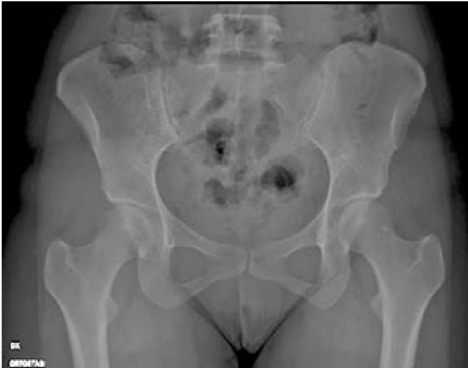
Normal acetabular and femoral head morphology.
Side concordance analysis for “cam”
Taking into consideration the “cam” type impingement findings for the right side, it emerged that both operators identified 75 patients negative and 19 positives. In the remaining 6 cases, the observers found discordant findings (4 positive cases for A were negative for B and 2 cases for A were negative and positive for B).
Cohen’s K analysis documented a value of 0.82 (SE 0.069; confidence interval 0.691 to 0.960).
Taking into consideration the “cam” type impingement findings for the left side, it emerged that both operators identified 76 patients negative and 20 positives. In the remaining 4 cases, the observers found discordant findings (2 positive cases for A were negative for B and 2 cases for A were negative and positive for B).
Cohen’s K analysis documented a value of 0.88 (SE 0.057; confidence interval 0.772 to 0.995). (Figure. 2).
Figure 2.
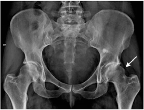
A “cam” type is when the impingement’s cause is a not perfectly spherical femoral head which causes abrasion of the acetabular cup when the aspheric portion engages the acetabular rim. The concordance in diagnosis in this case was high due to the presence of a “cam” type conflict on the left, while on the right a moderate to low concordance was obtained.
Side concordance analysis for “pincer”
Analyzing the normality of the findings, it was observed that on the right side both operators identified 87 patients as negative and 8 cases as pathological. In the remaining 5 cases, observers A and B found discordant findings (1 positive case for A was negative for B and 4 cases for A were negative and positive for B).
Cohen’s K analysis documented a value of 0.73 (SE 0.112; confidence interval 0.515 to 0.955).
Analyzing the normality of the findings, it was observed that on the left side both operators identified 89 patients as negative and 6 cases as pathological. In the remaining 5 cases, observers A and B found discordant findings (2 positive cases for A were negative for B and 3 cases for A were negative and positive for B). Cohen’s K analysis documented a value of 0.67 (SE 0.135; confidence interval 0.415 to 0.943). (Figure. 3)
Figure 3.
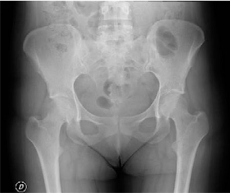
An example of “pincer” type; the acetabular excess coverage with early head-neck femoral and acetabulum contact with possible posteroinferior joint degeneration (contrecoup) due to a subclinical subluxation. The concordance in this case was moderate bilaterally because of the inherent difficulty in assessing a posterior-inferior conflict.
Side concordance analysis for “mixed”
By processing the data obtained by the two observers regarding the “mixed” type impingement for the right side, it turned out that both operators identified 82 patients as negative and 14 cases as pathological. In the remaining 4 cases, observers A and B found discordant findings (3 positive cases for A were negative for B and 1 case for A was negative and positive for B).
Cohen’s K analysis documented a value of 0.85 (SE 0.072; confidence interval 0.709 to 0.993).
By processing the data obtained by the two observers regarding the “mixed” type impingement for the left side, it turned out that both operators identified 80 patients as negative and 16 cases as pathological. In the remaining 4 cases, observers A and B found discordant findings (3 positive cases for A were negative for B and 1 case for A was negative and positive for B).
Cohen’s K analysis documented a value of 0.86 (SE 0.066; confidence interval 0.735 to 0.994). (Figure. 4)
Figure 4.
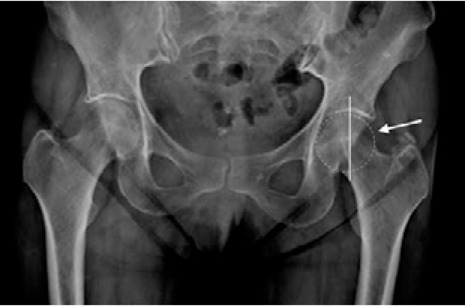
The “mixed” type shows the presence of both types of conflict (“cam” and “pincer”). The arrow indicates the bone bump while the medialization of the centre of the femoral head in relation to the bisector line of the acetabular roof is shown.
Side concordance analysis for “coxa profunda”
Analyzing the diagnostic findings for coxa profunda it was observed that on the right side both operators identified 54 patients as negative and 41 cases as pathological. In the remaining 4 cases, observers A and B found discordant findings (2 positive cases for A were negative for B and vice versa).
Cohen’s K analysis documented a value of 0.92 (SE 0.040; confidence interval 0.839 to 0.997).
Analyzing the diagnostic findings for coxa profunda it was observed that on the left side both operators identified 56 patients as negative and 38 cases as pathological. In the remaining 6 cases, observers A and B found discordant findings (3 positive cases for A were negative for B and vice versa).
Cohen’s K analysis documented a value of 0.88 (SE 0.049; confidence interval 0.780 to 0.972).
Figure 5.
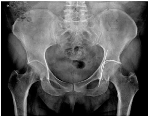
An example of coxa profunda. On the x-ray of the pelvis, it is noted that the acetabular fossa reaches or medially exceeds the ilio-ischial line. The finding is more evident on the left than on right and, as a result, a higher concordance in diagnosis was obtained on the left than on right.
Discussion and Conclusion
Femoral-acetabular impingement is a recognized cause of early hip arthrosis and its diagnosis, especially in the early phase in young and athletic patients which allows the prevention of irreversible cartilage damage and helps to address the patient to conservative therapy (4–9).
The international literature reports a prevalence of FAI in the general population of 10% -15%.
The consequences of FAI are constituted by morphological alterations of the proximal femur and the acetabulum with consequent anomalous contact between the two articular surfaces (6,10–12).
The diagnosis, as previously discussed, is mainly entrusted to the radiographic examination of the pelvis in the anteroposterior projection and in the axial projection of the hip, to which the Dunn projection can be added; it represents the first level examination to which patients with suspected FAI must be subjected, allowing the identification of subtle findings indicative of FAI even though the typical findings of coxarthrosis are not present yet (7,9,13).
The radiographic examination includes the evaluation of the femoral head-neck junction, the shape of the femoral head, the acetabular roof and its contour. The evaluation of the acetabular depth, the inclination and the antero- or retroversion of the acetabulum are important findings (13).
To our knowledge, in the scientific literature, the radiographic examination of the pelvis has a sensitivity of 60% and specificity of 81%, in the anterior-posterior projection; sensitivity of 70-74% and a specificity of 63% with the axial projection of the hip and sensitivity of 91-96% if the Dunn projection is added with 88% specificity (14).
Our study highlighted data in line with those reported in the international literature, according to which the diagnostic findings of FAI, whether “cam”, “pincer” or “mixed” type, are quite frequent in the general population (13).
It was found that the diagnostic findings for FAI “cam” type are more frequent in young male patients, while the diagnostic findings for FAI “pincer” type show greater frequency in middle-aged female patients. Our results are in line with the available literature. Gutiérrez-Ramos et al. conducted a multicentre study in the Mexican population and obtained comparable results: FAI “cam” type predominated in men on the right side and pincer type predominates in women on the left side (15).
The prevalence of cam, pincer, and combined pathological conditions was also studied in a meta-analysis following the Preferred Reporting Items for Systematic Reviews and Meta-Analyses (PRISMA) guidelines (14). Across different studies, significant heterogeneity exists in the anatomical landmarks used to define FAI, either “cam” or “pincer” (anterior focal coverage, acetabular retroversion, abnormal acetabular index, deep coxa, acetabular protrusion, ischial spine sign, crossover sign, and posterior wall sign). This makes the comparison between our study and the others in the literature difficult.
Our study showed a good agreement between the two observers regarding the diagnosis of “normality”, namely the absence of pathological diagnostic findings, with a Cohen’s k of 0.68 for the right side and 0.74 for the left side. To our knowledge, no studies evaluate the inter-reader coherence in the definition of “normal” findings on plain films.
The data showed good agreement considering the diagnosis of “pincer” type impingement, with a Cohen’s k of 0.73 for the right side and 0.67 for the left side.
In the analysis of the indicative findings for “cam” and “mixed” type FAI, an excellent agreement was obtained. The Cohen’s k were respectively, 0.82 for the right side and 0.88 for the left side in “cam”, and 0.86 for the right and 0.92 for the left side in “mixed”.
Results of similar significance emerged from the comparison of the findings indicative of coxa profunda, with a Cohen’s k of 0.92 for the right side and 0.88 for the left side.
It has been documented that the data of the two observers were more evident in the diagnosis of absence of pathology and in that of “pincer” type impingement.
This discrepancy between the two observers may be due to the different weight given by each of them to the clinical indication with which the patient arrived at the examination, sometimes misleading, but also to the difficulty of the non-dedicated radiologist to visualize some subtle diagnostic signs, indicative of early FAI. To our knowledge, the level of concordance in the diagnosis of FAI on plain radiographs has never been evaluated. Many recent studies focus the diagnosis on three-dimensional reconstructions after CT or MR imaging. Radiographic evaluation, however, is more accessible and indicated as the first imaging method in asymptomatic cases than the methods described above (16).
Our study shows a good to an optimal overall agreement between the two observers even if a fair heterogeneity of the obtained results is still present.
Our study presents some limitations: the sample size, despite being big enough to achieve statistical significance, should be implemented to evaluate if the lack of agreement is due to difficult FAI recognition in older patients. In fact, the prevalence of femoroacetabular conflict may increase with age. Further studies should be made to verify possible differences in concordance for different age groups.
Competing interests:
Each author declares that he or she has no commercial associations (e.g., consultancies, stock ownership, equity interest, patent/licensing arrangement etc.) that might pose a conflict of interest in connection with the submitted article.
References
- Genovese E, Spiga S, Vinci V, Aliprandi A, Di Pietto F, Coppolino F, et al. Femoroacetabular impingement: role of imaging. Musculoskelet Surg. 2013 Aug;97(S2):117–26. doi: 10.1007/s12306-013-0283-y. [DOI] [PubMed] [Google Scholar]
- O’Rourke RJ, El Bitar Y. Femoroacetabular Impingement. In: StatPearls [Internet] Treasure Island (FL): StatPearls Publishing; 2021. [cited 2021 Mar 18]. Available from: http://www.ncbi.nlm.nih.gov/books/NBK547699/ [PubMed] [Google Scholar]
- Mascarenhas VV, Castro MO, Rego PA, Sutter R, Sconfienza LM, Kassarjian A, et al. The Lisbon Agreement on Femoroacetabular Impingement Imaging—part 1: overview. Eur Radiol. 2020 Oct;30(10):5281–97. doi: 10.1007/s00330-020-06822-9. [DOI] [PubMed] [Google Scholar]
- Leunig M, Beaulé PE, Ganz R. The Concept of Femoroacetabular Impingement: Current Status and Future Perspectives. Clinical Orthopaedics & Related Research. 2009 Mar;467(3):616–22. doi: 10.1007/s11999-008-0646-0. [DOI] [PMC free article] [PubMed] [Google Scholar]
- Kappe T, Kocak T, Neuerburg C, Lippacher S, Bieger R, Reichel H. Reliability of radiographic signs for acetabular retroversion. Int Orthop. 2011 Jun;35(6):817–21. doi: 10.1007/s00264-010-1035-3. [DOI] [PMC free article] [PubMed] [Google Scholar]
- Laborie LB, Lehmann TG, Engesæter IØ, Eastwood DM, Engesæter LB, Rosendahl K. Prevalence of radiographic findings thought to be associated with femoroacetabular impingement in a population-based cohort of 2081 healthy young adults. Radiology. 2011 Aug;260(2):494–502. doi: 10.1148/radiol.11102354. [DOI] [PubMed] [Google Scholar]
- Griffin DR, Dickenson EJ, O’Donnell J, Agricola R, Awan T, Beck M, et al. The Warwick Agreement on femoroacetabular impingement syndrome (FAI syndrome): an international consensus statement. Br J Sports Med. 2016 Oct;50(19):1169–76. doi: 10.1136/bjsports-2016-096743. [DOI] [PubMed] [Google Scholar]
- Weir A, Brukner P, Delahunt E, Ekstrand J, Griffin D, Khan KM, et al. Doha agreement meeting on terminology and definitions in groin pain in athletes. Br J Sports Med. 2015 Jun;49(12):768–74. doi: 10.1136/bjsports-2015-094869. [DOI] [PMC free article] [PubMed] [Google Scholar]
- Sankar WN, Nevitt M, Parvizi J, Felson DT, Agricola R, Leunig M. Femoroacetabular impingement: defining the condition and its role in the pathophysiology of osteoarthritis. J Am Acad Orthop Surg. 2013;21(Suppl 1):S7–15. doi: 10.5435/JAAOS-21-07-S7. [DOI] [PubMed] [Google Scholar]
- Beall DP, Sweet CF, Martin HD, Lastine CL, Grayson DE, Ly JQ, et al. Imaging findings of femoroacetabular impingement syndrome. Skeletal Radiol. 2005 Nov;34(11):691–701. doi: 10.1007/s00256-005-0932-9. [DOI] [PubMed] [Google Scholar]
- Leunig M, Beck M, Kalhor M, Kim Y-J, Werlen S, Ganz R. Fibrocystic changes at anterosuperior femoral neck: prevalence in hips with femoroacetabular impingement. Radiology. 2005 Jul;236(1):237–46. doi: 10.1148/radiol.2361040140. [DOI] [PubMed] [Google Scholar]
- Nötzli HP, Wyss TF, Stoecklin CH, Schmid MR, Treiber K, Hodler J. The contour of the femoral head-neck junction as a predictor for the risk of anterior impingement. The Journal of Bone and Joint Surgery British volume. 2002 May;84-B(4):556–60. doi: 10.1302/0301-620x.84b4.12014. [DOI] [PubMed] [Google Scholar]
- Sutter R, Zanetti M, Pfirrmann CWA. New developments in hip imaging. Radiology. 2012 Sep;264(3):651–67. doi: 10.1148/radiol.12110357. [DOI] [PubMed] [Google Scholar]
- Frank JM, Harris JD, Erickson BJ, Slikker W, Bush-Joseph CA, Salata MJ, et al. Prevalence of Femoroacetabular Impingement Imaging Findings in Asymptomatic Volunteers: A Systematic Review. Arthroscopy. 2015 Jun;31(6):1199–204. doi: 10.1016/j.arthro.2014.11.042. [DOI] [PubMed] [Google Scholar]
- Gutiérrez-Ramos R, Ávalos-Calderón SA, Bahena-Peniche LA. Prevalence of X-ray signs of femoroacetabular impingement in Mexican population. Acta Ortop Mex. 2017 Jun;31(3):134–40. [PubMed] [Google Scholar]
- Lerch TD, Siegfried M, Schmaranzer F, Leibold CS, Zurmühle CA, Hanke MS, et al. Location of Intra-and Extra-articular Hip Impingement Is Different in Patients With Pincer-Type and Mixed-Type Femoroacetabular Impingement Due to Acetabular Retroversion or Protrusio Acetabuli on 3D CT-Based Impingement Simulation. Am J Sports Med. 2020 Mar;48(3):661–72. doi: 10.1177/0363546519897273. [DOI] [PubMed] [Google Scholar]


