Abstract
Background:
We performed a retrospective study of 67 patients and their data for radiological investigations by serial Xrays, computed tomography, magnetic resonance imaging, uniform surgical procedure of craniotomy. The results were analyzed to determine the natural course of the disease, anatomical changes at various intervals following trauma, and outcome of surgical procedure in terms of cranial reconstruction, seizures, and progress in neurological deficit.
Results:
Among 67 patients, 34 (50.74%) were male and 33 (49.26%) were female patients. About 86.67% of patients sustained the injury before the age of 3 years. Development of seizures in 28 patients (41.80%) is the most common symptom. In our study, 43.28% of patients (29 cases) had a combination of Type I and II of growing skull fracture. The dural defects confirmed in all cases were nearly twice (average 1.42) as large as the bone defects. All patients under the age of 3 years with diastatic skull fracture should be closely followed up and should be examined 2–3 months later to look for evidence of a growing skull fracture. Linear fractures and burst fractures in an infant with a scalp swelling must be corrected early to prevent a growing skull fracture.
Conclusion:
Early management can avoid difficult surgical dissection and progressive neurological sequelae seen with delayed intervention. Surgical correction results in the prevention of brain shift and increase in meningocerebral cicatrices. Meticulous surgery and vigilant postoperative care reduce the morbidity and mortality. In our opinion, the autologous material is the best choice because of its tissue compatibility, convenience, inexpensiveness, and rare rate of infection.
Keywords: Duroplasty, growing skull fracture, linear skull fracture
Introduction
Growing skull fracture is a rare complication of head injury in childhood. The incidence reported is only 0.05%–0.1% of skull fractures in childhood.[1,2] Although the development of growing skull fractures is multifactorial, the predominant factor in their causation is the presence of lacerated dura mater. The pulsatile force of the brain during its growth causes the fracture in the thin skull to enlarge. This interposition of tissue prevents osteoblasts from migrating to the fracture site and inhibiting healing. The resorption of the adjacent bone by the continuous pressure from tissue herniation through the bone gap adds to the progression of the fracture line.
The brain extrusion may be present shortly after diastatic linear fracture in neonates and young infants resulting in focal dilation of the lateral ventricle near the growing fracture.[3] This dilatation is said to be reversible and may normalize after surgical repair.[4] In addition, craniotomies performed without watertight closure of dural lacerations have been found to lead to growing skull fractures. These support dural tear as being the major risk factor in the development of a growing skull fracture.
Owing to the risk of neurological deterioration and development of seizure disorders, surgical correction of growing fractures is recommended by craniectomy and repair.[5]
The use of magnetic resonance imaging (MRI) studies in cases with growing fractures has changed our understanding of pathogenesis and surgical management of this lesion.[6]
Aims and objectives
To evaluate all the factors contributing toward the growing skull fractures
To evaluate the role of arachnoidal tear, brain contusion underlying a linear fracture, in the formation of growing skull fracture, thereby predicting which linear fracture may progress into growing fracture of the skull
To evaluate the outcome of neurosurgical intervention in terms of seizures and progress of neurological deficit in children with growing fractures of the skull
To elucidate the sequence of events contributing to the growth of skull fractures.
Materials and Methods
Study design
This was a retrospective study (1983–2012, 29 years).
Setting
This study was conducted at the Department of Neurosurgery, G. B. Pant Hospital, New Delhi.
Study population
The study population was 67 patients.
Inclusion criteria
All patients of growing skull fractures admitted in G. B. Pant Hospital New Delhi.
Methodology
Patients
A retrospective review of all patients presenting to our institution between 1983 and 2012 was undertaken. Detailed radiological investigations by serial X-rays, computed tomography (CT), MRI, and details of the uniform surgical procedure by the same surgeon were analyzed to determine the natural course of the disease. Other factors which were analyzed included calvarial defects, neurologic defects (static, progressive), incidence of convulsions, and state of ventricular dilatation after surgery. All patients had serial plain X-rays and CT scans before referral to us for surgery and then had MRI studies. Age of patient, mode and type of head trauma, presence of subgaleal swelling after injury, interval between time of initial trauma and development of growing fracture, presence or absence of convulsions, and progression of neurological deficits were recorded from hospital records. Follow-up imaging was also evaluated to study the effect of surgical management.
Operative steps
The principles of pediatric neurosurgery were meticulously and carefully followed when approaching growing skull fracture repair. Considerations included the timing of surgery after injury and the overall prognosis. An experienced pediatric neuroanesthesiologist is needed.
Incision Lazy S or C shaped according to the situation
Defining the edges of bone defect
Exposure of the dural defect
As the dura retracts under the bone edge for varying distances, the only way to do is to remove the overlying bone. This can be done in using two ways (a) using rongeurs the bone is nibbled until the dura is reached. The edge is then followed all around. (b) A single or multiple burr holes are made 1–2 cm away from the bone edge. Using a craniotome, the bone is cut in a circular fashion all around the bone defect [Figure 5]. This bone piece is elevated as a single piece separating it carefully from the underlying dura and soft tissue [Figure 4]. The latter technique has the advantage of being able to use the elevated bone to cover the bone defect, while in the former technique, the surgeon is left with only a small piece of bone which is of no use in performing a cranioplasty.
Figure 5.
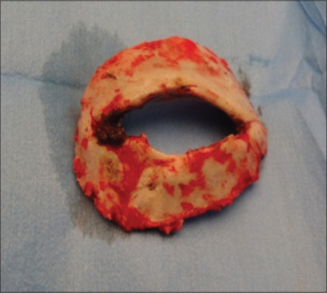
Photograph showing the craniotomy around the skull defect
Figure 4.
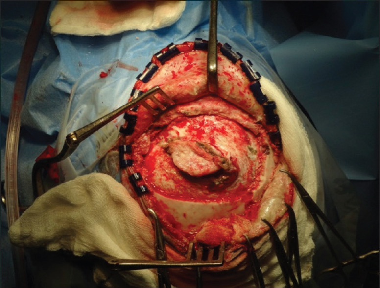
Photograph showing exposure of dural defect and meningocerebral cicatrix
Figure 2.
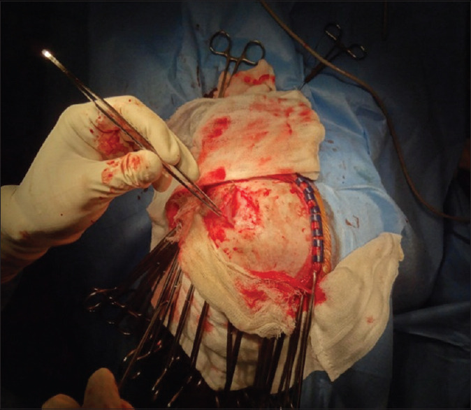
Photograph showing reflection of periosteum towards the bone defect
Microsurgical dissection and meningocerebral cicatrix excision
The underlying pia-arachnoid is often adherent to the dura for a few millimeters. As the separation is completed, it is often helpful to place a cottonoid between the dura and the separated cortex to prevent them from sticking to each other. The meningocerebral cicatrix is excised maintaining absolute hemostasis.
Duroplasty
Duroplasy is then performed using either locally harvested pericranium, galea, fascia lata, or dural substitute.[Figures 1-3,6,9-17] [Tables 1-2]
Figure 1.
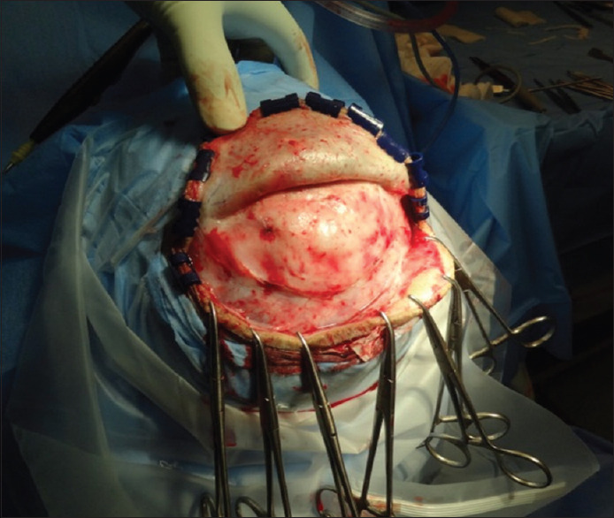
Photograph showing the exposed bone defect and periosteum surrounding the defect
Figure 3.
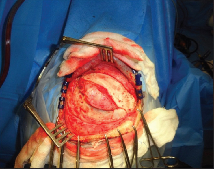
Photograph showing well defined bone defect
Figure 6.
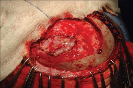
Photograph showing duroplasty using galea
Figure 9.
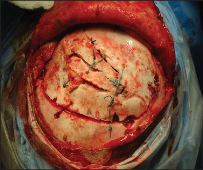
Photograph showing final appearance
Figure 17.
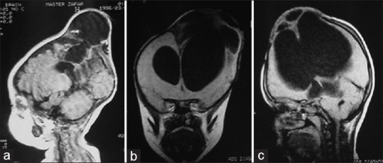
T1 weighted magnetic resonance imaging saggital (a and c), axial (b), showing porencephalic cyst
Table 1.
The types of growing skull fractures
| Types of growing skull fractures | Number of patients (%) |
|---|---|
| I | 9 (13.43) |
| II | 8 (11.94) |
| I, II | 29 (43.28) |
| I, III | 2 (2.98) |
| II, III | 5 (7.46) |
| I, II, III | 14 (20.89) |
Table 2.
Various types of cranioplasty
| Cranioplasty (n=55) | Number of patients | Patients |
|---|---|---|
| Split calvarial graft | 41 | 74.54 |
| Medpore | 14 | 25.45 |
| Acrylic | 3 | 5.45 |
| Tantalum | 1 | 1.81 |
| Bone morcellation | 1 | 1.81 |
Figure 10.
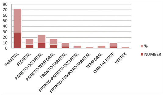
Showing location of growing skull fractures
Figure 11.
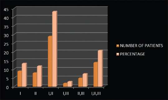
Shows the types of growing skull fractures
Figure 12.
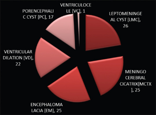
Shows the magnetic resonance imaging findings in patients with growing skull fractures
Figure 13.
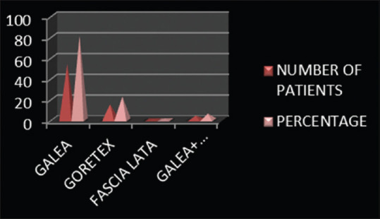
Shows various substitutes used for duroplasty
Figure 14.
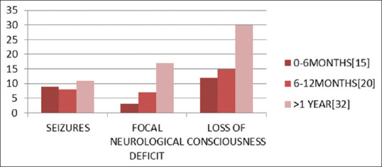
Shows the age at time of injury and development of clinical symptoms
Figure 15.
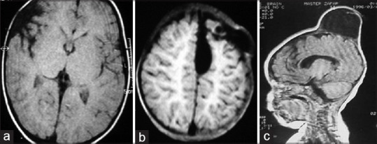
T1 and T2 weighted axial (a and b) and saggital magnetic resonance imaging (c) showing leptomeningeal cyst
Figure 16.
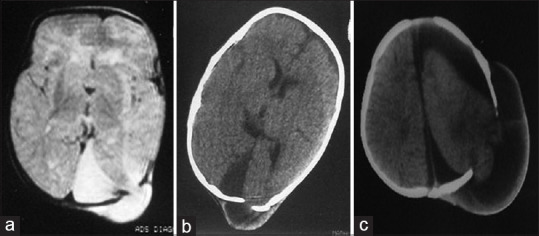
T2 axial magnetic resonance imaging (a), axial computed tomography (b and c) showing progressive cerebral migration and displacement
Table 3.
The localization of growing skull fractures without symptoms (seizures, focal neurological deficit, and loss of consciousness)
| Location | Absent (seizures, neurological deficit, and loss of consciousness) (n=12) |
|---|---|
| Parieto-occipital | 4 |
| Parietal | 3 |
| Orbital roof | 2 |
| Temporal | 1 |
| Fronto-temporo-parietal | 1 |
| Parietotemporal | 1 |
Cranioplasty
Cranioplasty is then done with the bone that has been removed. This can be then split and can be used to cover the defect [Figure 7]. Alternatively, acrylic implant or polymethylmethacrylate or porous polyethylene (Medpor) [Figure 8] can be used for cranioplasty.
Figure 7.
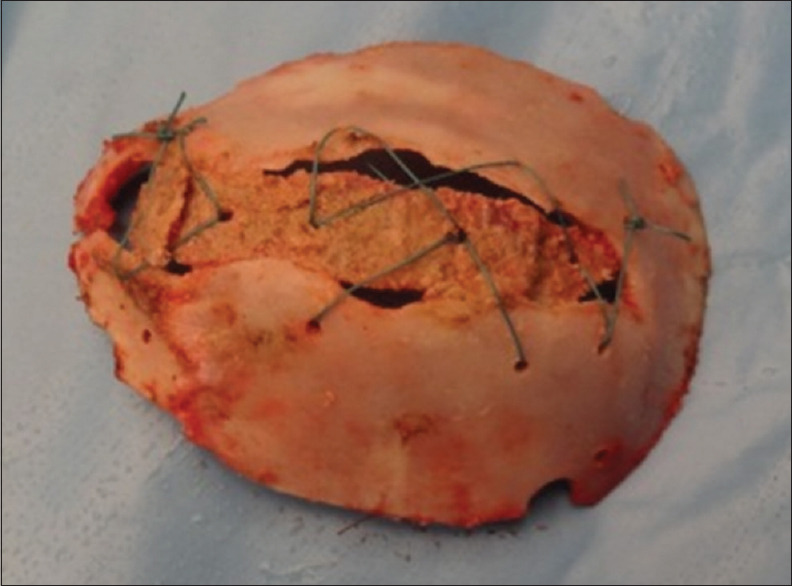
Photograph showing cranioplasty using split calvarial graft
Figure 8.
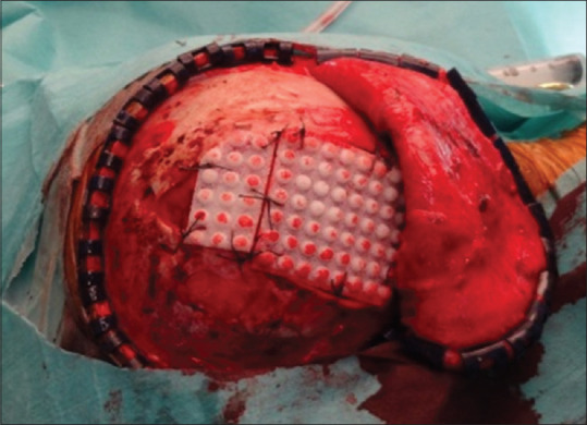
Photograph showing cranioplasty using porous polyethylene (Medpor)
Statistical analysis
The baseline demographics of the study population were analyzed using descriptive statistical parameters.
Results
-
Gender Distribution:
Among 67 patients, 34 (50.74%) were male and 33 (49.26%) were female patients
-
Age at the Time of Injury:
Among 67 patients, 15 (22.38%) were of <6 months age, 20 (29.85%) were of 7–12 months age, 17 (25.38%) were of 1–2 years age, 14 (20.90%) were of 2–5 years age, and 1 (1.49%) was of >5 years age.
-
Location of Growing Skull Fracture
The most common locations of growing skull fractures are parietal (43.28%), frontal (10.45%), parieto-occipital (14.92%), parieto-temporal (10.45%), fronto-parietal (5.97%), orbital roof (5.97%), fronto-parieto-occipital (2.98%), temporal (2.98%), fronto-emporo-parietal (1.49%), and vertex (1.49%). The parietal bone was involved overall in 79.10% of cases, whereas the frontal bone was involved overall in 20.89% of cases. The overall involvement of temporal bone and occipital bone was 14.92% and 17.91%, respectively.
-
Mode of injury
Fifty-nine patients (88.06%) had injuries due to a fall from height, whereas only six (8.9%) were involved in road traffic accidents. Two patients (2.9%) had other mechanisms of injury (bucket handle injury and fall of the box on the head).
-
Clinical presentation
Hemiparesis noted in 23 patients (34.32%) including cranial nerve palsy (3 presented with VIIth nerve, 1 with IVth nerve palsy) in 4 patients (5.97%).Headache, proptosis, and squint were present in 3 patients (4.47%); whereas diminution of vision, synkinesis, mental retardation, and quadriparesis were present in 1 (1.49%) patient each.
-
Time between injury and presentation of patients
Thirty patients presented within 1 year of injury. In 26 patients, the time period was between 1 and 5 years, whereas 3 patients were more than 5 years. The minimum time period was 10 days and the maximum was 20 years.
-
Type of growing skull fracture
A three-type classification has been suggested by Rahman et al. In our study, 43.28% of patients (29 cases) had a combination of type I and II. The next frequent types were type I, II, and III combined in 20.89% (14 patients), type I in 13.43% (9 patients), and type II in 11.94% (8 patients). The less common fractures were of type II and III in 7.46% (5 patients) and type I and III in 2.98% (2 patients).
-
Bone defect versus dural defect
It was observed that the bone defects located in parietal regions were larger than those in other regions. The smallest bone defect was 4 cm2 and the largest defect was 45 cm2. The dural defects confirmed in all cases were nearly twice (avg 1.42) as large as the bone defects.
-
Radiology of growing skull fractures. (X-ray, CT, and MRI)
In 13 patients of 67 patients, the X-ray at the time of injury was available. About 61.50% (8 patients) had linear fractures and 30.76% (4 patients) had depressed skull fracture. One (7.69%) patient had a burst fracture. The mean diastasis between the fracture lines was 4.53 mm in the X-rays analyzed
The patients underwent CT and MRI at various time points in the evolution of growing skull fractures. The findings in order of the frequency in the “index” CT scan (i.e., CT at the time of injury) were contusion 80% (20 patients), SAH 20% (4 patients), hematoma 10% (2 patients), and SDH and intraventricular hemorrhage in 5% (1 patient) each. The overall incidence in 53 of 67 patients who underwent CT scan was leptomeningeal cyst in 41.79% (28 patients), ventricular dilation toward the site of fracture in 29.85% (20 patients), porencephalic cyst in 13.43% (9 patients), hydrocephalus in 2.98% (2 patients), and intradiploic cyst was seen in 1.49% (1 patient).
Twenty-nine out of 67 patients underwent MRI. The findings in these patients were leptomeningeal cyst in 26 patients (89.65%), porencephalic cyst in 17 patients (58.62%), ventricular dilation toward the site of fracture in 22 patients (75.86%), ventriculocele in 1 patient (3.44%), and meningocerebral cicatrix and encephalomalacia in 25 patients (86.20%).
-
Treatment modality
In 1 patient (1.49%), VP shunt was done and the swelling disappeared. A dural repair alone was done in 25 patients (37.13%) and dural repair and cranioplasty were done in 42 patients (62.68%). In 6 patients (8.95%), a dural and cranial repair was followed by VP shunt.
In the earlier part of the study, cranioplasty was done using tantalum mesh (1 patient, 1.81%) and bone morcellation cranioplasty (1 patient, 1.81%). In 3 patients (5.45%), acrylic cranioplasty was done. Split calvarial graft cranioplasy was done in 41 patients (74.54%) of patients, whereas medpore was used in 14 patients (25.45%).
-
Postoperative complications
Out of 67 patients of growing skull fractures who were operated, there was one death (1.47%) due to status epileptics and pulomonary aspiration. Nineteen patients (28%) developed seroma, this was managed by aspiration and crepe bandage. Local wound infection occurred in 11 patients (16.41%) which were successfully treated by antibiotics. CSF leak and pseudomeningocele were encountered in 7.46% (5 patients) and 5.97% (4 patients), respectively, and were managed conservatively. Thirteen patients underwent multiple surgeries. Of these, 9 underwent twice and 3 patients underwent thrice. One patient underwent 5 surgeries for multiple shunt revisions.
Correlation between clinical presentation (seizures, focal neurological deficit, and loss of consciousness) and localization of growing skull defect, age at the time of injury, and interval time between head injury and presentation.
Regarding the site of growing skull fractures, we observed that frontal localization (7 patients) is associated with seizures in 42.85% (7 patients), focal neurological deficit in 14.28% (1 case), and loss of consciousness in all (7 patients).
A clinical presentation without seizures, loss of consciousness, and neurological deficit is more frequent if the lesion is parieto-occipital. These data are the same of the clinical evolution of the parietal localization (29 patients): 34.48% (10 patients) of patients develop seizures, 51.72% (15 patients) develop focal neurological deficit, and 89.65% (26 patients) develop loss of consciousness. In frontal localization, 42.85% (3 patients) developed seizures, 14.28% (1 patient) developed focal neurological deficit, and all developed loss of consciousness. In patients with parieto-occipital localization (10 patients), 40% developed seizures, focal neurological deficit, and loss of consciousness each. Three patients with frontoparietal localization developed seizures (75%) and loss of consciousness, whereas only half of them had focal neurological deficit. In parietotemporal localization, there is a major probability of seizures (85.71%), focal neurological deficit (42.85%), and loss of consciousness (85.71%).
In literature, there are no completed data regarding clinical follow-up and functional recovery of these patients. Among 15 patients up to 6 months of age 60% (9 patients) developed seizures, 20% (3 patients) presented with a neurological deficit and 80% (12 patients) with loss of consciousness. From 6 to 12 months of age, the probability of development of focal neurological deficit (35%) and loss of consciousness (35%) is quite the same as an asymptomatic course. After the age of 1 year, only 34.3% (11 patients) developed seizures, whereas 53% (17 patients) developed focal neurological deficit and 53.1% (30 patients) developed loss of consciousness. These differences of clinical presentation are connected to the physical development of the child; complete closure of skull sutures and definitive adjustment of cerebral functions cause an evident stronger brain damage in advanced age group of patients.
The interval time between the onset of injury and presentation of seizures is longer than 1 year in 11 patients (50% of those who developed seizures). In addition, neurological deficit and loss of consciousness are more frequent symptoms (respectively, 40% and 86%) in a late clinical presentation, but in 25% of patients, they are early symptoms appearing in 2 months from injury.
Follow-up
The mean duration of follow-up ranged from 1 month to 23 years.
There was no recurrence of growing skull fractures in our study.
Headache present in 3 patients (4.47%) resolved completely. Postoperatively, seizures were controlled in 18 patients (64.2%). The administration of anticonvulsant medication was continued for 3 months postoperatively for the patient who had a preexisting history of seizure and was then gradually reduced. In 7 patients (25%), seizures remained stable, administration of the same medication as that which he was receiving before surgery was continued. In 2 patients (7.14%), surgery did not result in any change in the seizure frequency.
In 20 patients (86.95%) who had hemiparesis preoperatively, the weakness remained static in follow-up evaluation. Proptosis presents in 3 patients (4.47%) resolved completely after surgery.
Discussion
The existence of growing skull fractures has been recognized since the 19th century, with the first report of an enlarging cranial defect secondary to a skull fracture being credited to John Halliday in 1876.[7] This entity has been reported under various names including expanding skull fractures, craniocerebral erosion, leptomeningeal cyst, posttraumatic bone absorption, traumatic ventricular cyst, and cephalhydrocele. There were 34 (50.74%) males and 33 (49.26%) female patients ranging in age from 6 months to 21 years in our study. The mean age at the time of injury was 14.26 months. About 86.67% of patients sustained the injury before the age of 3 years in Howship et al's study,[8] the mean age was 12.4 months. In Kyoshima et al.'s study,[9] 80.5% of patients were <5 years of age and also in Lende et al.'s study,[10] 9 of 10 patients were under 1 year of age.
The time interval between injury and presentation in our series ranged from <6 months to more than 20 years. We had one patient who presented to us in their adult life. One of these was a 21-year-old male who had her injury at the age of 6 months following which a defect was noticed in the left parietal region which remained asymptomatic till 21 years of age when the patient started experiencing pain over the defect. He achieved symptomatic improvement after surgery. Nine adult cases of growing skull defects have been reported.[1,11,12,13,14] Seven patients[1,11,12,13] had suffered from trauma in childhood. One patient had been asymptomatic for more than 60 years.[14] Diagnosis in older adults must exclude metastasis, multiple myelomas, and epidermoid. In most of the reports of adult growing skull fracture, the injury typically occurred in childhood as in our case.
Sixty-three patients in this study presented with scalp mass, 28 patients (41.80%) presented with seizures, 28 (41.79%) with neurological deficit, 55 (82.09%) with loss of consciousness, and cranial nerve palsy in 4 patients (5.97%) (3 presented with VIIth nerve, 1 with IVth nerve palsy). Headache, proptosis, and squint were present in 3 patients (4.47%), whereas diminution of vision, synkinesis, mental retardation, and quadriparesis were present in 1 (1.49%) patient each. In Howship et al.'s study[8] of 22 growing skull fracture patients, five patients presented with seizure (22%), four with hemiparesis (18%), and one with hydrocephalus (4.5%). In the study by Pezzotta et al.'s study[15] of 132 growing skull fractures, 46% developed seizures, 38% focal neurological deficit, and 21% loss of consciousness. The study also concluded that in the parietotemporal localization, there is a higher probability of seizures (62.5%) and loss of consciousness (62.5%). In our study, parietotemporal location was associated with seizures in 85.71% and loss of consciousness in 85.71%, this correlates to what has been established in the previous studies.
Growing skull fractures are frequently located in the parietal region. Often, the initial fracture is limited by bordering sutures, so the growth in the fracture is likewise limited. Fractures and, therefore, growing skull fractures can cross suture lines.[15,16] When such a fracture occurs, it most often affects the frontal or occipital areas, although all regions of the skull can be affected including the posterior fossa and skull base.[15] Rahman et al.[17] and Ersahin et al.[8] found that the most common site of growing skull fracture was parietal 50% and 56%, respectively. In our study, the incidence in the parietal site of GSF was (43.28%) as in other series.[6,8,17,18,19]
In this study, ventriculoperitoneal shunt was performed in 7 patients (10.44%), and in Ramamurthi[18] study of 15 patients, 2 patients had an accumulation of CSF requiring Ventriculoperitoneal shunt in 1 patient (6.5%) and repeated lumbar puncture in another patient. In Howship et al.[8] study, one patient out of 17 needs V-P shunt (5.9%). Gupta et al.[6] performed V-P shunt in 4 patients from 28 (14%). Sharma et al.[20] advocated that in such patients, shunt surgery should be considered as an initial or alternative procedure as it may result in the resolution of raised ICP, disappearance of scalp swelling, and regrowth of bone edges at the fracture site.
Ruberti et al.,[19] recommended that duroplasty alone with a flap of pericranium remains the simplest and least expensive method of treatment. Sharma et al.[20] also recommended duroplasty alone. In Sharma et al.'s study,[21] duroplasty alone was performed in 8 patients (19%) from 41 patients with no recurrence and 24 patients (58%) underwent a duro and cranioplasty. The material used for cranioplasty included acrylic, wire mesh, steel plates, or autologous bone. In Ersahin et al.'s study[8] duroplasty alone was performed in 21 patients from 22 (95%) with no recurrence. In Sharma et al.'s study,[6] duroplasty and cranioplasty were performed in all 28 patients with no recurrence. Smith et al.[22] and Tandon[23] recommended autogenous bone for cranioplasty with the following advantages: no additional skin incisions, no bone taken from other parts of the body, and physiological fusion can be expected; foreign body reaction is avoided. In our study, duroplasty alone was performed in 25 patients (35.13%). Duro- and cranioplasty was performed in 42 patients (62.68%).
Tandon et al.[21] described the use of local calvarial bone harvested from the edges of the defect and shaped the bony pieces to cover the defect. We have used this technique in 1 patient aged 2 years with good results. This technique is simple and provides an excellent scaffold for bony union to occur. Cranioplasty was performed using split calvarial graft in 41 patients (74.54%). Porous polyethylene (Medpor) was used in 14 patients but had a significant complication rate of 50% as compared to split calvarial grafts (26.19%). There was no postoperative recurrence of leptomeningeal cyst in patients after duroplasty or duro and cranioplasty with good results.
In our study, motor deficit is unlikely to improve but seizure disorder improved in 18 patients from 28 patients (64.28%), this agrees with Halliday et al.,[7] who reported a case with no postoperative improvement in motor deficit.
Regarding the site of growing skull fractures, we observed that in parietal localization (29 patients), 34.48% (10 patients) of patients developed seizures, 51.72% (15 patients) developed focal neurological deficit, and 89.65% (26 patients) developed loss of consciousness. In frontal localization: 42.85% (3 patients) developed seizures, 14.28% (1 patient) developed focal neurological deficit, and all developed loss of consciousness. In patients with parieto-occipital localization (10 patients), 40% developed seizures, focal neurological deficit, and loss of consciousness each. Three patients with frontoparietal localization developed seizures (75%) and loss of consciousness, whereas only half of them had focal neurological deficit. In parietotemporal localization, there is a major probability of seizures (85.71%), focal neurological deficit (42.85%), and loss of consciousness (85.71%). In the study conducted by Scarfo et al., parietotemporal localization was associated with seizures in 62.5%, focal neurological deficit in 37.5%, and loss of consciousness in 62.5%.
In literature, there are no completed data regarding clinical follow-up and functional recovery of these patients. Among 15 patients up to 6 months of age, 60% (9 patients) developed seizures, 20% (3 patients) presented with a neurological deficit, and 80% (12 patients) with loss of consciousness. From 6 to 12 months of age, the probability of development of focal neurological deficit (35%) and loss of consciousness (35%) is quite the same as an asymptomatic course. After the age of 1 year, only 34.3% (11 patients) developed seizures, whereas 53% (17 patients) developed focal neurological deficit and 53.1% (30 patients) developed loss of consciousness. These differences of clinical presentation were connected to the physical development of the child; the interval time between the onset of injury and presentation of seizures is longer than 1 year in 11 patients (50% of those who developed seizures). Furthermore, neurological deficit and loss of consciousness are more frequent symptoms (respectively, 40% and 86%) in a late clinical presentation, but in 25% of patients, there are early symptoms appearing in 2 months from injury.
It should be emphasized that there exists a higher probability of developing a neurological deficit in relation to the clinical presentation. Twenty-six patients who had seizures and 10 patients who did not developed a neurological deficit.
Radiological features
The plain X-ray findings in growing skull fractures are characteristic and diagnostic. There is an irregular oval or elliptical skull defect. The original fracture line may still be seen at one end of the defect. The margins are usually everted, scalloped, and sclerotic.[24] The inner table is usually affected more than the outer table.[25]
The CT scan picks up the bony defect and additional underlying brain changes.[26] In a study by Rahman et al[17], 80% of their patients had underlying contusion in the CT scan. In our study, CT at the time of injury showed underlying contusion seen in 80% (20 patients). Other findings included SAH 20%, hematoma 10%, and SDH and intraventricular hemorrhage in 5% each. The overall incidence in 53 of 67 patients who underwent CT scan was leptomeningeal cyst in 28 patients, ventricular dilation toward the site of fracture in 20 patients, porencephalic cyst in 9 patients, and hydrocephalus in 2 patients. CT also demonstrates the intradiploic cysts[11,15,27] which was seen in 1 patient in our study. In many patients, the bony defect encroaches upon either the transverse or superior sagittal sinus. In one of our cases, the dural defect extended up to the midline in the parietal region.
Recently, MRI findings in growing skull features have been demonstrated.[28] These authors evaluated 6 of their patients with MRI and reported three patterns of tissue herniation through the fracture site. Two patients had solely brain herniating through the fracture, three had leptomeningeal cysts and abnormal brain herniation, while one had only leptomeningeal cyst herniation.[28] In our study, 29 of 67 patients underwent MRI. The findings in these patients were leptomeningeal cyst in 26 patients, porencephalic cyst in 17 patients, ventricular dilation toward the site of fracture in 22 patients, and meningocerebral cicatrix and encephalomalacia in 25 patients. In one patient, there was ventriculocele where the ventricle was seen extending through the defect into the subgaleal space. Growing skull fractures progresses in a sequential pattern. Tear of the arachnoid and leptomeningeal hematoma under the dural tear results in a pseudomeningocele at the fracture site. Further growth of the fracture resulted from the formation of a leptomeningeal cyst and brain migration from the enlarged fracture site. Formation of meningocerebral cicatrice and porencephaly results in a further increase in the fracture and ventricular dilatation. The natural course of an untreated case is progressive in nature with progressive cranial and cerebral damage. In addition to the dural tear at the fracture site, local brain contusion and arachnoid reaction were observed in all cases and were the most important factor in the formation of growing skull fractures. Surgical correction results in the prevention of brain shift and increase in meningocerebral cicatrices.
Conclusion
The most common location of growing skull fracture is in the parietal region and 86.67% of patients sustained the injury before the age of 3 years. In our study, parietotemporal location was associated with maximum incidence of seizures in 85.71%. In our study, motor deficit is unlikely to improve, but seizure disorder improved in 64.28%. All patients under the age of 3 years with diastatic skull fracture should be closely followed up and should be examined 2–3 months later to look for evidence of a growing skull fracture. Linear fractures and burst fractures in an infant with a scalp swelling must be corrected early to prevent a growing skull fracture. Early management can avoid difficult surgical dissection and progressive neurological sequelae seen with delayed intervention. Surgical correction results in the prevention of brain shift and increase in meningocerebral cicatrices. Meticulous surgery and vigilant postoperative care reduce morbidity and mortality. In our opinion, the autologous material is the best choice because of its tissue compatibility, convenience, inexpensiveness, and rare rate of infection.
In patients who have a small defect, especially children below 5 years, cranioplasty may not be necessary if the dural rent is adequately repaired, but it increases the strength of the restraining dural membrane and provides additional protection from injury.
Neuropathology of surgical specimens and the outcome of the surgical intervention have helped us in a better understanding of the pathophysiology of this disorder.
There was no postoperative recurrence of leptomeningeal cyst in patients after duroplasty or duro and cranioplasty with good results.
Financial support and sponsorship
Nil.
Conflicts of interest
There are no conflicts of interest.
References
- 1.Djientcheu VD, Rilliet B, Delavelle J, Argyropoulo M, Gudinchet F, de Tribolet N. Leptomeningeal cyst in newborns due to vacuum extraction: Report of two cases. Childs Nerv Syst. 1996;12:399–403. doi: 10.1007/BF00395094. [DOI] [PubMed] [Google Scholar]
- 2.Ersahin Y, Gulmen V, Palali I, Multer S. Growing skull fractures. Neurosurg Rev. 2000;23:139–44. doi: 10.1007/pl00011945. [DOI] [PubMed] [Google Scholar]
- 3.Haar FL. Complication of linear skull fracture in young children. Am J Dis Child. 1975;129:1197–200. doi: 10.1001/archpedi.1975.02120470047013. [DOI] [PubMed] [Google Scholar]
- 4.Gruber FH. Posttraumatic leptomeningeal cyst. Am J Radiol. 1969;105:305–7. doi: 10.2214/ajr.105.2.305. [DOI] [PubMed] [Google Scholar]
- 5.Guilburd JN, Rakier A. Growing skull fracture: A clinical study in 15 children. Clin Neurol Neurosurg. 1997;99(1):S259. [Google Scholar]
- 6.Gupta SK, Reddy NM, Khosla VK, Mathuriya SN, Shama BS, Pathak A, et al. Growing skull fractures: A clinical study of 41 patients. Acta Neurochir (Wien) 1997;139:928–32. doi: 10.1007/BF01411301. [DOI] [PubMed] [Google Scholar]
- 7.Halliday AL, Chapman PH, Heros RC. Leptomeningeal cyst resulting from adulthood trauma. Neurosurgery. 1990;26:150–3. doi: 10.1097/00006123-199001000-00025. [DOI] [PubMed] [Google Scholar]
- 8.Howship PJ. London: Longman; 1816. Practical Observations in Surgery and Morbid Anatomy; p. 494. [Google Scholar]
- 9.Kyoshima K, Gibo H, Kobayashi S, Sugita K. Cranioplasty with inner table of bone flap. Technical note. J Neurosurg. 1985;62:607–9. doi: 10.3171/jns.1985.62.4.0607. [DOI] [PubMed] [Google Scholar]
- 10.Lende R, Erickson TC. Cranial defects developing at fracture sites in children. Trans Am Neurol Assoc. 1959;13:130–1. [PubMed] [Google Scholar]
- 11.Michael GM, John GP, Arnold HM. Pathogenesis and treatment of Growing Skull Fracture. Surg Neurol. 1995;43:367–73. doi: 10.1016/0090-3019(95)80066-p. [DOI] [PubMed] [Google Scholar]
- 12.Miranda P, Vila M, Alvarez-Garijo JA, Perez-Nunez A. Birth trauma and development of growing fracture after coronal suture disruption. Childs Nerv Syst. 2007;23:355–8. doi: 10.1007/s00381-006-0182-8. [DOI] [PubMed] [Google Scholar]
- 13.Numerow LM, Krcek JP, Wallace CJ, Tranmer BI, Auer RN, Fong TC. Growing skull fracture simulating a rounded lytic calvarial lesion. AJNR. 1991;12:783–4. [PMC free article] [PubMed] [Google Scholar]
- 14.Palaoglu S, Beskonakli E, Senel K, Taskin Y, Yaleinlar Y. Intraosseus location of posterior fossa posttraumatic leptomeningeal cyst. Neuroradiology. 1990;32:78. doi: 10.1007/BF00593951. [DOI] [PubMed] [Google Scholar]
- 15.Pezzotta S, Silvani V, Gaetani P, Spanu G, Rondini G. Growing skull fractures of childhood. Case report and review of 132 cases. J Neurosurg Sci. 1985;29:129–35. [PubMed] [Google Scholar]
- 16.Rahimizadeh A. Growing fracture of the skull in the elderly. Neurosurgery. 1986;19:675–6. [PubMed] [Google Scholar]
- 17.Rahman Nain VR, Abedeen B, Tanjoom ZA, Tanjoom HB, Murshid WR. Growing skull fractures. Classification and management. Br J Neurosurg. 1994;8:667–9. doi: 10.3109/02688699409101180. [DOI] [PubMed] [Google Scholar]
- 18.Ramamurthi B, Kalyanaraman S. Rationale for surgery in growing fractures of the skull. J Neurosurg. 1970;32:427–30. doi: 10.3171/jns.1970.32.4.0427. [DOI] [PubMed] [Google Scholar]
- 19.Ruberti RF. Cranioplasty with inner table of bone flap in children: Report of two cases. Afri J Neur Sci. 1997;16:2. [Google Scholar]
- 20.Sharma A, Bindra GS, Kaur R. Why some skull fractures in children grow: Observations on pathogenesis of growing fracture of skull. Journal of Pediatric Neurosciences. 2011;13:129–35. [Google Scholar]
- 21.Sharma A, Tatke M, Abraham M. Diastatic fracture of the skull with pseudomeningocele in childhood. Clin Neurol Neurosurg. 1997;99:S259. [Google Scholar]
- 22.Smith T. Traumatic cephalhydrocelc. St bartholomew'shospital reports. 1884;20:033–240. [Google Scholar]
- 23.Tandon PN, Banerji AK, Bhatia R, Goulatia RK. Cranio-cerebral erosion (growing fracture of the skull in children). Part II: Clinical and radiological observations. Acta Neurochir (Wien) 1987;88:1–9. doi: 10.1007/BF01400508. [DOI] [PubMed] [Google Scholar]
- 24.Taveras J, Ransohoff S. Leptomeningeal cysts of the brain following trauma with erosion of the skull: A study of seven cases treated by surgery. J Neurosurg. 1953;10:233–41. doi: 10.3171/jns.1953.10.3.0233. [DOI] [PubMed] [Google Scholar]
- 25.Thompson JB, Mason TH, Haines GL, Cassidy RJ. Surgical management of diastatic linear skull fractures in infants. J Neurosurg. 1973;39:493–7. doi: 10.3171/jns.1973.39.4.0493. [DOI] [PubMed] [Google Scholar]
- 26.Scarfo GB, Mariottini A, Tomoccini D, Palma L. Growing skull fractures: Progressive evolution of brain damage and effectiveness of surgical treatment. Childs Nerv Syst. 1989;5:163–7. doi: 10.1007/BF00272120. [DOI] [PubMed] [Google Scholar]
- 27.Zegers B, Jira P, Willemsen M, Grotenhuis J. The growing skull fracture, a rare complication of paediatric head injury. Eur J Pediatr. 2003;162:556–7. doi: 10.1007/s00431-003-1256-1. [DOI] [PubMed] [Google Scholar]
- 28.Ziyal IM, Aydin Y, Türkmen CS, Salas E, Kaya AR, Ozveren F. The natural history of late diagnosed or untreated growing skull fractures: Report on two cases. Acta Neurochir (Wien) 1998;140:651–4. doi: 10.1007/s007010050158. [DOI] [PubMed] [Google Scholar]


