



Abstract

Nature has employed heme proteins to execute a diverse set of vital life processes. Years of research have been devoted to understanding the factors which bias these heme enzymes, with all having a heme cofactor, toward distinct catalytic activity. Among them, axial ligation, distal super structure, and substrate binding pockets are few very vividly recognized ones. Detailed mechanistic investigation of these heme enzymes suggested that several of these enzymes, while functionally divergent, use similar intermediates. Furthermore, the formation and decay of these intermediates depend on proton and electron transfer processes in the enzyme active site. Over the past decade, work in this group, using in situ surface enhanced resonance Raman spectroscopy of synthetic and biosynthetic analogues of heme enzymes, a general idea of how proton and electron transfer rates relate to the lifetime of different O2 derived intermediates has been developed. These findings suggest that the enzymatic activities of all these heme enzymes can be integrated into one general cycle which can be branched out to different catalytic pathways by regulating the lifetime and population of each of these intermediates. This regulation can further be achieved by tuning the electron and proton transfer steps. By strategically populating one of these intermediates during oxygen reduction, one can navigate through different catalytic processes to a desired direction by altering proton and electron transfer steps.
Keywords: Bioinspired electrodes, Electron transfer and proton transfer, catalysis, monooxygenase and dioxygenase, oxidases, peroxidase and peroxygenase
Naturally occurring metalloenzymes catalyze crucial chemical steps in nature and have inspired chemists to design small molecule mimics of these enzymes.1−13 Among the known metallo-enzymes, the heme super family of enzymes comprises a large number of members which exhibit versatile activity.14 All these metallo-proteins contain heme as a prosthetic group covalently or noncovalently bound to the protein backbone. Heme enzymes are known to catalyze electron transfer (cytochrome c; Cytc, Figure 1A) between proteins15 and reduce O2 by four electrons to H2O (cytochrome c oxidase; CcO, Figure 1B) during mitochondrial respiration.16 It also transports and stores O2, e.g., hemoglobin (Hb) and myoglobin (Mb, Figure 1C).17,18 Additionally, the versatility of heme enzymes in different catalytic processes involving small molecule activation, helping to achieve useful chemical transformations, is well established.19 After the historical discovery of Mason and Hayashi, it took a few years to realize that heme enzymes can take part in oxygenation reaction, i.e., incorporation of oxygen atoms from molecular oxygen into inert organic substrates (monooxygenase and dioxygenase).20,21 The diverse reactivity of heme enzymes, essentially containing the same heme cofactor, have been attributed to the differences in their proximal ligand and distal super structure. Years of research have been invested to investigate the effect of these two factors to rationalize the diverse functional activity of these enzymes.14,22−26 The variation in these distal superstructures (e.g., hydrogen bonding, local electrostatics, proton transfer channels, etc.) may or may not affect the electronic structure of the metal center in the resting state substantially unlike the proximal axial ligation. However, the fact that both axial ligands and distal residues affect the reactivity of these active sites dramatically by tuning the transition states of different possible reaction pathways has been established beyond doubt.
Figure 1.

Active site structures of (A) cytochrome c (PDB ID: 1HRC), (B) cytochrome c oxidase (PDB ID: 1OCC), (C) myoglobin (PDB ID: 2 V1J), (D) cytochrome P450 (PDB ID: 1AKD), and (E) heme peroxidase (PDB ID: 2YLJ).
Apart from their different structural attributes, such as different proximal ligation and distal superstructure, heme enzymes involved in oxidative chemistry can be grouped in two broad classes based on the oxidants used, namely, oxygenases (e.g., cytochrome P450; Cyt P450, Figure 1D) and oxidases that use O2 and peroxidases (Figure 1E) and peroxygenases that use H2O2 to oxidize substrates.14,27 Molecular O2, while being a green and sustainable oxidant, is also difficult to activate. The spin forbidden electron transfer to the triplet ground state of O2 from the diamagnetic ground states of organic molecules/substrates makes this process kinetically uphill. To overcome this, nature has evolved to use transition metals like iron in the active site of the enzymes. Apart from the spin barrier, these reactions are challenging owing to the inertness of these organic substrates; and to activate the strong covalent bonds in the substrates, highly reactive oxidants are generated from O2 and H2O2 (Figure 2). The electron rich porphyrin systems as the ligand, provide reasonably stable ligand framework for this chemistry. Uncontrolled reaction between H2O2/O2 and transition metals results in reactive hydroxyl radicals, and the metallo-enzymes have evolved to circumvent such reactions in the enzymatic active site.28−31
Figure 2.

Oxidation of substrates by (A) peroxidase using H2O2 and (B) Cyt P450 using molecular O2
The diverse reactivity derived from iron porphyrin and dioxygen makes heme enzymes versatile catalysts, and decades of investigation into their mechanism has led to the identification of the reactive intermediates involved in these different reactions.1,14,32−36 Interestingly, several of these enzymes, while functionally divergent, go through similar intermediates. In fact, the O2 reduction process in CcO involves most of these intermediates (Figure 3, black line).16 In CcO, O2 reduction is initiated by the binding of O2 to a ferrous heme, formed after one electron reduction of the resting ferric state (E10, Figure 3), to afford a ferric-superoxo (FeIII–O2•–) species. The FeIII–O2•– species is also the reactive intermediate in dioxygenase.37 This FeIIIO2•– species after a one electron (E20) reduction and a single protonation generates a ferric–hydroperoxide species PFeIII–OOH. The O–O bond of this peroxide intermediate is heterolytically cleaved. One of these two electrons needed to heterolyze the O–O bond is provided by the ferric heme which gets oxidized to PFeIV=O, and the other electron is derived from another redox active site nearby (e.g., CuB or tyrosine residue).16,38 Stepwise reduction of these high-valent species leads to the elimination of a second molecule of water and regeneration of the resting ferric state.
Figure 3.

Consolidated reactivity scheme for heme enzymes involved in O2 activation. O2 binding proteins (orange arrow) use O2 to form FeIIIO2•– to carry and store O2. Heme dioxygenase (purple arrow) uses the same intermediate to incorporate both the oxygen atoms of the molecular O2 into organic substrates. CcO (black arrow) and Cyt P450 (golden arrow) enter into the cycle through the FeIIIO2•– intermediate but move forward in the cycle to form high valent intermediates and take part in the O2 reduction and oxygenase mechanism, respectively. The prominent bifurcation of the reactivity happens after the formation of the high valent intermediate, and this branching is schematically represented in the figure using color coded arrows. On the other hand, heme peroxidase (red arrow) enters cycle using H2O2 by forming the FeIII–OOH intermediate and then proceeds through the cycle to oxidize substrates. The formation of high valent intermediates with peroxides was also achieved using a similar technique for oxygen carrying enzymes and dioxygenase enzymes (dashed purple and orange arrows). Note that this does not always lead to any native enzymatic reaction.
The PFeIII–OOH species formed in CcO is also an intermediate in peroxidases and peroxygenases (Figure 3, red lines). In peroxidases, the formation of the reactive species, compound I, does not require additional electron but requires a protons for efficient O–O heterolysis.34 In peroxidases and monooxygenases, the two electrons needed for the heterolysis can be provided by the ferric porphyrin which, in turn, gets oxidized to compound I (described as P•+FeIV=O, i.e., a ferryl unit bound to a porphyrin cation-radical).32,39 The compound I species is the reactive intermediate in peroxidases which can oxidize 2 equiv of substrate by one electron oxidation to regenerate the resting ferric state. In monooxygenases, the compound I generated from O2 in a mechanism similar to CcO (i.e., via sequential ET and PT to a PFeIII–O2– species; Figure 3 brown line) oxidizes a substrate by two electrons to generate the resting ferric state which then requires an electron to be reduced to the active ferrous state.
The occurrence of similar intermediates in heme enzymes which are functionally different suggests that the enzymatic activities of all these heme enzymes can perhaps be integrated into one general reactivity cycle which can be branched out to different catalytic pathways by regulating the lifetime and population of each of these intermediates. Such a proposal was originally forwarded by Sligar et al..40 This regulation can further be achieved by tuning the electron and proton transfer steps as has been clearly demonstrated in peroxidases and cytP450 monooxygenases as has been discussed elsewhere.14,40 This idea gained support from the fact that several of these enzymes allows differences in their reactivity when the reaction conditions are changed (Figure 3, dashed lines).41−45 Good examples of these are the monooxygenases which can act as peroxygenasse when H2O2 is used as an oxidant42 or oxygen reductases when sufficient reducing equivalents are provided in the absence of specific substrates.41 In bioinspired analogues, strategically populating one of these intermediates during the oxygen reduction, via catalyst design, one may be able to navigate a catalytic process to a desired direction by controlling the electron and proton transfer steps.
In enzymatic systems, there are several factors which decisively affect electron and proton transfer steps. The proton transfer (PT) steps in the active site are determined by the nature and number of proton transfer residues. Generally, proton transfer channels shuttle protons from protein surface to the catalytic center which is often harbored deep inside a protein.44,46 The electron transfer (ET) rate, on the other hand, depends on factors like free energy of electron transfer (ΔG°ET), reorganization energy (λ), and electronic coupling between the donor and acceptor (HAB) centers.47,48 The axial ligand to the heme affects both the ET and PT steps. The thermodynamic potential of the heme center, the pKa of the transaxial ligands, as well as the HAB all depend on the axial ligand.22,49−53 The same have also been established in synthetic systems, and in general a thiolate ligand has been known to lower reduction potentials of the heme and raise the pKa of any transaxial ligand due to its strong covalent donation.54−59
The heme enzymes discussed above that react with O2 can be classified with respect to the number of electrons and protons it requires as well as the rates of ET and PT. As illustrated in Figure 3, hemoglobin and dioxygenases require no electrons or protons although their reactivities are different from each other (brown and purple lines, respectively).60 Peroxidases require only protons as the two oxidizing equivalents of peroxide are used to oxidize a substrate (red lines).39 Monooxygenase oxidizes the organic substrate but requires two electrons and two protons to do so (yellow lines).61 The rate of electron transfer (two steps, E1° and E2°) to the monooxygenase active site varies between 10 and 500 s–1.62 The catalytic cycle during the oxygen reduction (e.g., in CcO, black line) requires four electrons, having four different reduction potentials (E10, E20, E30, and E40, Figure 3) to be rapidly transferred to the reaction center (kET > 105 s–1).63,64 The FeIII–O2•– species is common to all O2 binding enzymes, the FeIII–OOH (compound 0), P•+FeIV=O (compound I), is common to peroxidases and monooxygenases, and FeIV=O (compound II, FeIV–OH in cytP450) is common to peroxidase and CcO. Due to similarities in the intermediates involved, it is possible for some of these heme enzymes to access intermediates that belong to the reaction cycles of different enzymes (dashed lines); e.g., myoglobin can react with H2O2 to form compound I,45 and Cyt P450 can react with H2O2 (peroxide shunt) like peroxidases.65 However, the reactivities of these species are different as they are strongly dependent on the axial ligand and/or distal environment of the heme and thus the ability to form these intermediates did not automatically result in facile reactivity.
Years of research have resulted in impressive structural models of several heme enzymes and helped to understand their chemistry.5,54,61,66−69 In addition to synthetic models, biosynthetic models using myoglobin and cytochrome c peroxidase scaffolds have provided a bridge between synthetic models and natural enzymes.70−79 However, creating functional synthetic models of these O2 activating enzymes in solutions is associated with several challenging limitations. In some cases, stable models are difficult to generate; for example, assembling thiolate bound heme complexes are synthetically challenging.68,80−82 In other cases, reaction of these complexes with the oxidant H2O2, for example, leads to self-degradation.83,84 Attempting to use O2 as an oxidant by activating it with ferrous porphyrin requires a chemical reducing agent to reinstate the reduced ferrous heme for catalysis.85 However, that inevitably leads to inefficient catalytic conditions owing to the adventitious partial reduction of O2 resulting in the formation of reactive oxygen species including radicals.86 At the same time, these reactivities are complicated by a bimolecular reaction between freely diffusing reactive iron porphyrin oxidant species. Proteins manage to avoid this by selectively controlling the electron transfer and proton transfer pathways and at the same time avoid direct reaction of O2 with the reductant which is generally an ET protein or an ET active site in the same protein. Recent developments have focused on including ET and PT processes in the design of biomimetic systems, relying substantially on the insight from in situ spectroscopy, which has resulted in several functional analogues of the heme proteins involved in O2 and H2O2 activation. These are discussed in this perspective.
Electrocatalytic O2 reduction by synthetic analogues of CcO has been extensively investigated where the electrode provides the electrons necessary for the reduction of O2 via ET.66,86−89 A reasonably detailed understanding of the species involved in this process has evolved from spectro-electrochemical investigations of iron porphyrins involved in O2 reduction both under heterogeneous and homogeneous electrocatalytic conditions.86,87 The mechanism evolving from these investigations points to distinct similarities with that of the naturally occurring metallo-enzymes involved in O2 reduction (Figure 3, CcO). A ferric porphyrin needs to be reduced to its ferrous state (Figure 4, E1), under both homogeneous and heterogeneous conditions, which in the presence of O2 should produce FeIII–O2•– (Figure 4). At E2, this FeIII–O2•– gets reduced by an electron to produce a ferric peroxo (Figure 4, FeIII–O22–) or hydroperoxo (Figure 4, FeIII–O2H) intermediate depending on the availability of protons. Nonetheless, for most cases, the potential range between E1 and E2 will be most likely be populated with the FeIII–O2•– intermediate during electrochemical O2 reduction, assuring O2 binding to ferrous porphyrin is fast. FeIII–O2•– can undergo different chemical transformations depending on the reaction conditions. The progress of the reaction in the forward direction depends on the proton sources that have been used. Depending on the relative pKa of the proton source used and the basicity of the FeIII–O2•– species (depends on axial ligand and distal hydrogen bonding interactions), it can undergo either proton transfer followed by electron transfer (PTET),90 electron transfer followed by proton transfer (ETPT),68,88 or proton coupled electron transfer (PCET)91 to form FeIII–O2H (Figure 4). Using in situ surface enhanced resonance Raman spectroscopy and electrochemistry, Chatterjee et al. demonstrated that for heterogeneous conditions a proton coupled electron transfer to this FeIII–O2•– species may be the rate-determining step for thiolate ligated iron porphyrins.67 This FeIII–O2H undergoes O–O bond scission to form compound I during the distal oxygen protonation followed by an electron transfer to form compound II, and each of these intermediates should exist in potential region E3. Alternatively, the protonation in the proximal oxygen of the FeIII–O2H leads to PROS generation.92
Figure 4.

Schematic representation of the branching pathways during the ORR by iron porphyrin systems. Here, L represents different axial ligands.
Several groups have been investigating the roles of proton transfer residues in O2 reduction by these heme enzymes. Functionalities like carboxylic acids,93,94 triazoles,95,96 pyridines,97 amines,98,99 phenols,100,101 guanidines,102 etc. have been appended to the porphyrin skeleton to facilitate proton transfer to the porphyrin active site. These modifications have produced enhancements in rate as well as selectivity in O2 reduction under both homogeneous and heterogeneous conditions and mimics the PT pathways present in heme enzymes involved in O2 activation. Alternatively, the ET process in nature has been modeled by providing electrons from the electrodes at controlled potentials using heterogeneous electrocatalysis (Figure 5B).103 In heterogeneous catalysis, reducing equivalents (electron) are provided from the electrode and substrates (O2, small molecules, and protons) are obtained from the solution, making the pathways of these components to the catalyst mutually exclusive (Figure 5A). The electrodes used for this purpose need to be modified with self-assembled monolayers (SAMs) of nonconducting organic molecules.104 ET through a SAM follows a tunnelling mechanism, and its rate can be tuned by altering the chain length of the thiols comprising the SAM.105 The most widely used form of SAMs is thiols absorbed on Au surfaces. Using these constructs, both the potential (driving force of ET) as well as the rate of ET can be independently addressed.
Figure 5.

Pictorial representation of the (A) the mutually exclusive delivery nature of the protons and electrons in the Cyt P450 active site and (B) electrodes used for heterogeneous electrocatalytic purposes where ‘L’ depicts the axial ligands to the Iron porphyrin.
Duly functionalized iron porphyrins mimicking the heme cofactor can be attached to the SAM impregnated with terminally functionalized thiols and can act as axial ligands to the iron porphyrin (Figure 5B). This results in electrodes with site-isolated bioinspired catalysts. Such constructs allow specific control of electron and proton transfer residues and allow the assembly of iron porphyrin active sites bound to specific axial ligands of choice. At the same time, thinly dispersing these “active sites” on the electrode (Figure 5B) rescinds all bimolecular pathways that lead to catalyst degradation in homogeneous solutions. Based on the pattern of reactivity of these heme enzymes described above (Figure 3), the reaction of their synthetic analogues with O2 can be classified in four categories: (i) fast proton and fast electron transfer, (ii) fast proton and slow electron transfer, (iii) no electron transfer and no proton transfer, and finally (iv) fast proton transfer and no electron transfer.
Fast Proton and Fast Electron Transfer: The Case of CcO
More than 90% of O2 consumption by living organisms is catalyzed by a superfamily of enzymes, the heme–copper oxidases, among which CcO is the most significant one.16,106−108 CcO is a large membrane bound enzyme (active site structure of CcO is depicted in Figure 6A and B) in the respiratory chain which catalyzes the 4e–/4H+ reduction of oxygen to water, and the free energy generated from this reaction is used to pump protons across the mitochondrial membranes. The resulting proton gradient drives the synthesis of adenosine 5′-triphosphate (ATP), an organic molecule that provides energy necessary to drive all processes in a living cell. However, these codependent processes which supply energy for the survival of all aerobic living organisms rely on efficient electron and proton transfer to the active site of CcO. Intraprotein electron transfer from the CuA and heme b ET sites to the binuclear O2 reduction site of CcO occurs at a very fast rate of ∼105–106 s–1.38 On the other hand, proton transfer happens via separate proton channels which are mediated by conserved residues, e.g., D91, E242. Apart from facile intramolecular proton and electron transfer, the 4e–/4H+ reduction of water in these proteins is also facilitated by conserved tyrosine residue.91
Figure 6.

(A) Active site structure of CcO in an enzyme environment (1OCC)109 and (B) synthetic model of the CcO active site. (C) Computer model of G65YCuBMb showing its catalytic center containing the distal CuB bound to three histidine and a tyrosine residue. Crystal structures of (D) V68ECuBMb and (E) I107E/V68ECuBMb showing the glutamate residues (yellow).
Myoglobin based biosynthetic models of CcO were constructed to investigate the role of proton transfer rate in the oxygen reduction reaction (ORR).110 In these models, the distal phenylalanine and leucine residues are mutated to histidine to create the CuB binding site111,112 and a glycine residue is mutated to tyrosine to emulate the Tyr244 residue in CcO (this construct is labeled G65YCuBMb, Figure 6C).113 In other constructs, glutamate residues were systematically introduced (V68ECuBMb and I107E V68ECuBMb; Figure 6D and E, respectively) by similar site directed mutagenesis of Mb to help facile proton transfer.74,114 The introduction of the tyrosine and glutamate residues helped achieve faster ORR rates in solution; however, ET from the reductant limited the rate of catalysis.71,113,115
To achieve facile catalysis not limited by ET from solution species, these biosynthetic models were directly attached to SAMs covered Au electrodes via 1,3-cycloaddition of the alkyne functionalized heme cofactor to azide terminated thiols thinly interspersed on the SAM surface (Figure 7).116 The short chain length of the SAM ensures fast ET rates from the electrode to the protein. All these biochemical electrodes demonstrated efficient 4e–/4H+ ORR with turnover rates in the order of 107 M–1 s–1. The introduction of tyrosine residue (G65YCuBMb) displayed a higher rate of ORR in heterogeneous conditions relative to the CuBMb (biosynthetic heme-Cu active site without tyrosine residue) and a substantial increase in the selectivity for 4e–/4H+ reduction of O2 to water. Systematic incorporation of proton transfer glutamate residues near the protein active site, via site-directed mutagenesis, resulted in an ORR rate that rivals that of native CcO. Importantly, while the rates of O2 reduction were all 107 M–1 s–1, the solvent kinetic isotope effect (KSIE) for O2 reduction changed from 16 in the G65YCuBMb mutant to 4.3 in V68ECuBMb and 2.4 in I107EV68ECuBMb (Table 1, rows 4); i.e., the KSIE decreases with increasing number of local proton transfer residues.117 In situ surface enhanced resonance Raman spectroscopy coupled to rotating disc electrochemistry (SERRS-RDE), a technique developed to probe electrocatalysts in operando, reveals that the O2 reduction mechanism was substantially perturbed on swapping the tyrosine with glutamate. Specifically, a ferric peroxide intermediate that accumulate under steady state catalytic conditions in the G65YCuBMb does not accumulate in either of the glutamate containing mutants. Avoiding the accumulation of the peroxide species also avoided the generation of the partially reduced product (2e–/2H+) H2O2 (resulting from hydrolysis of peroxide intermediate), resulting in increases selectivity of O2 reduction to water for these mutants. Similar enhancement in the rate and 4e–/4H+ selectivity for O2 reduction has been reported for iron porphyrin complexes appended with pendant basic residues like pyridine, guanidine, and amines (FeL2, Fe-MARG and FeL3 in Figure 8) that stay protonated at neutral pH.98,102,118 These complexes facilitate the rate determining O–O bond cleavage step by disposing of the FeIII–OOH intermediate rapidly, via O–O heterolysis, resulting in diffusion limited catalysis with high selectivity. Indeed, rapid kinetics experiments in organic solvents show that the pendant amine can catalyze heterolysis of the O–O bond of peracids forming compound I species having activation energies lower than that reported for native horseradish peroxidase (HRP). The hydrogen bonding between a protonated pendant amine and the distal oxygen of a bound −OOH group strongly polarizes the electron density of the peroxide, resulting in weakening of the O–O bond. This weakening lowers the energy of the O–O σ* orbital which in turn enhances the back bonding into it from the occupied t2 electrons in the low-spin FeIII center; all of these effects lead to a facile O–O bond heterolysis.98 In situ SERRS-RDE data on these systems indicated that these synthetic complexes show little or no accumulation of FeIII–OOH under catalytic condition as is observed for the V68ECuBMb and I107EV68ECuBMb and were thus, successful in emulating that protons transfer mechanism observed in the biochemical constructs.118
Figure 7.

Schematic representation of the fabrication of the bioinspired electrodes.119
Table 1. Rate and Selectivity of Different Biosynthetic and Synthetic Models of CcO toward Electrochemical O2 Reduction Reaction.
| models | synthetic model | ET rate (s–1) | proton transfer channel (numbers) | second order rate (M–1 s–1) | selectivity (4e–/4H+) | KSIE | ref |
|---|---|---|---|---|---|---|---|
| biosynthetic models | G65YCuBMb | ∼3.1 × 10⁴ | no | 1.98 × 10⁷ | 94% | 16.0 ± 1.0 | (116) |
| V68ECuBMb | ∼3.1 × 10⁴ | yes (1) | 2.20 × 10⁷ | 96% | 4.3 ± 0.5 | (117) | |
| I107E/V68E CuBMb | ∼3.1 × 10⁴ | yes (2) | 8.6 × 10⁶ | 93% | 2.4 ± 0.07 | (117) | |
| synthetic models | Fe–Cu heme Cu oxidase model | ∼106 | no | 1.2 × 10⁵ | 96% | (121) | |
| ∼1–10 | no | ND | 87% | (120) | |||
| 6L-FeCu | ∼106 | no | 4.49× 10⁶ | 96% | (67) | ||
| ∼1–10 | no | ND | 87% | ||||
| FeL2 | ∼106 | yes | 1.80 × 10⁷ | 99% | N.D | (98) | |
| ∼1–10 | ND | 95% | |||||
| FeL3 | ∼106 | yes | 1.35 × 10⁷ | 98% | N.D | (98) | |
| ∼1–10 | ND | 96% | |||||
| Fe-Marg | ∼106 | yes | 4.2 × 10⁶ | 98% | N.D | (118) | |
| ∼106 | ND | 82% |
Figure 8.
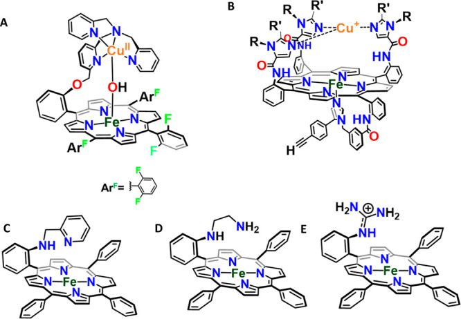
(A) L6-FeCu and (B) Fe–Cu heme Cu oxidase model. Porphyrins with basic residues in the distal super structure: (C) FeL2, (D) FeL3, and (E) Fe-Marg.
Under electrocatalytic conditions at neutral pH’s, a FeIII–OOH intermediate formed during O2 reduction needs to be reduced for the O–O bond to be cleaved.120,121 Failing to provide an electron rapidly results in its hydrolysis to generate H2O2.121 Alternatively, rapid heterolysis of the O–O bond would result in the formation of Compound I and avoid the release of H2O2. The effectiveness of these pendant groups in assisting the protonation and the ensuing heterolysis of the O–O bond in a FeIII–OOH intermediate, thus, can be judged better by slowing down the ET to the catalytic active site. ET rate constants for standard EPG electrodes are as high as ∼106 s–1, whereas electrodes modified with an octanethiol SAM (C8SH) and hexadecanethiol SAM (C16SH) show rate constants of 103 and 6–10 s–1, respectively.122−125 Under these slow ET fluxes, simple iron porphyrins, including ones having a distal hydrogen bonding cavity, which exhibit substantial selectivity for 4e–/4H+ ORR under rapid ET flux, show very high H2O2 production.67,120,126 Alternatively, these porphyrins with pendant basic residues show only a negligible decrease in selectivity even when the electron transfer was slowed down by several orders of magnitude, as they could heterolyze the O–O bond of this FeIII–OOH intermediate by rapid protonation, avoiding its hydrolysis as has been experimentally demonstrated in organic solution.98 The role of distal hydrogen bonding residues is also well established in binuclear synthetic models of CcO and mononuclear hangman porphyrins during the selective 4e–/4H+ reduction of O2.93,94,101,127
Slow Electron Transfer and Fast Proton Transfer: The Case of Cyt P450
The Cyt P450 superfamily of enzymes requires O2 binding and subsequent O–O bond scission during the ORR cycle but avoids reduction of these high valent compound I species.61,128 Cyt P450 (Figure 9A) is one of the largest known family of metallo-enzymes, which takes part in several catabolic and anabolic processes including but not limited to xenobiotic detoxification, steroids and hormone biosynthesis. Due to this diverse reactivity, Cyt P450 has always been a subject of interest to the scientific community, striving toward better understanding of the factors that govern their reactivity with an eventual goal of developing artificial catalysts mimicking the reactivity of these enzymes. The generation of the high valent intermediate compound I,32 responsible for the monooxygenation via a hydrogen atom transfer and rebound mechanism,129 from ferrous heme and O2 requires two protons and two electrons. Importantly further reduction of that compound I via ET would direct the reaction to the oxygen reduction pathway (Figure 9B, red arrow). The electrons required for generation of compound I from O2 are derived from a reductase protein or an ET active site in the same protein and, importantly, the ET rates in Cyt P450 are substantially lower than those of CcO. The reductase components of Cyt P450 systems involve various combinations of flavin and iron–sulfur clusters and these transfer electron to the Cyt P450 active site at rates of around 10–500 s–1.62 This is in contrast to the ET rates at the oxygen reduction site of CcO which are orders of magnitude greater (>105 s–1).
Figure 9.

(A) Active site of Cyt P450 (PDB ID: 1AKD)130 with thiolate bound heme. (B) Schematic representation of the competition between monooxygenation (blue arrow) and oxygen reduction (red arrow) by CytP450 family of enzymes.
A key toward achieving Cyt P450 reactivity is, thus, attenuating the rate of ET from the reductase component to the catalytic center to allow monooxygenase activity in preference to reductase activity (Figure 9B). Keeping this in mind, site isolated thiolate ligated Iron porphyrin active site which mimics of the active site of Cyt P450, were fabricated atop a SAM covered Au electrode and electrochemical O2 reduction was performed with that.69,131 An electrocatalytic O2 reduction (reductase activity) involves the same intermediates (Figure 9B) involved in substrate oxidation (oxidase reactivity) even in artificial systems as well. The only difference is that during electrocatalytic O2 reduction the high valent oxidants are reduced by ET from the electrode which acts as a mimic of the reductase component in the enzymatic systems. This raises the possibility of harnessing the oxidase activity of these high valent intermediates to oxidize organic molecules if their rates of formation via O–O heterolysis during O2 activation can be made much faster than their rates of decay (by electron transfer from the electrode), offering them a substantial lifetime to oxidize substrates present in the medium. The rate of electron transfer from the electrode to the iron porphyrin site was attenuated using SAMs of different chain lengths to derive monooxygenase reactivity from these constructs which otherwise show ORR activity at fast (∼106 s–1) ET rates.
Catalytic hydroxylation of strong C–H bonds (BDE > 100) to alcohol and epoxidation of alkenes, using molecular O2, with this electrochemical construct were demonstrated with turnover numbers > 104. Maximum yield was achieved when the gold electrode was modified with an octanethiol SAM which shows moderate electron transfer rate (103 s–1). Faster electron transfer than that with alkane thiols of shorter chain length resulted in reduced monooxygenase activity which was evident from the decreased yield of the oxidized product. Thus, tuning the electron transfer rate during the electrochemical ORR, molecular O2 can be efficiently harnessed for useful chemical transformation with synthetic molecules. Several Cyt P450 enzymes are known to exhibit kH/kD as high as 10 for HAT by compound I during the enzymatic process. The high H/D isotope effect (∼10) for cyclohexane (sp3 C–H) oxidation by this bioinspired electrocatalyst falls in the observed range expected for rate determining hydrogen atom transfer (HAT) reactions (Figure 10) by a compound I intermediate.132−136 Since these reactions were performed in an aqueous medium, it provided a unique opportunity to explore the H2O/D2O KSIE in these reactions in a synthetic system. The oxidation of cyclohexane by oxygen catalyzed by this electrochemical analogue has a H2O/D2O isotope effect of 12.8 ± 0.1. The large KSIE was associated with the heterolytic cleavage of the O–O bond in a FeIII–OOH intermediate owing to a lack of organized proton transfer channel mentioned in the previous section.67 Similar observation during the electrochemical ORR was also found in a previous investigation. For a combination of SERRS-RDE and H/D isotope effects on rate determining steps in that electrochemical O2 reduction, a KSIE as high as 18 was observed for an iron porphyrin complex with a covalently bound thiolate without any preorganized proton channel in the distal geometry.67 This suggests that the rate of O–O bond heterolysis, which depends on the availability of protons, becomes a decisive factor during the electrochemical ORR as well. Similarly, high KSIE of 15 was observed when the highly conserved aspartate residue asp251 in P450cam was mutated.137
Figure 10.

Schematic representation of the monooxygenase activity depicting both the kinetic solvent isotope effect (KSIE) and kinetic isotope effect (KIE) during the oxygenation of C–H bond.
Apart from that, this electrochemical construct was seen to exhibit an unusual selectivity. An apparent disparity with the erstwhile reported synthetic systems regarding the preferential oxidation of 2° C–H over 3° C–H and lack of overoxidation of the sterically demanding 2° alcohol produced was noted (Table 2).138−140 This was rationalized by the steric hindrance provided by the distal pocket as decorated by flanking, peripheral substituents of the synthetic porphyrin electrocatalyst. The four bulky pivaloyl groups in the distal structure provides a very narrow opening for the substrates to access the high valent intermediate (Figure 11). This narrow opening restricts the access of sterically demanding 3° C–H bonds over lesser sterically demanding 2° C–H bonds accounting for the preferential 2° C–H bond oxidation over 3° C–H bonds. Decreasing the steric hindrance by decreasing the number of peripheral substituents diminished the unusual selectivity of this electrocatalytic analogue of Cyt P450, supporting the steric control of selectivity proposed (Figure 11D).131
Table 2. Difference in Regioselectivity.

Figure 11.

(A) Top view of iron-picket fence porphyrin (FePf) showing a substrate access cavity with a diameter of 6 Å. (B) The access of tertiary H of adamantane demands a conelike geometry resulting in steric hindrance. This accounts for the lesser selectivity toward tertiary C–H bond oxidation. (C) Secondary H accesses the cavity with a lesser amount of steric hindrance because of its trigonal architecture, resulting in more selectivity toward secondary C–H oxidation despite the higher BDE. (D) Wider substrate access cavity of iron-half picket fence (FehPf) depicting the lesser amount of bulky pivolyl groups in the secondary geometry.
No Electron Transfer and No Proton Transfer: The Case of Heme Dioxygenase
Heme dioxygenases promote incorporation of both the oxygen atoms of molecular oxygen into organic substrates.37,141 Indoleamine 2,3-dioxygenase (IDO) and tryptophan 2,3-dioxygenase (TDO) are two heme based dioxygenases which play a crucial role in the physiological regulation of tryptophan (l-Trp) flux by catalyzing the initial and bottleneck step of l-Trp metabolism in the kynurenine (Kyn) pathway. This step involves the oxidative cleavage of the 2,3-double bond in the indole moiety of l-Trp, resulting in the formation of N-formyl Kyn. Both of the active sites of IDO and TDO contain the heme cofactor, proximally ligated to an imidazole residue and the active site is harbored inside a hydrophobic cavity (Figure 12A). Spectroscopic and analytical characterization of intermediates suggests that the mechanism of dioxygenation begins with the electrophilic attack of heme superoxide (FeIII–O2•–) to the C2 or the C3 position of the substrate, producing epoxides and a ferryl intermediate. This ferryl intermediate then continues further to produce a 4e– oxidized product (Figure 12B). Apart from the initial reduction from ferric heme to its ferrous state, unlike the monooxygenases, this reaction proceeds without any proton and electron as it regenerates the ferrous heme species in the end of the catalytic process. The active site is hydrophobic as indicated by ENDOR (electron–nuclear double resonance) data.142 The physiological reductase component for the dioxygenases is not known with certainty, but for IDO there is some evidence of it being cytochrome b5.143,144
Figure 12.

(A) O2 bound active site of heme dioxygenase (PDB ID: 5TI9).145 (B) Schematic representation of the formation of NFK by heme dioxygenase enzyme.
Replicating the reactivity of heme dioxygenase by generating FeIII–O2•– to achieve 4e– oxidation of organic substrates can be a lucrative chemical reaction. A major complication involved is that most reactions of ferrous porphyrins with O2 result in ferric porphyrin, and attempts to reduce the ferric porphyrin in the presence of O2 result in uncontrolled production of partially reduced O2 species (PROS) like O2–- and H2O2. However, in a recent report by Singha et al,. a FeIII–O2•– porphyrin was demonstrated to oxidize pendant quinols by 4 e– oxidized quinone molecules in homogeneous conditions (Figure 13A).146 The presence of FeIII–O2•–, FeIII–OOH, and FeIV=O intermediates is identified by employing EPR and resonance Raman spectroscopy during the course of reaction. A series of ferric heme superoxo oxidants was reported to oxidize an array of indole substrates into their corresponding 2,3-dioxygenated products in solution (Figure 13B).147 For the reasons described above, these transformations were not catalytic.
Figure 13.

Schematic representation of (A) reaction of a pendant quinol molecule with a heme superoxide complex and (B) reaction of a synthetic heme superoxide adduct with an array of indole substrates in solution.
Achieving the 4 e– oxidation of substrates using FeIII–O2•– in homogeneous conditions does not often ensure multiple turnovers, as the unreactive ferric porphyrin species is easily produced via the solvolysis of the FeIII–O2•– species.146 This can be remedied by performing the catalytic process in heterogeneous conditions. As stated earlier, an FeIII–O2•– species, the putative reactive intermediate of the heme dioxygenase enzyme, is an intermediate in heterogeneous electrochemical O2 reduction by iron porphyrins too and its population, under electrochemical conditions, can be tuned by controlling the applied potential. Recently, iron porphyrins having different axial ligands have been constructed on a self-assembled monolayer of thiols on a gold electrode, and electrochemical oxygen reduction reaction was performed with those constructs.148 The potential applied was held between E1 and E2 (Figure 3) to allow maximum population of the FeIII–O2•– species. During that process, dioxygenation of 3-methylindole and 4e– oxidation of a series of organic compounds having C–H bond dissociation energies (BDE) between 80 and 90 kcal/mol was achieved, with very high turnover, at potentials where FeIII–O2•– species are likely to be formed on the electrode (Figure 14A, Table 3). In contrast to the monooxygenation reaction, the involvement of the high valent compound-I-like species during this dioxygenation process was rescinded as evident from the fact that these electrochemical constructs were unable to oxidize substrates having BDE > 90 kcal/mol at the same potential. Mechanistic investigations through isotope effect calculations suggests that hydrogen atom transfer (HAT) by FeIII–O2•– is likely to be the rate determining step (Figure 14B). The lack of KSIE and insensitivity to ET rates (observed during monooxygenase activity) advocates for a 4e– oxidation catalyzed by these constructs using FeIII–O2•–. These results highlight a new reaction engineering approach to harness O2 as a green oxidant for efficient chemical oxidation.
Figure 14.

(A) Pictorial depiction of the electrochemical construct performing the dioxygenation of indole. (B) Schematic representation of the mechanism involved in the process of 4e- oxidation.
Table 3. Oxidation of the Substrates with Low BDE: Turnover Numbers (TON), Turnover Frequencies (TOF), Maximum TON Possible for Monooxygenase Reaction (TONmax), and ratio of TON and TONmax (μ) for the Products Obtained after the Oxidation of Substrates by Differently Ligated FePf Complexes.

L is the axial ligand.
TON implies turnover number.
Turnover frequency averaged over the entire duration of the experiment.
Maximum turnover expected from monooxygenase reactivity and.
The ratio of b and d. The >1 μ value clearly indicates a reaction mechanism other than monooxygenation.
Fast Proton Transfer and No Electron Transfer: The Case of Heme Peroxidase
Unlike heme oxygenases, heme peroxidases engage themselves in useful chemical transformations through oxidation and oxygenation by wielding the oxidizing power of peroxides.39,149 Among the various activities, biosynthesis of prostanoids,150 defending plants and animals against pathogens,151 and bioregulation of free peroxides to protect organisms from oxidative damage152 are a few worth mentioning. The prolific reactivity of peroxidase is due to the generation of the versatile high valent compound I intermediate upon the reaction of peroxides with resting ferric heme active site. Generally, reaction of heme enzymes with hydrogen peroxide generates a FeIII–OOH species which, after the heterolytic cleavage of the O–O bond, forms compound I.153 The facile generation of compound I in this process depends on the participation of the distal residues such as Arg and His (Figure 15).154,155 These residues contribute to the facile heterolytic cleavage of the O–O bond by an acid–base catalysis mechanism. This compound I oxidizes substrates by one electron, forming compound II which in turn participates in a second one electron oxidation process and regenerates the resting ferric species. Apart from accelerating the facile formation of compound I, the distal Arg residue is also proposed to stabilize both the compound I and II intermediates by hydrogen bonding to the ferryl oxygen. Thus, peroxidases participate in the oxidation procedure via the same intermediate as Cyt P450 without the requirement of any reducing equivalent. Note that a group of Cyt P450 enzymes, known as peroxygenases, use peroxides as a surrogate of O2 not only in the bacterial system but also in several developed organisms like human beings primarily if the electron transfer to the active site becomes unfavorable. Though the use of peroxides can lead to high inefficiency and inactivation of enzymes, there are many examples where peroxides have been used effectively in α–β hydroxylation, decarboxylation, and oxidation of the cyano groups.42,156,157
Figure 15.

(A) Active site of heme peroxidase (PDB ID: 2CYP).149 (B) Interaction of the distal Arg and His with FeIII–OOH.
The critical role of the Arg residue in the enzymatic activity of heme peroxidase enzymes has always been a matter of interest to the scientific community.14,155 Recently, a synthetic iron porphyrin (Fe-MARG) was reported where a pendant guanidine residue, mimicking the Arg residue in the enzymatic site, was strategically placed on top of the active site.102 This synthetic design achieves the goal of incorporating a distal guanidinium residue in an iron porphyrin mimicking the Arg in the active site of HRP. The crystal structure of the free ligand indicates a suitable orientation of the guanidinium group with respect to the metal binding site in the porphyrin. The Fe-MARG forms compound I (Figure 16), verified by mechanistic and spectroscopic analysis, using organic peroxide which rapidly oxidizes phenolic and amine substrates with high rates. Unsubstituted iron TPP does not show either rapid peroxidase activity or formation of compound I in the same time scales, illustrating the advantage of the pendant guanidinium (protonated at neutral pH) group. The guanidinium group is a strong hydrogen bond donor and can hydrogen bond to the substrates used. This strong H-bonding interaction is responsible for enzyme-like Michalis-Menten kinetics exhibited by this complex. However, the activation of peroxide is not limited to a guanidium group in the distal pocket, any pendant basic residue that are protonated at neutral pH and potent to form second sphere hydrogen bonding interactions, results in facile formation of compound I from peracids. Short-lived Compound I intermediates are formed from peroxides and characterized at cryogenic temperatures using UV–vis electronic absorption spectroscopy and EPR spectroscopy with synthetic iron porphyrins having pendant aliphatic amine in the distal position (FeL3, Figure 16B) having pKa close to that of guanidine.99 The advantage of distal basic residue which remain protonated in the neutral pH was assessed with Density Functional Theory (DFT) calculations. DFT calculations indicate that these protonated residues result in the polarization of the electron density of the O–O σ-bond and thus results in facile O–O bond heterolysis. These artificial constructs could catalyze the oxidation of Ferrocene, 2,4,6-Tri tertiarybutyl phenol (TBPH), o-Phenylenediamine, 3,3′,5,5′-Tetramethylbenzidine with TOF of 2.7, 1.4, 0.85, and 25 s–1 respectively.99,102
Figure 16.

Peroxidase activity of synthetic heme (A) Fe-MARG and (B) Fe-L3.
Formation of compound I was envisaged during the H2O2 disproportionation reaction with phenolate ligated iron porphyrin complexes.158 Compound I has been detected using in situ SERRS during the H2O2 disproportionation (HPD) reaction by phenolate ligated iron porphyrin immobilized on self-assembled monolayer modified Au electrodes where Fe–O vibration of the compound I species were detected at 796 and 803 cm–1 for porphyrins containing hydrogen bonding (Fe-tetraester porphyrin, FeEs4) and non-hydrogen bonding (Fe-“picket fence” porphyrin, FePf) distal structures, respectively. Compound I, so generated, was used to oxidize range organic substrates in aqueous medium.159 The in situ generated compound I, with each of the axially ligated constructs, via a peroxide shunt, abstracts a hydrogen atom from the C–H bond as is evident from the recorded KIEs. Variation of the axial ligand from thiolate to phenolate to imidazole during the oxidation not only result in the decrease in the yield but also result in significantly higher isotope effect than thiolate ligation. These higher KIE values in phenolate and imidazole ligated constructs likely reflect the involvement of HAT transition states (TS) having different polarity.160 For example, a higher KIE for FePf with an anionic axial ligand (phenolate) than for that with a neutral axial ligand (imidazole) is consistent with the hypothesis of Shaik et al.160 The barrier of hydrogen atom tunneling depends on the electrostatic stabilization of the Hδ+ as it moves between the Cδ− and Oδ− (Figure 17B) moieties in the transition state (TS).160 The neutral imidazole axial ligand results in a lesser electrostatic stabilization in the TS than the anionic phenolate axial ligand in the rate-limiting step. Thus, the lesser electrostatically stabilized transition state for imidazole bound compound I goes through a wider tunneling barrier and thus results in lower KIE values than the more electrostatically stabilized TS of phenolate bound compound I.
Figure 17.

(A) Tunneling lowers the observed barrier relative to the semi classical TS. (B) Schematic representation of the TS where L represents the different axial ligations.
Electron Transfer and No Proton Transfer: The Case of Cytochrome c
Heme enzymes that only have a high electron transfer rate to the active site are the electron transfer proteins like cytochrome c.15 The Fe-porphyrin active site on top of a SAM modified gold electrode was demonstrated by Bandyopadhyay et al. where spectroscopic characterization coupled to electrochemical and theoretical calculation provided a detailed understanding of the factors affecting the ET kinetics in the synthetic Fe-porphyrin systems.161 Detailed discussion on this topic is mindfully avoided here as it is not the focus of this Article.
Outlook
The ability to control the ET and PT pathways to iron porphyrins which mimic the active site of different heme enzymes has led to the realization that by manipulating the ET and PT supply the function of the same iron porphyrin site may be controlled. Such control of branching of different reactive pathways has been demonstrated in heme enzymes, with monooxygenase vs peroxygenase being a great example of that.40,162 In these heterogeneous constructs, SAM chain length and porphyrin ligand design allow attenuation of ET and PT pathways, respectively. The branching or reactivity by controlling the ET and PT is also enacted in these constructs. For example, a thiolate bound iron porphyrin can reduce O2 to H2O when ET and PT are fast. The same system acts as a monooxygenase when the ET is slowed down and dioxygenase when ET is utilized only to reduce the ferric state to the ferrous state. Similarly, iron porphyrins with active proton shuttles can reduce O2 to H2O when ET is fast while they act as peroxidases when H2O2 is used and ET is blocked. Thus, in addition to the generally held convictions regarding the role of axial ligands and distal environment, biomimetic chemistry should be cognizant of the fact that the ET and PT steps are factors that contribute to the diversity of reactivity exhibited by heme active sites as well. While the individual oxygenase, oxidase, peroxidase, and peroxygenase activities are optimal only when proper combinations of axial ligands and distal residues are used, the ability to use these heterogeneous bioinspired constructs to catalyze O2 reduction, monooxygenase, dioxygenase, catalase, and peroxidase (all with TON > 103 at room temperature and in aqueous solvent) simply by controlling the ET rate, nonetheless, opens up a new versatile reaction engineering approach toward O2 activation for useful chemical transformations.
Acknowledgments
The work is supported by Department of Science and Technology, Government of India Grant SERB/EMR-0008063 and STR/2019/000081.
The authors declare no competing financial interest.
References
- Huang X.; Groves J. T. Oxygen Activation and Radical Transformations in Heme Proteins and Metalloporphyrins. Chem. Rev. 2018, 118, 2491–2553. 10.1021/acs.chemrev.7b00373. [DOI] [PMC free article] [PubMed] [Google Scholar]
- Lee D.; Pierce B.; Krebs C.; Hendrich M. P.; Huynh B. H.; Lippard S. J. Functional Mimic of Dioxygen-Activating Centers in Non-Heme Diiron Enzymes: Mechanistic Implications of Paramagnetic Intermediates in the Reactions between Diiron(II) Complexes and Dioxygen. J. Am. Chem. Soc. 2002, 124, 3993–4007. 10.1021/ja012251t. [DOI] [PubMed] [Google Scholar]
- Que L.; Tolman W. B. Biologically inspired oxidation catalysis. Nature 2008, 455, 333–340. 10.1038/nature07371. [DOI] [PubMed] [Google Scholar]
- Garcia-Bosch I.; Ribas X.; Costas M. Electrophilic Arene Hydroxylation and Phenol O-H Oxidations Performed by an Unsymmetric μ-η1:η1-O2-Peroxo Dicopper(II) Complex. Chem. - Eur. J. 2012, 18, 2113–2122. 10.1002/chem.201102372. [DOI] [PubMed] [Google Scholar]
- Collman J. P.; Fu L. Synthetic Models for Hemoglobin and Myoglobin. Acc. Chem. Res. 1999, 32, 455–463. 10.1021/ar9603064. [DOI] [Google Scholar]
- Collman J. P.; Boulatov R.; Sunderland C. J.; Fu L. Functional Analogues of Cytochrome c Oxidase, Myoglobin, and Hemoglobin. Chem. Rev. 2004, 104, 561–588. 10.1021/cr0206059. [DOI] [PubMed] [Google Scholar]
- Costas M.; Mehn M. P.; Jensen M. P.; Que L. Dioxygen Activation at Mononuclear Nonheme Iron Active Sites: Enzymes, Models, and Intermediates. Chem. Rev. 2004, 104, 939–986. 10.1021/cr020628n. [DOI] [PubMed] [Google Scholar]
- Kim E.; Chufán E. E.; Kamaraj K.; Karlin K. D. Synthetic Models for Heme-Copper Oxidases. Chem. Rev. 2004, 104, 1077–1134. 10.1021/cr0206162. [DOI] [PubMed] [Google Scholar]
- Lewis E. A.; Tolman W. B. Reactivity of Dioxygen-Copper Systems. Chem. Rev. 2004, 104, 1047–1076. 10.1021/cr020633r. [DOI] [PubMed] [Google Scholar]
- Mirica L. M.; Ottenwaelder X.; Stack T. D. P. Structure and Spectroscopy of Copper-Dioxygen Complexes. Chem. Rev. 2004, 104, 1013–1046. 10.1021/cr020632z. [DOI] [PubMed] [Google Scholar]
- Tshuva E. Y.; Lippard S. J. Synthetic Models for Non-Heme Carboxylate-Bridged Diiron Metalloproteins: Strategies and Tactics. Chem. Rev. 2004, 104, 987–1012. 10.1021/cr020622y. [DOI] [PubMed] [Google Scholar]
- Venkateswara Rao P.; Holm R. H. Synthetic Analogues of the Active Sites of Iron-Sulfur Proteins. Chem. Rev. 2004, 104, 527–560. 10.1021/cr020615+. [DOI] [PubMed] [Google Scholar]
- Cady C. W.; Crabtree R. H.; Brudvig G. W. Functional models for the oxygen-evolving complex of photosystem II. Coord. Chem. Rev. 2008, 252, 444–455. 10.1016/j.ccr.2007.06.002. [DOI] [PMC free article] [PubMed] [Google Scholar]
- Poulos T. L. Heme Enzyme Structure and Function. Chem. Rev. 2014, 114, 3919–3962. 10.1021/cr400415k. [DOI] [PMC free article] [PubMed] [Google Scholar]
- Bertini I.; Cavallaro G.; Rosato A. Cytochrome c: Occurrence and Functions. Chem. Rev. 2006, 106, 90–115. 10.1021/cr050241v. [DOI] [PubMed] [Google Scholar]
- Wikström M.; Krab K.; Sharma V. Oxygen Activation and Energy Conservation by Cytochrome c Oxidase. Chem. Rev. 2018, 118, 2469–2490. 10.1021/acs.chemrev.7b00664. [DOI] [PMC free article] [PubMed] [Google Scholar]
- Lukin J. A.; Ho C. The Structure-Function Relationship of Hemoglobin in Solution at Atomic Resolution. Chem. Rev. 2004, 104, 1219–1230. 10.1021/cr940325w. [DOI] [PubMed] [Google Scholar]
- Silverstein T. P.; Kirk S. R.; Meyer S. C.; Holman K. L. M. Myoglobin structure and function: A multiweek biochemistry laboratory project. Biochem. Mol. Biol. Educ. 2015, 43, 181–188. 10.1002/bmb.20845. [DOI] [PubMed] [Google Scholar]
- Sono M.; Roach M. P.; Coulter E. D.; Dawson J. H. Heme-Containing Oxygenases. Chem. Rev. 1996, 96, 2841–2888. 10.1021/cr9500500. [DOI] [PubMed] [Google Scholar]
- Hayaishi O.; Rothberg S.; Mehler A. H.; Saito Y. STUDIES ON OXYGENASES: ENZYMATIC FORMATION OF KYNURENINE FROM TRYPTOPHAN. J. Biol. Chem. 1957, 229, 889–896. 10.1016/S0021-9258(19)63693-8. [DOI] [PubMed] [Google Scholar]
- Mason H. S. Mechanisms of Oxygen Metabolism. Science 1957, 125, 1185. 10.1126/science.125.3259.1185. [DOI] [PubMed] [Google Scholar]
- Rydberg P.; Sigfridsson E.; Ryde U. On the role of the axial ligand in heme proteins: a theoretical study. JBIC, J. Biol. Inorg. Chem. 2004, 9, 203–223. 10.1007/s00775-003-0515-y. [DOI] [PubMed] [Google Scholar]
- Gross Z. The effect of axial ligands on the reactivity and stability of the oxoferryl moiety in model complexes of Compound I of heme-dependent enzymes. JBIC, J. Biol. Inorg. Chem. 1996, 1, 368–371. 10.1007/s007750050067. [DOI] [Google Scholar]
- Collman J. P.; Decréau R. A.; Lin H.; Hosseini A.; Yang Y.; Dey A.; Eberspacher T. A. Role of a distal pocket in the catalytic O2 reduction by cytochrome c oxidase models immobilized on interdigitated array electrodes. Proc. Natl. Acad. Sci. U. S. A. 2009, 106, 7320. 10.1073/pnas.0902285106. [DOI] [PMC free article] [PubMed] [Google Scholar]
- Lee H. J.; Reimann J.; Huang Y.; Ädelroth P. Functional proton transfer pathways in the heme-copper oxidase superfamily. Biochim. Biophys. Acta, Bioenerg. 2012, 1817, 537–544. 10.1016/j.bbabio.2011.10.007. [DOI] [PubMed] [Google Scholar]
- The Porphyrin Handbook. Volumes 1–10 Edited by Karl M. Kadish (Univeristy of Houston), Kevin M. Smith (University of California, Davis), and Roger Guilard (Universite de Bourgogne). Academic Press: San Diego, CA. 2000. $2975.00 (set). ISBN 0-12-393200-7 (set). J. Am. Chem. Soc. 2000, 122, 3984–3984. 10.1021/ja0047065 [DOI] [Google Scholar]
- deVisser S. P.; Shaik S.; Sharma P. K.; Kumar D.; Thiel W. Active Species of Horseradish Peroxidase (HRP) and Cytochrome P450: Two Electronic Chameleons. J. Am. Chem. Soc. 2003, 125, 15779–15788. 10.1021/ja0380906. [DOI] [PubMed] [Google Scholar]
- Fenton H. J. H. LXXIII.—Oxidation of tartaric acid in presence of iron. J. Chem. Soc., Trans. 1894, 65, 899–910. 10.1039/CT8946500899. [DOI] [Google Scholar]
- Goldstein S.; Meyerstein D.; Czapski G. The Fenton reagents. Free Radical Biol. Med. 1993, 15, 435–445. 10.1016/0891-5849(93)90043-T. [DOI] [PubMed] [Google Scholar]
- Akhtar M.; Pechet M. M. The Mechanism of the Barton Reaction. J. Am. Chem. Soc. 1964, 86, 265–268. 10.1021/ja01056a035. [DOI] [Google Scholar]
- Bogan D. J.; Havel J. J.; Coveleskie R. A.; Celii F.; Stammerjohn B. J.; Sanders W. A.; Williams F. W.; Carhart H. W. Quantitative product study of russell termination reactions of peroxy radicals in the gas phase. Symp. (Int.) Combust., [Proc.] 1981, 18, 843–844. 10.1016/S0082-0784(81)80087-2. [DOI] [Google Scholar]
- Rittle J.; Green M. T. Cytochrome P450 Compound I: Capture, Characterization, and C-H Bond Activation Kinetics. Science 2010, 330, 933. 10.1126/science.1193478. [DOI] [PubMed] [Google Scholar]
- Green M. T. The Structure and Spin Coupling of Catalase Compound I: A Study of Noncovalent Effects. J. Am. Chem. Soc. 2001, 123, 9218–9219. 10.1021/ja010105h. [DOI] [PubMed] [Google Scholar]
- Li H.; Poulos T. L. Structural variation in heme enzymes: a comparative analysis of peroxidase and P450 crystal structures. Structure 1994, 2, 461–464. 10.1016/S0969-2126(00)00046-0. [DOI] [PubMed] [Google Scholar]
- Groves J. T. Key elements of the chemistry of cytochrome P-450: The oxygen rebound mechanism. J. Chem. Educ. 1985, 62, 928. 10.1021/ed062p928. [DOI] [Google Scholar]
- Derat E.; Cohen S.; Shaik S.; Altun A.; Thiel W. Principal Active Species of Horseradish Peroxidase, Compound I: A Hybrid Quantum Mechanical/Molecular Mechanical Study. J. Am. Chem. Soc. 2005, 127, 13611–13621. 10.1021/ja0534046. [DOI] [PubMed] [Google Scholar]
- Efimov I.; Basran J.; Thackray S. J.; Handa S.; Mowat C. G.; Raven E. L. Structure and Reaction Mechanism in the Heme Dioxygenases. Biochemistry 2011, 50, 2717–2724. 10.1021/bi101732n. [DOI] [PMC free article] [PubMed] [Google Scholar]
- Ferguson-Miller S.; Babcock G. T. Heme/Copper Terminal Oxidases. Chem. Rev. 1996, 96, 2889–2908. 10.1021/cr950051s. [DOI] [PubMed] [Google Scholar]
- Poulos T. L. Thirty years of heme peroxidase structural biology. Arch. Biochem. Biophys. 2010, 500, 3–12. 10.1016/j.abb.2010.02.008. [DOI] [PMC free article] [PubMed] [Google Scholar]
- Makris T. M.; Denisov I.; Schlichting I.; Sligar S. G.. Activation of Molecular Oxygen by Cytochrome P450. In Cytochrome P450: Structure, Mechanism, and Biochemistry; Ortiz de Montellano P. R., Ed.; Springer US: Boston, MA, 2005; pp 149–182. [Google Scholar]
- Grinkova Y. V.; Denisov I. G.; McLean M. A.; Sligar S. G. Oxidase uncoupling in heme monooxygenases: Human cytochrome P450 CYP3A4 in Nanodiscs. Biochem. Biophys. Res. Commun. 2013, 430, 1223–1227. 10.1016/j.bbrc.2012.12.072. [DOI] [PMC free article] [PubMed] [Google Scholar]
- Munro A. W.; McLean K. J.; Grant J. L.; Makris T. M. Structure and function of the cytochrome P450 peroxygenase enzymes. Biochem. Soc. Trans. 2018, 46, 183–196. 10.1042/BST20170218. [DOI] [PMC free article] [PubMed] [Google Scholar]
- Hersleth H.-P.; Hsiao Y.-W.; Ryde U.; Gorbitz C. H.; Andersson K. K. The crystal structure of peroxymyoglobin generated through cryoradiolytic reduction of myoglobin compound III during data collection. Biochem. J. 2008, 412, 257–264. 10.1042/BJ20070921. [DOI] [PubMed] [Google Scholar]
- Kuo H. H.; Mauk A. G. Indole peroxygenase activity of indoleamine 2,3-dioxygenase. Proc. Natl. Acad. Sci. U. S. A. 2012, 109, 13966. 10.1073/pnas.1207191109. [DOI] [PMC free article] [PubMed] [Google Scholar]
- Egawa T.; Shimada H.; Ishimura Y. Formation of Compound I in the Reaction of Native Myoglobins with Hydrogen Peroxide*. J. Biol. Chem. 2000, 275, 34858–34866. 10.1074/jbc.M004026200. [DOI] [PubMed] [Google Scholar]
- Vidakovic M.; Sligar S. G.; Li H.; Poulos T. L. Understanding the Role of the Essential Asp251 in Cytochrome P450cam Using Site-Directed Mutagenesis, Crystallography, and Kinetic Solvent Isotope Effect. Biochemistry 1998, 37, 9211–9219. 10.1021/bi980189f. [DOI] [PubMed] [Google Scholar]
- Gray H. B.; Winkler J. R. Annu. Rev. Biochem. 1996, 65, 537–561. 10.1146/annurev.bi.65.070196.002541. [DOI] [PubMed] [Google Scholar]
- Makinen M. W.; Schichman S. A.; Hill S. C.; Gray H. B. Heme-heme orientation and electron transfer kinetic behavior of multisite oxidation-reduction enzymes. Science 1983, 222, 929. 10.1126/science.6415814. [DOI] [PubMed] [Google Scholar]
- Kamachi T.; Kouno T.; Nam W.; Yoshizawa K. How axial ligands control the reactivity of high-valent iron(IV)-oxo porphyrin π-cation radicals in alkane hydroxylation: A computational study. J. Inorg. Biochem. 2006, 100, 751–754. 10.1016/j.jinorgbio.2006.01.007. [DOI] [PubMed] [Google Scholar]
- Yoshioka S.; Takahashi S.; Ishimori K.; Morishima I. Roles of the axial push effect in cytochrome P450cam studied with the site-directed mutagenesis at the heme proximal site. J. Inorg. Biochem. 2000, 81, 141–151. 10.1016/S0162-0134(00)00097-0. [DOI] [PubMed] [Google Scholar]
- Green M. T. CH bond activation in heme proteins: the role of thiolate ligation in cytochrome P450. Curr. Opin. Chem. Biol. 2009, 13, 84–88. 10.1016/j.cbpa.2009.02.028. [DOI] [PubMed] [Google Scholar]
- Dawson J. H.; Sono M. Cytochrome P-450 and chloroperoxidase: thiolate-ligated heme enzymes. Spectroscopic determination of their active-site structures and mechanistic implications of thiolate ligation. Chem. Rev. 1987, 87, 1255–1276. 10.1021/cr00081a015. [DOI] [Google Scholar]
- Vogel K. M.; Kozlowski P. M.; Zgierski M. Z.; Spiro T. G. Determinants of the FeXO (X = C, N, O) Vibrational Frequencies in Heme Adducts from Experiment and Density Functional Theory. J. Am. Chem. Soc. 1999, 121, 9915–9921. 10.1021/ja990042r. [DOI] [Google Scholar]
- Das P. K.; Chatterjee S.; Samanta S.; Dey A. EPR, Resonance Raman, and DFT Calculations on Thiolate-and Imidazole-Bound Iron(III) Porphyrin Complexes: Role of the Axial Ligand in Tuning the Electronic Structure. Inorg. Chem. 2012, 51, 10704–10714. 10.1021/ic3016035. [DOI] [PubMed] [Google Scholar]
- Amanullah S.; Singha A.; Dey A. Tailor made iron porphyrins for investigating axial ligand and distal environment contributions to electronic structure and reactivity. Coord. Chem. Rev. 2019, 386, 183–208. 10.1016/j.ccr.2019.01.021. [DOI] [Google Scholar]
- Dey A.; Okamura T.-a.; Ueyama N.; Hedman B.; Hodgson K. O.; Solomon E. I. Sulfur K-Edge XAS and DFT Calculations on P450 Model Complexes: Effects of Hydrogen Bonding on Electronic Structure and Redox Potentials. J. Am. Chem. Soc. 2005, 127, 12046–12053. 10.1021/ja0519031. [DOI] [PMC free article] [PubMed] [Google Scholar]
- Chottard G.; Schappacher M.; Ricard L.; Weiss R. Resonance Raman spectra of iron(II) cytochrome P450 model complexes: influence of the thiolate ligand. Inorg. Chem. 1984, 23, 4557–4561. 10.1021/ic00194a030. [DOI] [Google Scholar]
- Tani F.; Matsu-ura M.; Nakayama S.; Naruta Y. Synthetic models for the active site of cytochrome P450. Coord. Chem. Rev. 2002, 226, 219–226. 10.1016/S0010-8545(01)00444-1. [DOI] [Google Scholar]
- Safo M. K.; Walker F. A.; Raitsimring A. M.; Walters W. P.; Dolata D. P.; Debrunner P. G.; Scheidt W. R. Axial Ligand Orientation in Iron(III) Porphyrinates: Effect of Axial.pi.-Acceptors. Characterization of the Low-Spin Complex [Fe(TPP)(4-CNPy)2]ClO4. J. Am. Chem. Soc. 1994, 116, 7760–7770. 10.1021/ja00096a037. [DOI] [Google Scholar]
- Shin I.; Ambler B. R.; Wherritt D.; Griffith W. P.; Maldonado A. C.; Altman R. A.; Liu A. Stepwise O-Atom Transfer in Heme-Based Tryptophan Dioxygenase: Role of Substrate Ammonium in Epoxide Ring Opening. J. Am. Chem. Soc. 2018, 140, 4372–4379. 10.1021/jacs.8b00262. [DOI] [PMC free article] [PubMed] [Google Scholar]
- Denisov I. G.; Makris T. M.; Sligar S. G.; Schlichting I. Structure and Chemistry of Cytochrome P450. Chem. Rev. 2005, 105, 2253–2278. 10.1021/cr0307143. [DOI] [PubMed] [Google Scholar]
- Guengerich F. P.; Johnson W. W. Kinetics of Ferric Cytochrome P450 Reduction by NADPH-Cytochrome P450 Reductase: Rapid Reduction in the Absence of Substrate and Variations among Cytochrome P450 Systems. Biochemistry 1997, 36, 14741–14750. 10.1021/bi9719399. [DOI] [PubMed] [Google Scholar]
- Varotsis C.; Zhang Y.; Appelman E. H.; Babcock G. T. Resolution of the reaction sequence during the reduction of O2 by cytochrome oxidase. Proc. Natl. Acad. Sci. U. S. A. 1993, 90, 237. 10.1073/pnas.90.1.237. [DOI] [PMC free article] [PubMed] [Google Scholar]
- von Ballmoos C.; Ädelroth P.; Gennis R. B.; Brzezinski P. Proton transfer in ba3 cytochrome c oxidase from Thermus thermophilus. Biochim. Biophys. Acta, Bioenerg. 2012, 1817, 650–657. 10.1016/j.bbabio.2011.11.015. [DOI] [PubMed] [Google Scholar]
- Matsui T.; Ozaki S.-i.; Watanabe Y. On the Formation and Reactivity of Compound I of the His-64 Myoglobin Mutants*. J. Biol. Chem. 1997, 272, 32735–32738. 10.1074/jbc.272.52.32735. [DOI] [PubMed] [Google Scholar]
- Collman J. P.; Decréau R. A. Functional biomimetic models for the active site in the respiratory enzyme cytochrome c oxidase. Chem. Commun. 2008, 5065–5076. 10.1039/b808070b. [DOI] [PubMed] [Google Scholar]
- Chatterjee S.; Sengupta K.; Hematian S.; Karlin K. D.; Dey A. Electrocatalytic O2-Reduction by Synthetic Cytochrome c Oxidase Mimics: Identification of a “Bridging Peroxo” Intermediate Involved in Facile 4e-/4H+ O2-Reduction. J. Am. Chem. Soc. 2015, 137, 12897–12905. 10.1021/jacs.5b06513. [DOI] [PMC free article] [PubMed] [Google Scholar]
- Samanta S.; Das P. K.; Chatterjee S.; Sengupta K.; Mondal B.; Dey A. O2 Reduction Reaction by Biologically Relevant Anionic Ligand Bound Iron Porphyrin Complexes. Inorg. Chem. 2013, 52, 12963–12971. 10.1021/ic4020652. [DOI] [PubMed] [Google Scholar]
- Sengupta K.; Chatterjee S.; Samanta S.; Bandyopadhyay S.; Dey A. Resonance Raman and Electrocatalytic Behavior of Thiolate and Imidazole Bound Iron Porphyrin Complexes on Self Assembled Monolayers: Functional Modeling of Cytochrome P450. Inorg. Chem. 2013, 52, 2000–2014. 10.1021/ic302369v. [DOI] [PubMed] [Google Scholar]
- Reed C. J.; Lam Q. N.; Mirts E. N.; Lu Y. Molecular understanding of heteronuclear active sites in heme-copper oxidases, nitric oxide reductases, and sulfite reductases through biomimetic modelling. Chem. Soc. Rev. 2021, 50, 2486–2539. 10.1039/D0CS01297A. [DOI] [PMC free article] [PubMed] [Google Scholar]
- Yu Y.; Cui C.; Liu X.; Petrik I. D.; Wang J.; Lu Y. A Designed Metalloenzyme Achieving the Catalytic Rate of a Native Enzyme. J. Am. Chem. Soc. 2015, 137, 11570–11573. 10.1021/jacs.5b07119. [DOI] [PMC free article] [PubMed] [Google Scholar]
- Mirts E. N.; Bhagi-Damodaran A.; Lu Y. Understanding and Modulating Metalloenzymes with Unnatural Amino Acids, Non-Native Metal Ions, and Non-Native Metallocofactors. Acc. Chem. Res. 2019, 52, 935–944. 10.1021/acs.accounts.9b00011. [DOI] [PMC free article] [PubMed] [Google Scholar]
- Mirts E. N.; Petrik I. D.; Hosseinzadeh P.; Nilges M. J.; Lu Y. A designed heme-[4Fe-4S] metalloenzyme catalyzes sulfite reduction like the native enzyme. Science 2018, 361, 1098. 10.1126/science.aat8474. [DOI] [PMC free article] [PubMed] [Google Scholar]
- Yeung N.; Lin Y.-W.; Gao Y.-G.; Zhao X.; Russell B. S.; Lei L.; Miner K. D.; Robinson H.; Lu Y. Rational design of a structural and functional nitric oxide reductase. Nature 2009, 462, 1079–1082. 10.1038/nature08620. [DOI] [PMC free article] [PubMed] [Google Scholar]
- Liu J.; Chakraborty S.; Hosseinzadeh P.; Yu Y.; Tian S.; Petrik I.; Bhagi A.; Lu Y. Metalloproteins Containing Cytochrome, Iron-Sulfur, or Copper Redox Centers. Chem. Rev. 2014, 114, 4366–4469. 10.1021/cr400479b. [DOI] [PMC free article] [PubMed] [Google Scholar]
- Pfister T. D.; Ohki T.; Ueno T.; Hara I.; Adachi S.; Makino Y.; Ueyama N.; Lu Y.; Watanabe Y. Monooxygenation of an Aromatic Ring by F43W/H64D/V68I Myoglobin Mutant and Hydrogen Peroxide: MYOGLOBIN MUTANTS AS A MODEL FOR P450 HYDROXYLATION CHEMISTRY *. J. Biol. Chem. 2005, 280, 12858–12866. 10.1074/jbc.M410853200. [DOI] [PubMed] [Google Scholar]
- Sabuncu S.; Reed J. H.; Lu Y.; Moënne-Loccoz P. Nitric Oxide Reductase Activity in Heme-Nonheme Binuclear Engineered Myoglobins through a One-Electron Reduction Cycle. J. Am. Chem. Soc. 2018, 140, 17389–17393. 10.1021/jacs.8b11037. [DOI] [PMC free article] [PubMed] [Google Scholar]
- Sigman J. A.; Kwok B. C.; Gengenbach A.; Lu Y. Design and Creation of a Cu(II)-Binding Site in Cytochrome c Peroxidase that Mimics the CuB-heme Center in Terminal Oxidases. J. Am. Chem. Soc. 1999, 121, 8949–8950. 10.1021/ja991195h. [DOI] [Google Scholar]
- Pfister T. D.; Mirarefi A. Y.; Gengenbach A. J.; Zhao X.; Danstrom C.; Conatser N.; Gao Y.-G.; Robinson H.; Zukoski C. F.; Wang A. H. J.; Lu Y. Kinetic and crystallographic studies of a redesigned manganese-binding site in cytochrome c peroxidase. JBIC, J. Biol. Inorg. Chem. 2006, 12, 126. 10.1007/s00775-006-0171-0. [DOI] [PubMed] [Google Scholar]
- Collman J. P.; Sorrell T. N.; Hoffman B. M. Models for cytochrome P-450. J. Am. Chem. Soc. 1975, 97, 913–914. 10.1021/ja00837a050. [DOI] [PubMed] [Google Scholar]
- Collman J. P.; Sorrell T. N. Model for the carbonyl adduct of ferrous cytochrome P 450. J. Am. Chem. Soc. 1975, 97, 4133–4134. 10.1021/ja00847a046. [DOI] [PubMed] [Google Scholar]
- Sono M.; Andersson L. A.; Dawson J. H. Sulfur donor ligand binding to ferric cytochrome P-450-CAM and myoglobin. Ultraviolet-visible absorption, magnetic circular dichroism, and electron paramagnetic resonance spectroscopic investigation of the complexes. J. Biol. Chem. 1982, 257, 8308–8320. 10.1016/S0021-9258(18)34332-1. [DOI] [PubMed] [Google Scholar]
- Matsui T.; Nakajima A.; Fujii H.; Matera K. M.; Migita C. T.; Yoshida T.; Ikeda-Saito M. O2-and H2O2-dependent Verdoheme Degradation by Heme Oxygenase: REACTION MECHANISMS AND POTENTIAL PHYSIOLOGICAL ROLES OF THE DUAL PATHWAY DEGRADATION*. J. Biol. Chem. 2005, 280, 36833–36840. 10.1074/jbc.M503529200. [DOI] [PubMed] [Google Scholar]
- Uchida T.; Sekine Y.; Dojun N.; Lewis-Ballester A.; Ishigami I.; Matsui T.; Yeh S.-R.; Ishimori K. Reaction intermediates in the heme degradation reaction by HutZ from Vibrio cholerae. Dalton Trans. 2017, 46, 8104–8109. 10.1039/C7DT01562C. [DOI] [PMC free article] [PubMed] [Google Scholar]
- Halime Z.; Kotani H.; Li Y.; Fukuzumi S.; Karlin K. D. Homogeneous catalytic O2 reduction to water by a cytochrome c oxidase model with trapping of intermediates and mechanistic insights. Proc. Natl. Acad. Sci. U. S. A. 2011, 108, 13990. 10.1073/pnas.1104698108. [DOI] [PMC free article] [PubMed] [Google Scholar]
- Chatterjee S.; Sengupta K.; Mondal B.; Dey S.; Dey A. Factors Determining the Rate and Selectivity of 4e-/4H+ Electrocatalytic Reduction of Dioxygen by Iron Porphyrin Complexes. Acc. Chem. Res. 2017, 50, 1744–1753. 10.1021/acs.accounts.7b00192. [DOI] [PubMed] [Google Scholar]
- Pegis M. L.; Wise C. F.; Martin D. J.; Mayer J. M. Oxygen Reduction by Homogeneous Molecular Catalysts and Electrocatalysts. Chem. Rev. 2018, 118, 2340–2391. 10.1021/acs.chemrev.7b00542. [DOI] [PubMed] [Google Scholar]
- Kostopoulos N.; Achaibou C.; Noël J.-M.; Kanoufi F.; Robert M.; Fave C.; Anxolabéhère-Mallart E. Electrocatalytic O2 Activation by Fe Tetrakis(pentafluorophenyl)porphyrin in Acidic Organic Media. Evidence of High-Valent Fe Oxo Species. Inorg. Chem. 2020, 59, 11577–11583. 10.1021/acs.inorgchem.0c01379. [DOI] [PubMed] [Google Scholar]
- Oliveira R.; Zouari W.; Herrero C.; Banse F.; Schöllhorn B.; Fave C.; Anxolabéhère-Mallart E. Characterization and Subsequent Reactivity of an Fe-Peroxo Porphyrin Generated by Electrochemical Reductive Activation of O2. Inorg. Chem. 2016, 55, 12204–12210. 10.1021/acs.inorgchem.6b01804. [DOI] [PubMed] [Google Scholar]
- Pegis M. L.; Martin D. J.; Wise C. F.; Brezny A. C.; Johnson S. I.; Johnson L. E.; Kumar N.; Raugei S.; Mayer J. M. Mechanism of Catalytic O2 Reduction by Iron Tetraphenylporphyrin. J. Am. Chem. Soc. 2019, 141, 8315–8326. 10.1021/jacs.9b02640. [DOI] [PMC free article] [PubMed] [Google Scholar]
- Kaila V. R. I.; Verkhovsky M. I.; Wikström M. Proton-Coupled Electron Transfer in Cytochrome Oxidase. Chem. Rev. 2010, 110, 7062–7081. 10.1021/cr1002003. [DOI] [PubMed] [Google Scholar]
- Mukherjee S.; Bandyopadhyay S.; Chatterjee S.; Dey A. Electrocatalytic O2 reduction by a monolayer of hemin: the role of pKa of distal and proximal oxygen of a FeIII-OOH species in determining reactivity. Chem. Commun. 2014, 50, 12304–12307. 10.1039/C4CC03886J. [DOI] [PubMed] [Google Scholar]
- Chang C. J.; Chng L. L.; Nocera D. G. Proton-Coupled O-O Activation on a Redox Platform Bearing a Hydrogen-Bonding Scaffold. J. Am. Chem. Soc. 2003, 125, 1866–1876. 10.1021/ja028548o. [DOI] [PubMed] [Google Scholar]
- McGuire Jr R.; Dogutan D. K.; Teets T. S.; Suntivich J.; Shao-Horn Y.; Nocera D. G. Oxygen reduction reactivity of cobalt(ii) hangman porphyrins. Chem. Sci. 2010, 1, 411–414. 10.1039/c0sc00281j. [DOI] [Google Scholar]
- Samanta S.; Sengupta K.; Mittra K.; Bandyopadhyay S.; Dey A. Selective four electron reduction of O2 by an iron porphyrin electrocatalyst under fast and slow electron fluxes. Chem. Commun. 2012, 48, 7631–7633. 10.1039/c2cc32832a. [DOI] [PubMed] [Google Scholar]
- Mittra K.; Chatterjee S.; Samanta S.; Dey A. Selective 4e-/4H+ O2 Reduction by an Iron(tetraferrocenyl)Porphyrin Complex: From Proton Transfer Followed by Electron Transfer in Organic Solvent to Proton Coupled Electron Transfer in Aqueous Medium. Inorg. Chem. 2013, 52, 14317–14325. 10.1021/ic402297f. [DOI] [PubMed] [Google Scholar]
- Matson B. D.; Carver C. T.; Von Ruden A.; Yang J. Y.; Raugei S.; Mayer J. M. Distant protonated pyridine groups in water-soluble iron porphyrin electrocatalysts promote selective oxygen reduction to water. Chem. Commun. 2012, 48, 11100–11102. 10.1039/c2cc35576k. [DOI] [PubMed] [Google Scholar]
- Bhunia S.; Rana A.; Roy P.; Martin D. J.; Pegis M. L.; Roy B.; Dey A. Rational Design of Mononuclear Iron Porphyrins for Facile and Selective 4e-/4H+ O2 Reduction: Activation of O-O Bond by 2nd Sphere Hydrogen Bonding. J. Am. Chem. Soc. 2018, 140, 9444–9457. 10.1021/jacs.8b02983. [DOI] [PubMed] [Google Scholar]
- Bhunia S.; Rana A.; Dey S. G.; Ivancich A.; Dey A. A designed second-sphere hydrogen-bond interaction that critically influences the O-O bond activation for heterolytic cleavage in ferric iron-porphyrin complexes. Chem. Sci. 2020, 11, 2681–2695. 10.1039/C9SC04388H. [DOI] [PMC free article] [PubMed] [Google Scholar]
- Singha A.; Mondal A.; Nayek A.; Dey S. G.; Dey A. Oxygen Reduction by Iron Porphyrins with Covalently Attached Pendent Phenol and Quinol. J. Am. Chem. Soc. 2020, 142, 21810–21828. 10.1021/jacs.0c10385. [DOI] [PubMed] [Google Scholar]
- Liu J.-G.; Naruta Y.; Tani F. Synthetic Models of the Active Site of Cytochrome c Oxidase: Influence of Tridentate or Tetradentate Copper Chelates Bearing a His-Tyr Linkage Mimic on Dioxygen Adduct Formation by Heme/Cu Complexes. Chem. - Eur. J. 2007, 13, 6365–6378. 10.1002/chem.200601884. [DOI] [PubMed] [Google Scholar]
- Bhakta S.; Nayek A.; Roy B.; Dey A. Induction of Enzyme-like Peroxidase Activity in an Iron Porphyrin Complex Using Second Sphere Interactions. Inorg. Chem. 2019, 58, 2954–2964. 10.1021/acs.inorgchem.8b02707. [DOI] [PubMed] [Google Scholar]
- Chidsey C. E. D. Free Energy and Temperature Dependence of Electron Transfer at the Metal-Electrolyte Interface. Science 1991, 251, 919. 10.1126/science.251.4996.919. [DOI] [PubMed] [Google Scholar]
- Prime K. L.; Whitesides G. M. Self-assembled organic monolayers: model systems for studying adsorption of proteins at surfaces. Science 1991, 252, 1164. 10.1126/science.252.5009.1164. [DOI] [PubMed] [Google Scholar]
- Chidsey C. E. D.; Bertozzi C. R.; Putvinski T. M.; Mujsce A. M. Coadsorption of ferrocene-terminated and unsubstituted alkanethiols on gold: electroactive self-assembled monolayers. J. Am. Chem. Soc. 1990, 112, 4301–4306. 10.1021/ja00167a028. [DOI] [Google Scholar]
- Yoshikawa S.; Shimada A. Reaction Mechanism of Cytochrome c Oxidase. Chem. Rev. 2015, 115, 1936–1989. 10.1021/cr500266a. [DOI] [PubMed] [Google Scholar]
- Adam S. M.; Wijeratne G. B.; Rogler P. J.; Diaz D. E.; Quist D. A.; Liu J. J.; Karlin K. D. Synthetic Fe/Cu Complexes: Toward Understanding Heme-Copper Oxidase Structure and Function. Chem. Rev. 2018, 118, 10840–11022. 10.1021/acs.chemrev.8b00074. [DOI] [PMC free article] [PubMed] [Google Scholar]
- Tsukihara T.; Aoyama H.; Yamashita E.; Tomizaki T.; Yamaguchi H.; Shinzawa-Itoh K.; Nakashima R.; Yaono R.; Yoshikawa S. Structures of metal sites of oxidized bovine heart cytochrome c oxidase at 2.8 A. Science 1995, 269, 1069. 10.1126/science.7652554. [DOI] [PubMed] [Google Scholar]
- Tsukihara T.; Aoyama H.; Yamashita E.; Tomizaki T.; Yamaguchi H.; Shinzawa-Itoh K.; Nakashima R.; Yaono R.; Yoshikawa S. The Whole Structure of the 13-Subunit Oxidized Cytochrome c Oxidase at 2.8 Å. Science 1996, 272, 1136. 10.1126/science.272.5265.1136. [DOI] [PubMed] [Google Scholar]
- Yeung N.; Lu Y. One Heme, Diverse Functions: Using Biosynthetic Myoglobin Models to Gain Insights into Heme-Copper Oxidases and Nitric Oxide Reductases. Chem. Biodiversity 2008, 5, 1437–1454. 10.1002/cbdv.200890134. [DOI] [PMC free article] [PubMed] [Google Scholar]
- Sigman J. A.; Kwok B. C.; Lu Y. From Myoglobin to Heme-Copper Oxidase: Design and Engineering of a CuB Center into Sperm Whale Myoglobin. J. Am. Chem. Soc. 2000, 122, 8192–8196. 10.1021/ja0015343. [DOI] [Google Scholar]
- Zhao X.; Yeung N.; Wang Z.; Guo Z.; Lu Y. Effects of Metal Ions in the CuB Center on the Redox Properties of Heme in Heme-Copper Oxidases: Spectroelectrochemical Studies of an Engineered Heme-Copper Center in Myoglobin. Biochemistry 2005, 44, 1210–1214. 10.1021/bi0479151. [DOI] [PubMed] [Google Scholar]
- Miner K. D.; Mukherjee A.; Gao Y.-G.; Null E. L.; Petrik I. D.; Zhao X.; Yeung N.; Robinson H.; Lu Y. A Designed Functional Metalloenzyme that Reduces O2 to H2O with Over One Thousand Turnovers. Angew. Chem., Int. Ed. 2012, 51, 5589–5592. 10.1002/anie.201201981. [DOI] [PMC free article] [PubMed] [Google Scholar]
- Lin Y.-W.; Yeung N.; Gao Y.-G.; Miner K. D.; Tian S.; Robinson H.; Lu Y. Roles of glutamates and metal ions in a rationally designed nitric oxide reductase based on myoglobin. Proc. Natl. Acad. Sci. U. S. A. 2010, 107, 8581. 10.1073/pnas.1000526107. [DOI] [PMC free article] [PubMed] [Google Scholar]
- Petrik I. D.; Davydov R.; Kahle M.; Sandoval B.; Dwaraknath S.; Ädelroth P.; Hoffman B.; Lu Y. An Engineered Glutamate in Biosynthetic Models of Heme-Copper Oxidases Drives Complete Product Selectivity by Tuning the Hydrogen-Bonding Network. Biochemistry 2021, 60, 346–355. 10.1021/acs.biochem.0c00852. [DOI] [PMC free article] [PubMed] [Google Scholar]
- Mukherjee S.; Mukherjee A.; Bhagi-Damodaran A.; Mukherjee M.; Lu Y.; Dey A. A biosynthetic model of cytochrome c oxidase as an electrocatalyst for oxygen reduction. Nat. Commun. 2015, 6, 8467. 10.1038/ncomms9467. [DOI] [PMC free article] [PubMed] [Google Scholar]
- Mukherjee S.; Mukherjee M.; Mukherjee A.; Bhagi-Damodaran A.; Lu Y.; Dey A. O2 Reduction by Biosynthetic Models of Cytochrome c Oxidase: Insights into Role of Proton Transfer Residues from Perturbed Active Sites Models of CcO. ACS Catal. 2018, 8, 8915–8924. 10.1021/acscatal.8b02240. [DOI] [PMC free article] [PubMed] [Google Scholar]
- Ghatak A.; Bhunia S.; Dey A. Effect of Pendant Distal Residues on the Rate and Selectivity of Electrochemical Oxygen Reduction Reaction Catalyzed by Iron Porphyrin Complexes. ACS Catal. 2020, 10, 13136–13148. 10.1021/acscatal.0c02836. [DOI] [Google Scholar]
- Mukherjee S.; Sengupta K.; Das M. R.; Jana S. S.; Dey A. Site-specific covalent attachment of heme proteins on self-assembled monolayers. JBIC, J. Biol. Inorg. Chem. 2012, 17, 1009–1023. 10.1007/s00775-012-0915-y. [DOI] [PubMed] [Google Scholar]
- Collman J. P.; Devaraj N. K.; Decréau R. A.; Yang Y.; Yan Y.-L.; Ebina W.; Eberspacher T. A.; Chidsey C. E. D. A Cytochrome c Oxidase Model Catalyzes Oxygen to Water Reduction Under Rate-Limiting Electron Flux. Science 2007, 315, 1565. 10.1126/science.1135844. [DOI] [PMC free article] [PubMed] [Google Scholar]
- Boulatov R.; Collman J. P.; Shiryaeva I. M.; Sunderland C. J. Functional Analogues of the Dioxygen Reduction Site in Cytochrome Oxidase: Mechanistic Aspects and Possible Effects of CuB. J. Am. Chem. Soc. 2002, 124, 11923–11935. 10.1021/ja026179q. [DOI] [PubMed] [Google Scholar]
- Chattopadhyay S.; Bandyopadhyay S.; Dey A. Kinetic Isotope Effects on Electron Transfer Across Self-Assembled Monolayers on Gold. Inorg. Chem. 2021, 60, 597–605. 10.1021/acs.inorgchem.0c02185. [DOI] [PubMed] [Google Scholar]
- Smalley J. F.; Feldberg S. W.; Chidsey C. E. D.; Linford M. R.; Newton M. D.; Liu Y.-P. The Kinetics of Electron Transfer Through Ferrocene-Terminated Alkanethiol Monolayers on Gold. J. Phys. Chem. 1995, 99, 13141–13149. 10.1021/j100035a016. [DOI] [Google Scholar]
- Devaraj N. K.; Decreau R. A.; Ebina W.; Collman J. P.; Chidsey C. E. D. Rate of Interfacial Electron Transfer through the 1,2,3-Triazole Linkage. J. Phys. Chem. B 2006, 110, 15955–15962. 10.1021/jp057416p. [DOI] [PMC free article] [PubMed] [Google Scholar]
- Collman J. P.; Devaraj N. K.; Eberspacher T. P. A.; Chidsey C. E. D. Mixed Azide-Terminated Monolayers: A Platform for Modifying Electrode Surfaces. Langmuir 2006, 22, 2457–2464. 10.1021/la052947q. [DOI] [PMC free article] [PubMed] [Google Scholar]
- Sengupta K.; Chatterjee S.; Mukherjee S.; Dey S. G.; Dey A. Heme bound amylin self-assembled monolayers on an Au electrode: an efficient bio-electrode for O2 reduction to H2O. Chem. Commun. 2014, 50, 3806–3809. 10.1039/c3cc49571j. [DOI] [PubMed] [Google Scholar]
- Ehudin M. A.; Schaefer A. W.; Adam S. M.; Quist D. A.; Diaz D. E.; Tang J. A.; Solomon E. I.; Karlin K. D. Influence of intramolecular secondary sphere hydrogen-bonding interactions on cytochrome c oxidase inspired low-spin heme-peroxo-copper complexes. Chem. Sci. 2019, 10, 2893–2905. 10.1039/C8SC05165H. [DOI] [PMC free article] [PubMed] [Google Scholar]
- Shaik S.; Cohen S.; Wang Y.; Chen H.; Kumar D.; Thiel W. P450 Enzymes: Their Structure, Reactivity, and Selectivity—Modeled by QM/MM Calculations. Chem. Rev. 2010, 110, 949–1017. 10.1021/cr900121s. [DOI] [PubMed] [Google Scholar]
- Huang X.; Groves J. T. Beyond ferryl-mediated hydroxylation: 40 years of the rebound mechanism and C-H activation. JBIC, J. Biol. Inorg. Chem. 2017, 22, 185–207. 10.1007/s00775-016-1414-3. [DOI] [PMC free article] [PubMed] [Google Scholar]
- Schlichting I.; Jung C.; Schulze H. Crystal structure of cytochrome P-450cam complexed with the (1S)-camphor enantiomer. FEBS Lett. 1997, 415, 253–257. 10.1016/S0014-5793(97)01135-6. [DOI] [PubMed] [Google Scholar]
- Mukherjee M.; Dey A. Electron Transfer Control of Reductase versus Monooxygenase: Catalytic C-H Bond Hydroxylation and Alkene Epoxidation by Molecular Oxygen. ACS Cent. Sci. 2019, 5, 671–682. 10.1021/acscentsci.9b00046. [DOI] [PMC free article] [PubMed] [Google Scholar]
- Mandal D.; Mallick D.; Shaik S. Kinetic Isotope Effect Determination Probes the Spin of the Transition State, Its Stereochemistry, and Its Ligand Sphere in Hydrogen Abstraction Reactions of Oxoiron(IV) Complexes. Acc. Chem. Res. 2018, 51, 107–117. 10.1021/acs.accounts.7b00442. [DOI] [PubMed] [Google Scholar]
- Guengerich F. P.Chapter Nine-Kinetic Deuterium Isotope Effects in Cytochrome P450 Reactions. In Methods in Enzymology; Harris M. E., Anderson V. E., Eds.; Academic Press, 2017; Vol. 596; pp 217–238. [DOI] [PMC free article] [PubMed] [Google Scholar]
- Miwa G. T.; Walsh J. S.; Kedderis G. L.; Hollenberg P. F. The use of intramolecular isotope effects to distinguish between deprotonation and hydrogen atom abstraction mechanisms in cytochrome P-450-and peroxidase-catalyzed N-demethylation reactions. J. Biol. Chem. 1983, 258, 14445–14449. 10.1016/S0021-9258(17)43882-8. [DOI] [PubMed] [Google Scholar]
- Guengerich F. P. Kinetic deuterium isotope effects in cytochrome P450 oxidation reactions. J. Labelled Compd. Radiopharm. 2013, 56, 428–431. 10.1002/jlcr.3031. [DOI] [PMC free article] [PubMed] [Google Scholar]
- Chowdhury G.; Calcutt M. W.; Guengerich F. P. Oxidation of N-Nitrosoalkylamines by Human Cytochrome P450 2A6: SEQUENTIAL OXIDATION TO ALDEHYDES AND CARBOXYLIC ACIDS AND ANALYSIS OF REACTION STEPS*. J. Biol. Chem. 2010, 285, 8031–8044. 10.1074/jbc.M109.088039. [DOI] [PMC free article] [PubMed] [Google Scholar]
- Gerber N. C.; Sligar S. G. A role for Asp-251 in cytochrome P-450cam oxygen activation. J. Biol. Chem. 1994, 269, 4260–4266. 10.1016/S0021-9258(17)41772-8. [DOI] [PubMed] [Google Scholar]
- Groves J. T.; Nemo T. E. Aliphatic hydroxylation catalyzed by iron porphyrin complexes. J. Am. Chem. Soc. 1983, 105, 6243–6248. 10.1021/ja00358a009. [DOI] [Google Scholar]
- Costas M.; Chen K.; Que L. Biomimetic nonheme iron catalysts for alkane hydroxylation. Coord. Chem. Rev. 2000, 200–202, 517–544. 10.1016/S0010-8545(00)00320-9. [DOI] [Google Scholar]
- Nam W. High-Valent Iron(IV)-Oxo Complexes of Heme and Non-Heme Ligands in Oxygenation Reactions. Acc. Chem. Res. 2007, 40, 522–531. 10.1021/ar700027f. [DOI] [PubMed] [Google Scholar]
- Raven E. L. A short history of heme dioxygenases: rise, fall and rise again. JBIC, J. Biol. Inorg. Chem. 2017, 22, 175–183. 10.1007/s00775-016-1412-5. [DOI] [PMC free article] [PubMed] [Google Scholar]
- Batabyal D.; Yeh S.-R. Substrate-Protein Interaction in Human Tryptophan Dioxygenase: The Critical Role of H76. J. Am. Chem. Soc. 2009, 131, 3260–3270. 10.1021/ja807969a. [DOI] [PMC free article] [PubMed] [Google Scholar]
- Vottero E.; Mitchell D. A.; Page M. J.; MacGillivray R. T. A.; Sadowski I. J.; Roberge M.; Mauk A. G. Cytochrome b5 is a major reductant in vivo of human indoleamine 2,3-dioxygenase expressed in yeast. FEBS Lett. 2006, 580, 2265–2268. 10.1016/j.febslet.2006.03.034. [DOI] [PubMed] [Google Scholar]
- Maghzal G. J.; Thomas S. R.; Hunt N. H.; Stocker R. Cytochrome b5, Not Superoxide Anion Radical, Is a Major Reductant of Indoleamine 2,3-Dioxygenase in Human Cells*. J. Biol. Chem. 2008, 283, 12014–12025. 10.1074/jbc.M710266200. [DOI] [PubMed] [Google Scholar]
- Lewis-Ballester A.; Forouhar F.; Kim S.-M.; Lew S.; Wang Y.; Karkashon S.; Seetharaman J.; Batabyal D.; Chiang B.-Y.; Hussain M.; Correia M. A.; Yeh S.-R.; Tong L. Molecular basis for catalysis and substrate-mediated cellular stabilization of human tryptophan 2,3-dioxygenase. Sci. Rep. 2016, 6, 35169. 10.1038/srep35169. [DOI] [PMC free article] [PubMed] [Google Scholar]
- Singha A.; Dey A. Hydrogen atom abstraction by synthetic heme ferric superoxide and hydroperoxide species. Chem. Commun. 2019, 55, 5591–5594. 10.1039/C9CC01423C. [DOI] [PubMed] [Google Scholar]
- Mondal P.; Wijeratne G. B. Modeling Tryptophan/Indoleamine 2,3-Dioxygenase with Heme Superoxide Mimics: Is Ferryl the Key Intermediate?. J. Am. Chem. Soc. 2020, 142, 1846–1856. 10.1021/jacs.9b10498. [DOI] [PubMed] [Google Scholar]
- Mukherjee M.; Dey A. Catalytic C-H Bond Oxidation Using Dioxygen by Analogues of Heme Superoxide. Inorg. Chem. 2020, 59, 7415–7425. 10.1021/acs.inorgchem.9b03767. [DOI] [PubMed] [Google Scholar]
- Finzel B. C.; Poulos T. L.; Kraut J. Crystal structure of yeast cytochrome c peroxidase refined at 1.7-A resolution. J. Biol. Chem. 1984, 259, 13027–13036. 10.1016/S0021-9258(18)90651-4. [DOI] [PubMed] [Google Scholar]
- Caughey G. E.; Cleland L. G.; Penglis P. S.; Gamble J. R.; James M. J. Roles of Cyclooxygenase (COX)-1 and COX-2 in Prostanoid Production by Human Endothelial Cells: Selective Up-Regulation of Prostacyclin Synthesis by COX-2. J. Immunol. 2001, 167, 2831. 10.4049/jimmunol.167.5.2831. [DOI] [PubMed] [Google Scholar]
- Zamocky M.; Jakopitsch C.; Furtmüller P. G.; Dunand C.; Obinger C. The peroxidase-cyclooxygenase superfamily: Reconstructed evolution of critical enzymes of the innate immune system. Proteins: Struct., Funct., Genet. 2008, 72, 589–605. 10.1002/prot.21950. [DOI] [PubMed] [Google Scholar]
- Muller F. L.; Lustgarten M. S.; Jang Y.; Richardson A.; Van Remmen H. Trends in oxidative aging theories. Free Radical Biol. Med. 2007, 43, 477–503. 10.1016/j.freeradbiomed.2007.03.034. [DOI] [PubMed] [Google Scholar]
- Derat E.; Shaik S. The Poulos-Kraut Mechanism of Compound I Formation in Horseradish Peroxidase: A QM/MM Study. J. Phys. Chem. B 2006, 110, 10526–10533. 10.1021/jp055412e. [DOI] [PubMed] [Google Scholar]
- Groves J. T.; Boaz N. C. Fishing for peroxidase protons. Science 2014, 345, 142. 10.1126/science.1256754. [DOI] [PMC free article] [PubMed] [Google Scholar]
- Berglund G. I.; Carlsson G. H.; Smith A. T.; Szöke H.; Henriksen A.; Hajdu J. The catalytic pathway of horseradish peroxidase at high resolution. Nature 2002, 417, 463–468. 10.1038/417463a. [DOI] [PubMed] [Google Scholar]
- Fujishiro T.; Shoji O.; Nagano S.; Sugimoto H.; Shiro Y.; Watanabe Y. Crystal Structure of H2O2-dependent Cytochrome P450SPα with Its Bound Fatty Acid Substrate: INSIGHT INTO THE REGIOSELECTIVE HYDROXYLATION OF FATTY ACIDS AT THE α POSITION*. J. Biol. Chem. 2011, 286, 29941–29950. 10.1074/jbc.M111.245225. [DOI] [PMC free article] [PubMed] [Google Scholar]
- Shoji O.; Watanabe Y. Peroxygenase reactions catalyzed by cytochromes P450. JBIC, J. Biol. Inorg. Chem. 2014, 19, 529–539. 10.1007/s00775-014-1106-9. [DOI] [PubMed] [Google Scholar]
- Sengupta K.; Chatterjee S.; Dey A. Catalytic H2O2 Disproportionation and Electrocatalytic O2 Reduction by a Functional Mimic of Heme Catalase: Direct Observation of Compound 0 and Compound I in Situ. ACS Catal. 2016, 6, 1382–1388. 10.1021/acscatal.5b02668. [DOI] [Google Scholar]
- Mukherjee M.; Dey A. A heterogeneous bio-inspired peroxide shunt for catalytic oxidation of organic molecules. Chem. Commun. 2020, 56, 11593–11596. 10.1039/D0CC03468A. [DOI] [PubMed] [Google Scholar]
- Klein J. E. M. N.; Mandal D.; Ching W.-M.; Mallick D.; Que L.; Shaik S. Privileged Role of Thiolate as the Axial Ligand in Hydrogen Atom Transfer Reactions by Oxoiron(IV) Complexes in Shaping the Potential Energy Surface and Inducing Significant H-Atom Tunneling. J. Am. Chem. Soc. 2017, 139, 18705–18713. 10.1021/jacs.7b11300. [DOI] [PubMed] [Google Scholar]
- Bandyopadhyay S.; Rana A.; Mittra K.; Samanta S.; Sengupta K.; Dey A. Effect of Axial Ligand, Spin State, and Hydrogen Bonding on the Inner-Sphere Reorganization Energies of Functional Models of Cytochrome P450. Inorg. Chem. 2014, 53, 10150–10158. 10.1021/ic501112a. [DOI] [PubMed] [Google Scholar]
- Hrycay E. G.; Bandiera S. M.. Monooxygenase, Peroxidase and Peroxygenase Properties and Reaction Mechanisms of Cytochrome P450 Enzymes. In Monooxygenase, Peroxidase and Peroxygenase Properties and Mechanisms of Cytochrome P450; Hrycay E. G., Bandiera S. M., Eds.; Springer International Publishing: Cham, 2015; pp 1–61. [DOI] [PubMed] [Google Scholar]