

CASE PRESENTATION
A 77-year-old obese man presented with a 4-day history of scrotal enlargement following 2 weeks of frequent urination and dysuria. He had no fever. Physical examination revealed left scrotal swelling without tenderness. Computed tomography (CT) revealed a prolapsed urinary bladder into the scrotum (Fig. 1) and bilateral hydronephrosis (Fig. 2). He was diagnosed with inguinal bladder hernia (IBH). His symptoms improved after bladder catheterization.
Figure 1.
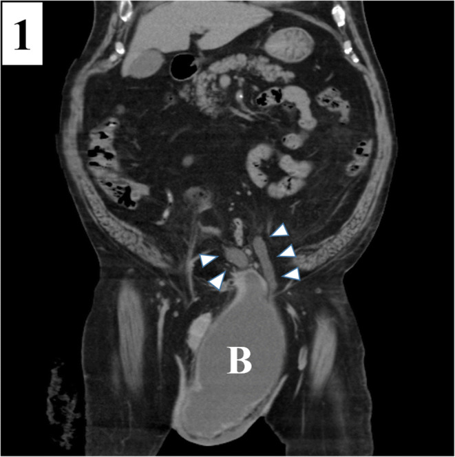
Computed tomography demonstrating a left inguinal hernia and prolapse of the bladder and bilateral ureters (arrowheads) into the scrotum. B, bladder.
Figure 2.
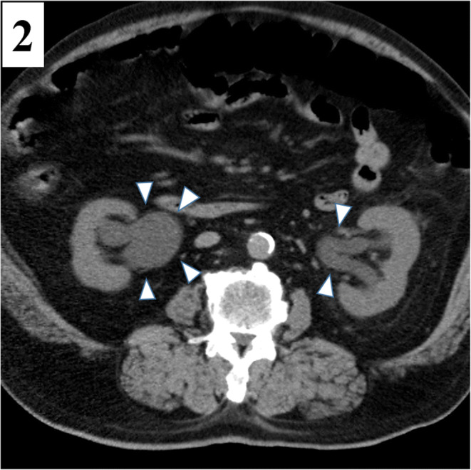
Axial computed tomography revealing bilateral hydronephrosis (arrowheads) at the kidney level.
IBH occurs in 1–3% of inguinal hernias.1 Although most patients with IBH are asymptomatic; they can develop lower urinary tract symptoms including incontinence and dysuria. Reduction in hernia size after urination is a characteristic sign of IBH.1 Reduced bladder wall tension, obesity, and pelvic floor muscle weakness contribute to IBH pathogenesis.2,3 The patient’s obesity and profession (a baseball umpire) may have contributed to IBH development.
Delayed diagnosis of IBH can lead to necrosis at the hernia site, a complication which may require bladder resection.2,4 IBH can be diagnosed by CT, ultrasound, retrograde cystography, or magnetic resonance imaging. When IBH is suspected, imaging should be performed to guide surgical intervention and avoid complications.
Acknowledgements
The authors would like to thank Editage (https://www.editage.jp/) for the English language review.
Declarations
Conflict of Interest
The authors declare that they do not have a conflict of interest.
Footnotes
Publisher’s Note
Springer Nature remains neutral with regard to jurisdictional claims in published maps and institutional affiliations.
References
- 1.Bacigalupo LE, Bertolotto M, Barbiera F, Pavlica P, Lagalla R, Pozzi Mucelli RS, Derchi LE. Imaging of urinary bladder hernias. AJR Am J Roentgenol. 2005;184(2):546–551. doi: 10.2214/ajr.184.2.01840546. [DOI] [PubMed] [Google Scholar]
- 2.Okauchi H, Nitta N, Kojima M, Mekata E. Three cases of inguinal bladder hernia diagnosed preoperatively by CT. J Jpn Surg Assoc. 2016;77:1854–8. doi: 10.3919/jjsa.77.1854. [DOI] [Google Scholar]
- 3.Taskovska M, Janež J. Inguinal hernia containing urinary bladder-a case report. Int J Surg Case Rep. 2017;40:36–8. doi: 10.1016/j.ijscr.2017.08.046. [DOI] [PMC free article] [PubMed] [Google Scholar]
- 4.Akashi S, Sugimori S, Sasaki Y, Yamada Y. Six cases of inguinal bladder hernia diagnosed preoperatively. J Jpn Coll Surg. 2017;42:624–631. [Google Scholar]