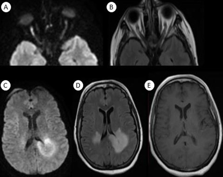
Figure 2. Orbital and cerebral magnetic resonance imaging after four weeks showed extension of previous lesions with involvement of the corpus callosum with diffusion restriction and peripheral enhancement, and appearance of bilateral optic neuritis more obvious on the right optic nerve than on the left one.
(A) Axial diffusion view of orbital magnetic resonance imaging; (B) axial T2 fluid-attenuated inversion recovery (FLAIR) view of orbital magnetic resonance imaging; (C) axial diffusion view of cerebral magnetic resonance imaging; (D) axial T2 FLAIR view of cerebral magnetic resonance imaging; (E) axial T1 Gado of cerebral magnetic resonance imaging.

