αvβ6-Integrin is exclusively expressed by epithelial cells and plays an important role for invasion and metastasis of carcinomas. We found a high expression of β6 on tumor cells in 88% of nearly 400 specimens of pancreatic ductal adenocarcinoma (PDAC) primaries and in virtually all metastases [1]. We earlier reported a series of 68 Ga- and 177Lu-labeled αvβ6-integrin-specific cyclic nonapeptides, but found that despite some showed a good tumor-to-background contrast in rodent models, tumor accumulation was ultimately too low for a successful clinical transfer [2]. We hypothesized that trimerization might result in elevated target-specific uptake and prolonged retention and thus elaborated a trimerized αvβ6-specific 68 Ga-peptide named 68 Ga-Trivehexin.
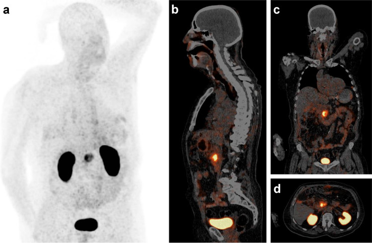
The image shows a 68 Ga-Trivehexin PET/CT of a male patient (82 y, 89 kg) with a histologically confirmed PDAC in the pancreatic head (87 MBq, 70 min p.i., acquisition time 25 min, 0.7 mm/s, 3 min/bed position; anterior MIP (a) scaled to SUV 12; PET in slices (b–d) to SUV 10). Apart from the PDAC lesion (SUVmax = 13.1), prominent signals are observed only in kidneys and urinary bladder due to renal excretion. No relevant uptake is seen in lungs, stomach, liver, and intestines. In light of a limited value of [18F]FGD-PET for early detection of PDAC [3], we anticipate that 68 Ga-Trivehexin will have a clinical value in this setting, besides the potential applications for fibrosis and other carcinomas (head-and-neck squamous cell, lung adenocarcinoma, colon, cervical, mammary) which have been addressed previously by αvβ6-integrin targeted PET-radiopharmaceuticals [4–6].
Author contributions
Conceived and designed the experiment: NGQ, NC, WS, JN. Performed the experiments: NC, WS. Analyzed the data: NGQ, JN. Wrote the original manuscript: JN. All authors approved the final version of the manuscript.
Funding
Open Access funding enabled and organized by Projekt DEAL. This work was funded by the Deutsche Forschungsgemeinschaft (SFB 824, project A10).
Data availability
The datasets used and/or analysed during the current study are available from the corresponding author on reasonable request.
Declarations
Ethics approval
Not applicable.
Consent to participate
The authors affirm that the patient provided written informed consent prior to the investigation.
Consent for publication
The authors affirm that the patient provided written informed consent for publication of the images.
Competing interests
N.G.Q. and J.N. are co-inventors of patents related to 68 Ga-Trivehexin. J.N. is shareholder of TRIMT GmbH (Radeberg, Germany), which is active in the field of radiopharmaceutical development.
Footnotes
This article is part of the Topical Collection on Image of the month.
Publisher's note
Springer Nature remains neutral with regard to jurisdictional claims in published maps and institutional affiliations.
References
- 1.Steiger K, Schlitter AM, Weichert W, Esposito I, Wester HJ, Notni J. Perspective of αvβ6-Integrin Imaging for Clinical Management of Pancreatic Carcinoma and Its Precursor Lesions. Mol Imaging. 2017;16:1536012117709384. doi: 10.1177/1536012117709384. [DOI] [PMC free article] [PubMed] [Google Scholar]
- 2.Färber SF, Wurzer A, Reichart F, Beck R, Kessler H, Wester HJ, et al. Therapeutic radiopharmaceuticals targeting integrin αvβ6. ACS Omega. 2018;3:2428–2436. doi: 10.1021/acsomega.8b00035. [DOI] [PMC free article] [PubMed] [Google Scholar]
- 3.Strobel O, Büchler MW. Pancreatic cancer: FDG-PET is not useful in early pancreatic cancer diagnosis. Nat Rev Gastroenterol Hepatol. 2013;4:203–205. doi: 10.1038/nrgastro.2013.42. [DOI] [PubMed] [Google Scholar]
- 4.Roesch S, Lindner T, Sauter M, Loktev A, Flechsig P, Müller M, et al. Comparison of the RGD motif-containing αvβ6 integrin-binding peptides SFLAP3 and SFITGv6 for diagnostic application in HNSCC. J Nucl Med. 2018;59:1679–1685. doi: 10.2967/jnumed.118.210013. [DOI] [PubMed] [Google Scholar]
- 5.Hausner SH, Bold RJ, Cheuy LY, Chew HK, Daly ME, Davis RA, et al. Preclinical development and first-in-human imaging of the integrin αvβ6 with [18F]αvβ6-binding peptide in metastatic carcinoma. Clin Cancer Res. 2019;25:1206–1215. doi: 10.1158/1078-0432.CCR-18-2665. [DOI] [PMC free article] [PubMed] [Google Scholar]
- 6.Kimura RH, Wang L, Shen B, Huo L, Tummers W, Filipp FV, et al. Evaluation of integrin αvβ6 cystine knot PET tracers to detect cancer and idiopathic pulmonary fibrosis. Nat Commun. 2019;10:4673. doi: 10.1038/s41467-019-11863-w. [DOI] [PMC free article] [PubMed] [Google Scholar]
Associated Data
This section collects any data citations, data availability statements, or supplementary materials included in this article.
Data Availability Statement
The datasets used and/or analysed during the current study are available from the corresponding author on reasonable request.


