
Figure 2. SFL Is a Broad-Spectrum Inhibitor of −1PRF.
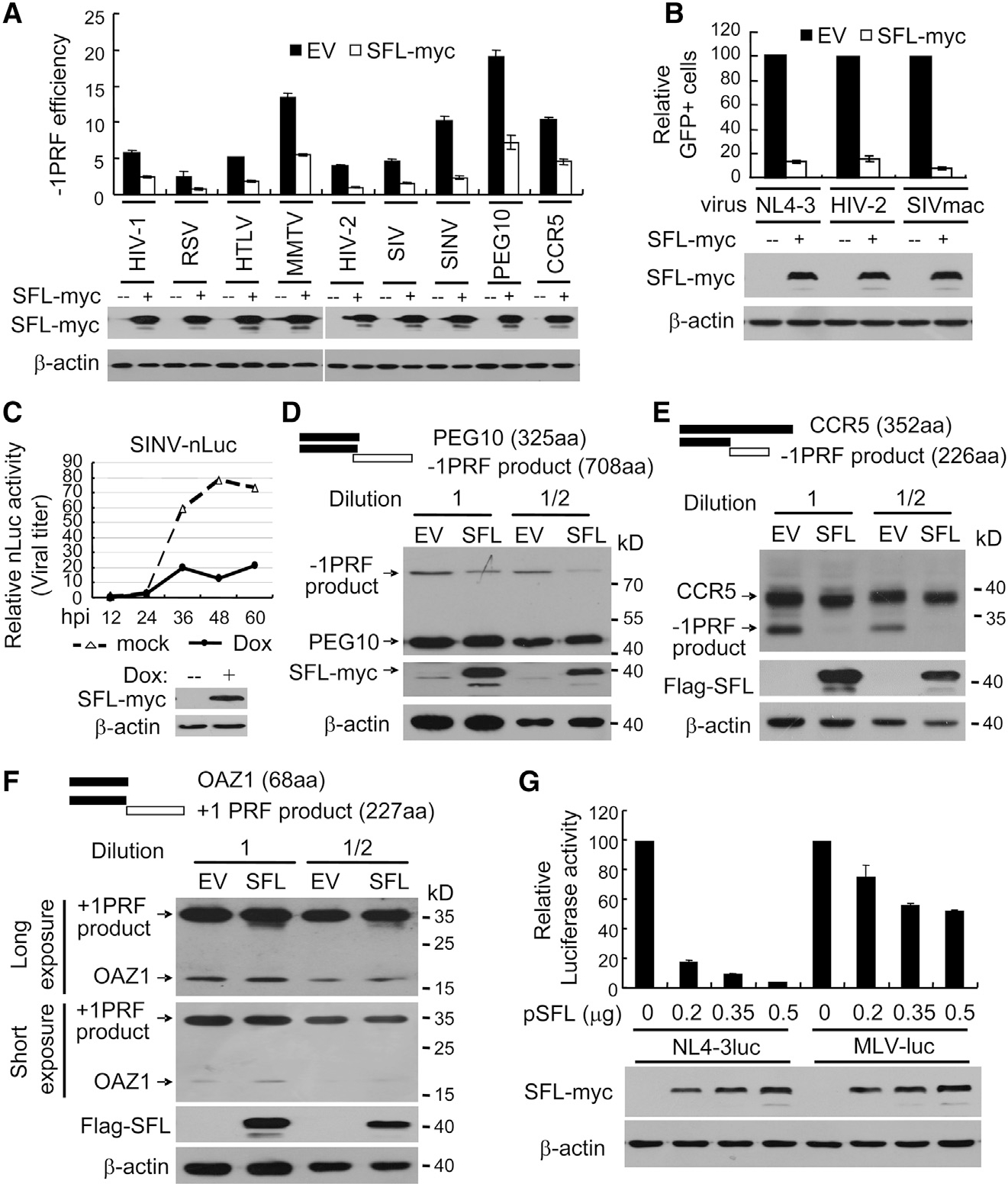
(A) Dual-luciferase reporters were expressed in 293T cells with or without SFL-Myc. The −1PRF efficiency of each reporter was calculated as the Fluc/Rluc ratio divided by that of the HIV(0) reporter.
(B) VSV-G-pseudotyped GFP-expressing lentivectors were produced in 293T cells with or without SFL-Myc and used to infect HeLa cells. The relative number of GFP-positive HeLa cells without SFL was set as 100.
(C) 293TREx-SFL-Myc cells were infected with SVNI-nLuc virus and mock treated or treated with doxycycline to induce SFL-Myc expression. At time points indicated, viral titers in culture supernatants were measured. hpi, hours post infection.
(D–F) N-terminally Myc-tagged PEG-10 (D), CCR5 (E), or OAZ1 (F) was transiently expressed in 293T cells, with or without SFL. Cell lysates were analyzed by western blotting.
(G) Plasmids producing VSV-G-pseudotyped NL4–3luc or MLV-luc were transfected into 293T cells with increasing amounts of a plasmid expressing SFL-Myc. The viruses were collected to infect 293T cells followed by luciferase activity measurement. The relative luciferase activity without SFL was set as 100. Data presented are means ± SD of three independent measurements, representative of three independent experiments.
See also Table S2.