Abstract
Supplemental Digital Content is available in the text.
Generally, a low-density, irregular-shaped area that appears outside the tunica media on intravascular ultrasound (IVUS) after coronary debulking, such as rotational atherectomy or coronary orbital atherectomy, is an indication of coronary perforation. Over time the perforation can be susceptible to enlargement and patients become hemodynamically unstable.
Although very rare, acute diffuse thickening of the coronary artery wall which consists of a low-density circle around the media on IVUS images after rotational atherectomy have been observed. The low-density circle is clearly demarcated and appears like a ‘halo’ around the vessel without any angiographical signs of coronary perforation and hemodynamic collapse. Recently, this phenomenon observed by IVUS has been reported as a ‘perivascular or extra-media hematoma’ typically seen in percutaneous intervention for coronary chronic total obstructions [1].
Aggressive dilatation is typically avoided because it is unknown whether this indicates coronary perforation or not. As limited information on the tunica adventitia (outside the media) could be obtained from IVUS, it is difficult to differentiate whether it is within the wall (Ellis TypeI) or outside the wall (TypeII) [2].
To our knowledge, this is the first report on observation of the ‘halo’-like IVUS findings with optical frequency domain imaging (OFDI, Terumo, Japan). These phenomena were observed in two rotational atherectomy cases using both OFDI and IVUS imaging.
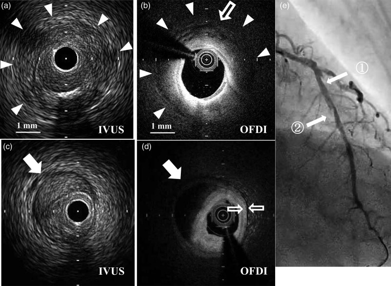
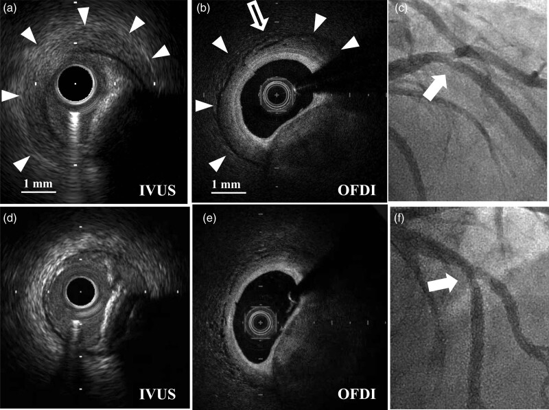
OFDI showed that these phenomena were diffuse, low-density thickening of the tunica adventitia with a clear outside border (Figs. 1b and 2b. See video Case1, Supplemental digital content 1, http://links.lww.com/MCA/A415), which differs from the findings of common hematoma (Fig. 1d) viewed at the distal site of the vessel (Fig. 1e2). In general, near-infrared (NIR) light is attenuated by blood and lipid plaque. As common hematoma consists of only blood pooled in the torn space within the tunica media, OFDI findings of hematoma appear as a homogenous pitch-black low-density area with strong attenuation (Fig. 1d). On the other hand, this ‘halo’-like phenomenon has less attenuation of NIR light with a clear demarcated border. A component of the phenomena appeared like ‘cobblestoning,’ which may be aggregates of vasa vasorum or edema of tunica adventitia (Fig. 1b and 2b).
Fig. 1.
Findings in case 1. (a) After rotational atherectomy, intravascular ultrasound (IVUS) (ViewIT 40 MHz, Terumo, Japan) showed a clearly demarcated low-density circle around the media (triangle arrow), which appears like a ‘halo’ around the vessel. These findings were obtained at the mid-left anterior descending artery just distal to the diagonal branch (E white arrow 1). (b) The optical frequency domain imaging (OFDI) findings of the same site. The arrow head showed clear demarcation of the thickening tunica adventitia. Unlike common hematoma, there was less attenuation of near-infrared (NIR) light and cobblestoning appearance of small low-density aggregates was clearly observed (white blank arrow) at the outermost position of thickened adventitia. (c,d) Common hematoma (white arrow) was observed distally to the lesion (e white arrow 2). IVUS (c) and OFDI (d) images showed very different findings from the images in a and b. First, a common hematoma was clearly demarcated with a low-density band, which may indicate plaque dissection extending into the media (c). Second, due to being rich in pooled blood cells (seen in c), OFDI findings of the hematoma appeared to be a homogenous pitch-black low-density area with strong attenuation of NIR light (d). (e) Angiographic image immediately after rotational atherectomy did not show any signs of coronary perforation (white arrow 1). See video 1, Supplemental digital content 1, http://links.lww.com/MCA/A415.
Fig. 2.
Findings in case 2. (a,b) After rotational atherectomy, the same images were obtained in case 2. intravascular ultrasound (IVUS) images (AltaView 40-60 MHz, used set at 50 MHz, Terumo, Japan) and OFDI images (b) closely correlated with case 1. White blank arrow in B indicates a small cobblestoning appearance. (c) Angiographic image immediately after rotational atherectomy did not show any signs of coronary perforation (white arrow). (d) Final IVUS images after the dilatation of drug-coated balloons (DCB 3.5 mm in diameter). The ‘halo’ phenomenon had significantly diminished. (e) OFDI images after the inflation of balloon dilatation (3.0 mm in diameter) before drug-coated ballooning. Unfortunately, final OFDI examination after DCB was not performed, though the thickness of tunica adventitia had normalized at this point, compared with the thickness of adventitia shown in Fig. 1c (white blank arrow). (f) Final angiography. See video 2, Supplemental digital content 2, http://links.lww.com/MCA/A416).
The second case involved balloon and drug-coated balloon dilatation; thus, the imaging findings obtained were in response to balloon dilatation. After multiple dilatations of balloons, these observations predominantly disappeared in IVUS (Fig. 2d), and OFDI showed that the tunica adventitia had become normal in thickness (Fig. 2e. See video Case2, Supplemental digital content 2, http://links.lww.com/MCA/A416).
Even with OFDI findings, the mechanism of this phenomenon could not be explained clearly. However, as OFDI apparently showed no disruption of the adventitia during the procedures, these phenomena were considered to occur within the adventitia in both cases. As a result, adjunctive coronary intervention was performed safely.
Although it has recently been reported that hemorrhage of intraplaque neovessels arising from vasa vasorum is associated with coronary atherosclerosis [3], this phenomenon has less attenuation of NIR light which means less blood or lipid plaque in the thickening of the adventitia. Additionally, the thickening normalized after dilatation with balloons. It might be feasible to speculate that the thickening is acute edema of the tunica adventitia attributed by heat injury associated with rotablator.
Without pathological validation, it is inconclusive whether this phenomenon indicates ‘adventitia with blood or edema’ or ‘some disappearance of adventitia and blood stasis in the perivascular tissue’ and is a limitation of this investigation. Further, validation studies are required.
Acknowledgements
Conflicts of interest
There are no conflicts of interest.
Supplementary Material
Footnotes
Supplemental Digital Content is available for this article. Direct URL citations appear in the printed text and are provided in the HTML and PDF versions of this article on the journal's website, www.coronary-artery.com.
Reference
- 1.Song L, Maehara A, Finn MT, Kalra S, Moses JW, Parikh MA, et al. Intravascular ultrasound analysis of intraplaque versus subintimal tracking in percutaneous intervention for coronary chronic total occlusions and association with procedural outcomes. JACC Cardiovasc Interv 2017; 10:1011–1021. [DOI] [PMC free article] [PubMed] [Google Scholar]
- 2.Ellis SG, Ajluni S, Arnold AZ, Popma JJ, Bittl JA, Eigler NL, et al. Increased coronary perforation in the new device era. Incidence, classification, management, and outcome. Circulation 1994; 90:2725–2730. [DOI] [PubMed] [Google Scholar]
- 3.Nishimiya K, Matsumoto Y, Shimokawa H. Viewpoint: recent advances in intracoronary imaging for vasa vasorum visualisation. Eur Cardiol 2017; 12:121–123. [DOI] [PMC free article] [PubMed] [Google Scholar]
Associated Data
This section collects any data citations, data availability statements, or supplementary materials included in this article.


