CELL BIOLOGY Correction for “Dual functions for OVAAL in initiation of RAF/MEK/ERK prosurvival signals and evasion of p27-mediated cellular senescence,” by Ben Sang, Yuan Yuan Zhang, Su Tang Guo, Ling Fei Kong, Qiong Cheng, Guang Zhi Liu, Rick F. Thorne, Xu Dong Zhang, Lei Jin, and Mian Wu, which was first published November 26, 2018; 10.1073/pnas.1805950115 (Proc. Natl. Acad. Sci. U.S.A. 115, E11661–E11670).
The authors note that Fig. 4 appeared incorrectly. The corrected figure and its legend appear below. The online version has been corrected.
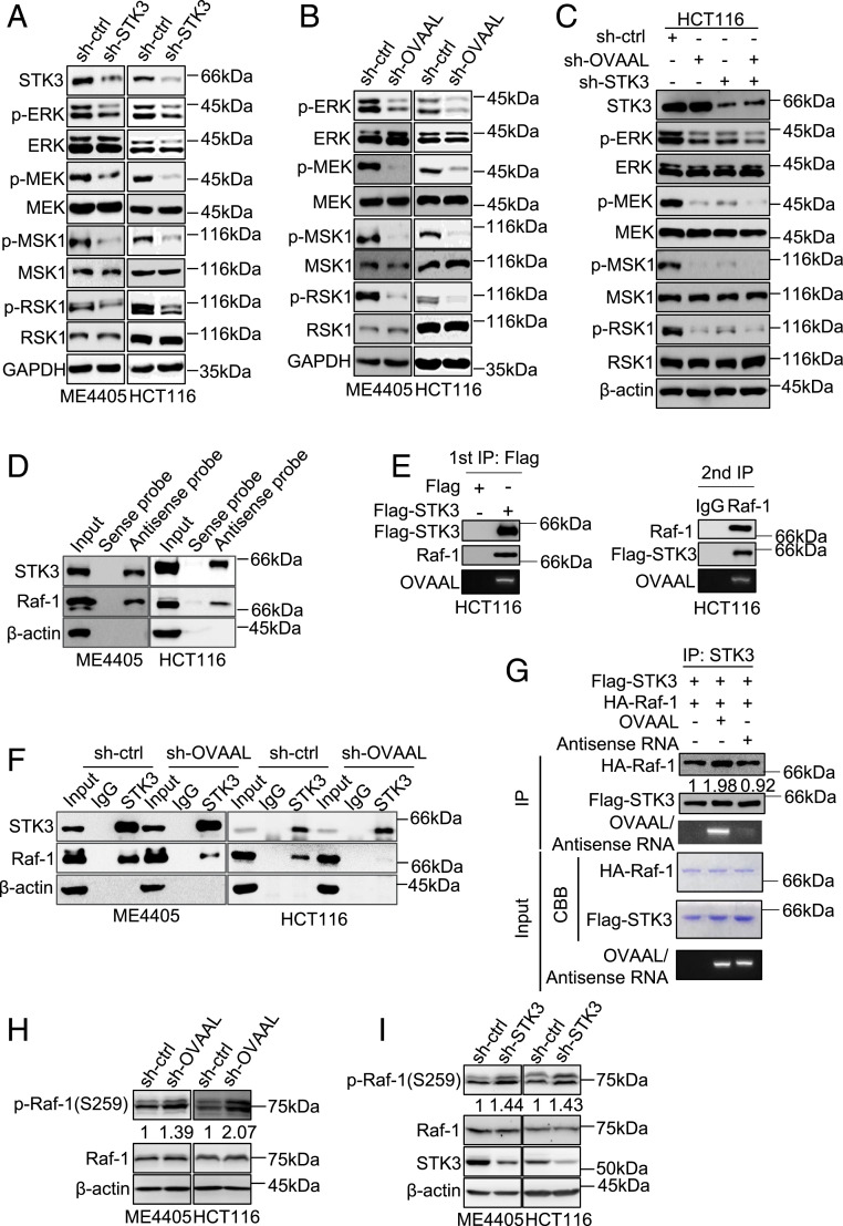
Fig. 4.
OVAAL activates RAF/MEK/ERK cascade through facilitating the binding between STK3 and Raf-1. (A) STK3 shRNA decreased levels of p-ERK, p-MEK, p-MSK1, and p-RSK1 in ME4405 and HCT116 cells as shown in Western blotting. Analysis of total ERK, MEK, MSK1, and RSK1, along with a GAPDH loading control (ctrl), is shown for comparison (n = 3). (B) OVAAL shRNA decreased levels of p-ERK, p-MEK, p-MSK1, and p-RSK1 in ME4405 and HCT116 cells as shown in Western blotting (n = 3). (C) Cosilencing of OVAAL with STK3 in HCT116 cells did not impart further reductions in the phosphorylated levels of ERK, MEK, MSK1, or RSK1 levels in comparison to STK3 knockdown alone as measured by Western blotting (n = 3). (D) Biotin-labeled antisense probes used to capture OVAAL from total cellular lysates of ME4405 and HCT116 cells also pulled down STK3, along with Raf-1, as shown by Western blotting. β-Actin was used as a negative ctrl here and in the following experiments (n = 3). (E) Flag-STK3, Raf-1, and OVAAL were coprecipitated with an anti-Flag antibody in whole-cell lysates of HCT116 cells transfected with Flag-STK3 (Left), and after elution with Flag peptide, all were further coprecipitated with an anti–Raf-1 antibody in the resultant precipitates (Right). IP, immunoprecipitation. (F) OVAAL depletion strongly diminished the levels of Raf-1 associating with STK3. Endogenous STK3 was immunoprecipitated from sh-ctrl– or sh-OVAAL–treated ME4405 and HCT116 cells, with results shown by Western blotting (n = 3). (G) Presence of in vitro-transcribed OVAAL, but not an antisense RNA, enhanced the binding between recombinant HA–Raf-1 and Flag-STK3. Assay inputs were visualized by Coomassie Brilliant Blue (CBB) and ethidium bromide staining (n = 3). OVAAL shRNA (H) and STK3 shRNA (I) increased levels of p-Raf-1 (Ser259) in ME4405 and HCT116 cells as shown in Western blotting (n = 3).


