Abstract
The aim of this case is to illustrate the18F-fluorodeoxyglucose (FDG) positron emission tomography-computed tomography findings of a patient who was admitted in AIIMS, Raipur, for the preoperative evaluation of Marjolin ulcer and was later diagnosed with COVID-19 infection. Apart from the primary lesion in the right foot and pelvic lymph nodes, the scan revealed mild FDG-avid basal ground-glass opacities in bilateral lung fields with mediastinal and hilar lymph nodal involvement, in an otherwise asymptomatic patient.
Keywords: Asymptomatic, coronavirus, COVID-19, fluorodeoxyglucose positron emission tomography-computed tomography, ground-glass opacification, mediastinal lymph nodes
INTRODUCTION
In the present era of the COVID-19 pandemic, nuclear medicine departments must be prepared for handling COVID patients. This is one such diagnosed case of Marjolin ulcer who underwent positron emission tomography-computed tomography (PET/CT) in our department with subtle pulmonary findings, and later turned out to COVID positive.
CASE REPORT
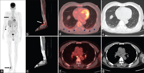
A 63-year-old male, with biopsy-proven Marjolin ulcer (keratinizing squamous cell carcinoma) of the right lateral aspect of the foot of 1-year duration associated with pain and bleeding, was referred for PET/CT for metastatic workup. PET/CT scan revealed a fluorodeoxyglucose (FDG)-avid ulceroproliferative lesion measuring approximately 5.4 AP × 4.3 TR cm with SUVmax– 7.4, involving the dorsolateral aspect of the right foot involving the subcutaneous and muscular planes. The lesion is also noted to involve the periosteum of the distal end of the right tibia and fibula. An incidental note of mild FDG-avid ground-glass opacification with reticulations was noted in the basal aspect of the bilateral lung fields with FDG-avid, right lower paratracheal, bilateral hilar lymph nodes [Figure 1]. The referring physician was informed of the lung findings immediately. Within a day of PET/CT, COVID testing was done in view of the findings on the18F-FDG PET/CT and as a part of routine surgical workup by reverse transcriptase-polymerase chain reaction method which is a gold standard investigation for COVID-19. The patient's sample was reported as COVID positive within a day of PET/CT scan. The patient was started on hydroxychloroquine 400 mg twice a day with azithromycin and zinc supplements and remained asymptomatic during the course of admission.
Figure 1.
18F-labeled fluoro-2-deoxyglucose positron emission tomography-computed tomography; maximum-intensity projection image (a) of positron emission tomography showing increased fluorodeoxyglucose uptake in the right foot and mediastinal lymph nodes (arrows). Positron emission tomography-computed tomography fused (coronal [b, white arrow] and computed tomography image [c]) revealing fluorodeoxyglucose-avid ulceroproliferative lesion in the dorsal aspect of the right foot. Positron emission tomography-computed tomography fused ([d]: arrowhead) and computed tomography images (e) revealing subtle ground-glass opacification with reticulations with mild fluorodeoxyglucose uptake in the basal segments of the bilateral lung fields. Positron emission tomography-computed tomography fused transaxial images (f) and computed tomography images (g) revealing fluorodeoxyglucose-avid mediastinal and hilar lymph nodes
DISCUSSION
The authors wish to highlight the lung findings of asymptomatic COVID-19 patients. Although COVID-19 patients may have a varied presentation on contrast-enhanced CT (CECT) such as ground-glass opacification (especially in the peripheral and posterior parts of the lung), consolidation, and mediastinal lymphadenopathy,[1,2,3] our patient had subtle ground-glass opacities with reticulations in the basal segments with mild FDG uptake and FDG-avid mediastinal lymphadenopathy. However, it may be noted that the lung parenchyma findings are not specific for COVID-19 infection, thus should be differentiated from other infective/inflammatory causes of lung diseases. High-resolution CT thorax is the investigation for evaluation of diffuse parenchymal lung disease such as interstitial lung diseases and other airway diseases such as chronic obstructive pulmonary disease and bronchial asthma. Contrast CT is mainly useful for the evaluation of lung masses, nodules, or lymph nodes within the thorax to look for mitotic activity or blood supply, and to look for postcontrast enhancement. Hence, CECT is not useful for the evaluation of lung parenchyma and exposes patients to hazardous effects of radiocontrast. There have been only a few case studies of PET/CT published in the literature so far.[4,5] It is also to be noted that in the present scenario any suspicious lung finding with FDG uptake may be reported to the referring physician to rule out COVID-19 infection. This case also highlights that the nuclear medicine staff needs to be aware of the possibility of contact with asymptomatic COVID-19 patients. Therefore, necessary safety measures need to be adopted for other patients and hospital staff in order to block the spread of infection. At our tertiary care center, all the patients and attendants (preferably one) are advised to wear at least triple-layered surgical masks; hand sanitization for patients as well as doctors is performed after any contact with patient's case sheets and reports, etc. All high-touch areas are sanitized every 2 h with hypochlorite solution. Single-use disposable sheets are used for scanning. The sheets are changed after a single use. Transparent screens are used in outpatient department areas; proper social distancing is maintained. Furthermore, the room is closed for 2 days, and equipment is disinfected on the 3rd day. In the present case, after the scan of the patient, proper contact tracing was done, and all primary contacts were divided into high and low risks. One of the doctors had examined the patient and one ward boy helped the patient on the scanner. Both were sent for home quarantine and tested negative for COVID on 7 and 14 days. Thus, by following proper measures, none of the staff got infected. All the necessary infection control measures were in place in our department in the wake of the present COVID-19 pandemic as per our hospital control committee instructions, which saved the staff from getting infected.
CONCLUSION
Suspicious lung findings on PET/CT must be communicated to the referring physician in view of the current COVID-19 pandemic. However, it is to be kept in mind that FDG PET-CT findings are nonspecific and cannot differentiate COVID infection from other pneumonias.
Declaration of patient consent
The authors certify that they have obtained all appropriate patient consent forms. In the form the patient(s) has/have given his/her/their consent for his/her/their images and other clinical information to be reported in the journal. The patients understand that their names and initials will not be published and due efforts will be made to conceal their identity, but anonymity cannot be guaranteed.
Financial support and sponsorship
Nil.
Conflicts of interest
There are no conflicts of interest.
REFERENCES
- 1.Xu X, Yu C, Qu J, Zhang L, Jiang S, Huang D, et al. Imaging and clinical features of patients with 2019 novel coronavirus SARS-CoV-2. Eur J Nucl Med Mol Imaging. 2020;47:1275–80. doi: 10.1007/s00259-020-04735-9. [DOI] [PMC free article] [PubMed] [Google Scholar]
- 2.Yang W, Cao Q, Qin L, Wang X, Cheng Z, Pan A, et al. Clinical characteristics and imaging manifestations of the 2019 novel coronavirus disease (COVID-19):A multi-center study in Wenzhou city, Zhejiang, China. J Infect. 2020;80:388–93. doi: 10.1016/j.jinf.2020.02.016. [DOI] [PMC free article] [PubMed] [Google Scholar]
- 3.Li B, Li X, Wang Y, Han Y, Wang Y, Wang C, et al. Diagnostic value and key features of computed tomography in Coronavirus Disease 2019. Emerg Microbes Infect. 2020;9:787–93. doi: 10.1080/22221751.2020.1750307. [DOI] [PMC free article] [PubMed] [Google Scholar]
- 4.Qin C, Liu F, Yen TC, Lan X. 18F-FDG PET/CT findings of COVID-19: A series of four highly suspected cases. Eur J Nucl Med Mol Imaging. 2020;47:1281–6. doi: 10.1007/s00259-020-04734-w. [DOI] [PMC free article] [PubMed] [Google Scholar]
- 5.Setti L, Kirienko M, Dalto SC, Bonacina M, Bombardieri E. FDG-PET/CT findings highly suspicious for COVID-19 in an Italian case series of asymptomatic patients. Eur J Nucl Med Mol Imaging. 2020;47:1649–56. doi: 10.1007/s00259-020-04819-6. [DOI] [PMC free article] [PubMed] [Google Scholar]


