

Abstract

Photophysical and photochemical processes are ruled by the interplay between transient vibrational and electronic degrees of freedom, which are ultimately determined by the multidimensional potential energy surfaces (PESs). Differences between ground and excited PESs are encoded in the relative intensities of resonant Raman bands, but they are experimentally challenging to access, requiring measurements at multiple wavelengths under identical conditions. Herein, we perform a two-color impulsive vibrational scattering experiment to launch nuclear wavepacket motions by an impulsive pump and record their coupling with a targeted excited-state potential by resonant Raman processes with a delayed probe, generating in a single measurement background-free vibrational spectra across the entire sample absorption. Building on the interference between the multiple pathways resonant with the excited-state manifold that generate the Raman signal, we show how to experimentally tune their relative phase by varying the probe chirp, decoding nuclear displacements along different normal modes and revealing the multidimensional PESs. Our results are validated against time-dependent density functional theory.
The investigation of light-induced processes is essential for the understanding of a variety of complex phenomena at the interface between physics, chemistry, and biology, in which excited-state dynamics causes the transient modification of molecular properties and atomic configurations. The latter are ultimately determined by the multidimensional potential energy surfaces (PESs) describing how the excited-state potential changes along the different normal modes.1 Spontaneous Raman spectroscopy plays a pivotal role for accessing the vibrational fingerprints of solid-state systems or complex molecular compounds. Tuning the excitation of the Raman pulse in resonance with an electronic transition enables selective enhancement of the Raman cross section.2 Because the resonance Raman (RR) cross section depends on the displacement between ground and excited potential energy surfaces along the normal coordinates, the relative intensities of the measured RR bands encode information on the potential energy surfaces. Critically, in order to extract such molecular information, several spectra have to be recorded scanning the Raman pump wavelength across the absorption profile, performing a sequence of measurements at multiple wavelengths under identical conditions, and with the detection of the experimental signals that is typically hampered by the overwhelming fluorescent background. Most importantly, spontaneous Raman spectroscopy can provide only differential information on excited-state geometries relative to the equilibrium configuration on the ground state.
Herein, we introduce a method to determine the nuclear displacements between different PESs, based on an impulsive stimulated Raman scattering (ISRS) experiment, circumventing the limitations of spontaneous Raman spectroscopy. ISRS exploits the joint action of two ultrashort laser fields to measure vibrational excitations in the time domain.3−5 A femtosecond Raman pump (RP) coherently stimulates nuclear wavepackets of Raman active modes,6,7 which modulate the transmissivity of the sample and are detected by monitoring the transmission of a temporally delayed probe pulse (PP) as a function of the time delay ΔT between the RP–PP pair: Fourier transforming over ΔT enables retrieving the Raman information in the frequency domain.8,9 Because the ISRS spectra are recorded for temporally separated Raman and probe fields, the experimental signals are not affected by nonlinear background processes generated during the overlap of the ultrashort pulses, which in contrast can plague frequency domain coherent Raman techniques.10−14 In addition, thanks to the heterodyne detection, the vibrational information is engraved on the PP; hence, the fluorescence and the incoherent background signals are efficiently suppressed. Importantly, when the Raman pulse is longer than the period of a normal mode, it cannot efficiently stimulate vibrational coherences, making ISRS less effective in probing high energetic modes. In order to isolate the vibrational fingerprints on the initially populated electronic level, we employ a two-color ISRS experimental configuration, with an off-resonant Raman pulse ER and a broadband probe EP, which covers the molecular response across its absorption profile, enabling the study of the couplings with a targeted electronically excited level, by monitoring the frequency-dispersed ISRS signal as a function of the probe wavelength λP. We demonstrate how to assign the complex dependence (as a function of λP) of the measured mode-dependent Raman excitation profiles to the corresponding nuclear displacements. The relative intensities of the ISRS modes are studied, including chirp effects, which can critically modulate the amplitude of the time domain Raman response even in the fully off-resonance regime.15,16 The addition of an actinic pump, temporally longer than the period of the normal modes under investigation, to the scheme proposed here allows photoexcitation of the sample on a targeted electronic level17−22 without promoting vibrational coherences. Hence, because of the femtosecond resolution probing of the RP–PP pair,23−27 probing the ISRS response across the excited-state absorption activated upon the photoexcitation enables the extension to the mapping of the relative orientation and displacements between arbitrary excited transient PESs. Within such a scheme the RP may be tuned in resonance with the stimulated emission to enhance the ISRS cross section. The measurement of excited-state orientations by ISRS offers the chance to map complex PESs and to identify the vibrational degrees of freedom responsible for the ultrafast relaxation of the system along excited-state potentials, as in the presence of dynamic Stokes shift,28 where measuring the time-dependent REPs would offer the chance to follow the photoexcited chromophore PES relaxation along the involved vibrational degrees of freedom, or photoinduced charge-transfer events.29
The two-color ISRS response is measured for cresyl violet (CV)30−34 dissolved in methanol, a highly fluorescent oxazine dye with a long-lived excited electronic state. Because of the strong fluorescence background, with a small (∼500 cm–1) Stokes shift, the CV spontaneous resonant Raman response cannot be explored around the system absorption profile and can be obtained only at excitation wavelengths far to the blue side of the absorption maximum.31 Hence, CV represents an ideal candidate for testing the capabilities of our two-color ISRS setup. Our experimental results are reported in Figure 1: the temporal traces recorded in the time domain are shown in the top panels (a and b) for two values of the probe chirp C2 (360 fs2 and 40 fs2), while the corresponding spectra in the frequency domain, obtained upon truncating the coherent artifact (highlighted by the blue boxes) and fast Fourier transforming (FFT), are reported in the bottom panels (d and e). Because the ISRS signal is dominated by the mode centered at 592 cm–1, the spectral region around the 592 cm–1 peak has been scaled by a factor 0.1 in order to enhance the visibility of the other weaker Raman bands (at 345, 470, 493, 526 675, 752, and 833 cm–1). It is worth stressing that in view of the nonresonant nature of the Raman pump employed in the present scheme, only the Raman modes with a nonvanishing polarizability derivative can be efficiently stimulated. As expected, the amplitude of the ISRS oscillations is enhanced around the sample absorption profile, which is reported in Figure 1c. The intensities of the different Raman peaks show complex profiles, which vary as a function of the PP wavelength and depend on both PP chirp as well as on the specific Raman mode under consideration. For example, the 592 cm–1 mode shows a maximum intensity slightly red-shifted with respect to the absorption maximum for C2 = 360 fs2 and turns to a broad bilobed profile for C2 = 40 fs2. In contrast, the 525 cm–1 mode shows similar bilobed profiles for both C2 = 40 fs2 and C2 = 360 fs2. The traces in the time domain reveal a π phase shift between the oscillations red and blue-shifted with respect to the node with vanishing signal.
Figure 1.

Broadband two-color ISRS spectra of cresyl violet recorded in the time domain (top panels, a and b) as a function of the probe wavelength λP and the RP–PP relative delay ΔT, acquired for two different values of the probe chirp (360 and 40 fs2). The blue box marks the region covered by the coherent artifact. The corresponding spectra in the frequency domain (d and e) can be obtained by truncating the coherent artifact and Fourier transforming over ΔT and are reported in the bottom panels as a function of the wavenumber ν̃ (the central region around 592 cm–1 has been scaled by a factor 0.1 to enhance the visibility of the weaker Raman modes). The absorption spectrum of the methanol dissolved sample is shown in the right panel (c) with the RP and PP spectral profiles and is compared with the one (yellow dashed line) simulated from the molecular parameters reported in Table 1. Low (300 cm–1) and a high (1400 cm–1) frequency contributions have been included in the simulation, taking into account low-frequency contributions with vanishing polarizability derivative not stimulated by the nonresonant pump and the high-frequency modes out of reach for the ISRS technique. The contributions originated by the vibronic progression are reported as magenta lines. The black dashed line indicates the transition to the vibrationally ground level of the excited electronic state.
To interpret this complex behavior and extract
structural information
from the measurements of mode-dependent intensity profiles in CV,
we studied the role of the resonant PP chirp and detection wavelength
in the generation of the nonlinear signal. The third-order nonlinear
polarization associated with the ISRS process can be evaluated through
a perturbative expansion of the molecular density matrix in powers
of the electric fields ER/P = ER/P0(t)e–iωR/Pt + c.c., and the different pathways that contribute to the
total response can be identified by taking advantage of a diagrammatic
approach.6,35−38 Considering the pulse scheme
and the energy levels reported in Figure 2a,b, with vibrational manifolds in the electronic
ground and resonant excited state indicated as |g⟩, |g′⟩, ... and |e⟩, |e′⟩, ..., respectively,
the Feynman diagrams that take into account the system response are
shown in Figure 2c.
Because the RP is tuned off resonant with respect to the sample absorption,
it selectively generates vibrational coherences only in the initially
populated electronic level, the ground state in the present case (|g′⟩ ⟨g| and |g⟩⟨g′| states, corresponding
to diagrams Ak and Bk, respectively). Then the
system evolves unperturbed until an interaction with the PP and a
free induction decay (that leaves the system in a population state)
enabling us to probe the vibrational coherences after the tunable
time delay ΔT. Importantly, while in the off-resonant
PP regime the state e is a virtual level and Ak–Bk destructively interfere resulting in
small or vanishing signals,15 in the resonant
case the ket side of the density matrix is promoted to the excited
state and, taking advantage of the broadband nature of the PP, it
can be projected to the entire vibrational manifold, with a distribution
ruled by the transition dipole moments μnm = ⟨ψn|μ̂|ψm⟩. Assuming the Condon approximation,22,39 the intensities of the transition dipole moments are determined
by the Franck–Condon (FC) factors and can be evaluated by computing
the FC overlap integrals between the initial and final vibrational
wave functions μgjek = ⟨gj|ek⟩, which depend on the displacement of the
PES along the considered normal mode. We adopt the harmonic approximation
for the molecular potentials, according to which the vibrational wave
functions are expressed by products of orthogonal shifted harmonic
oscillators. In the left panel of Figure 3, we report some values of the transition
dipole moments as a function of the dimensionless displacement factor d =  .
.
Figure 2.

Sketch of the two-color ISRS experimental setup used in this work (a). The electronic and vibrational levels involved in the generation of the measured signal are reported in panel b: the vibrational progression on the ground electronic state is indicated as |g⟩, |g′⟩, |g″⟩, while the corresponding manifold in the electronic excited state is termed |e⟩, |e′⟩, |e″⟩. The Feynman diagrams describing the ISRS process are reported in panel c.
Figure 3.

(a) Transition dipole moments as a function of the dimensionless displacement along the normal modes. In the top panel the absolute value of single transition dipole moments are shown (the vibrational progression on the ground electronic state is indicated as |g⟩, |g′⟩; the corresponding manifold in the electronic excited state is termed |e⟩, |e′⟩, |e″⟩), while in the bottom panel we report the intensity factors associated with the various ISRS pathways shown in panel b. Each factor corresponds to the product of two dipole moments. The corresponding signals in the time domain are reported in panel c, with the horizontal dashed line indicating the PP wavelength matching the e–g transition.
The third-order polarizations, PAg′(3)(ω, ΔT) and PBg′(ω, ΔT), taking into account the signal induced by a given vibrational mode g′, can be directly computed from the diagrams in Figure 2 and are40,41
 |
1 |
and
 |
2 |
ω̃ij = ωij – iγij, ωij = ωi – ωj indicates the energy difference between i–j levels; Δ is an integration variable (with the maximum contribution centered at ω̃g′g); G(ωD) is the inhomogeneous broadening Gaussian function, and γij = τij–1 is the dephasing rate of the |i⟩ ⟨j| coherence. The IRA(Δ, ΔT) and IRB(Δ, ΔT) terms in eqs 1 and 2 represent the preparation function of the |g′⟩ ⟨g| and |g⟩ ⟨g′| vibrational coherences generated by the Raman pulse, and under off-resonant RP excitation, they are purely real functions. Importantly, Raman modes with a period much shorter than the pump duration will exhibit an inefficient stimulation of the vibrational coherences, resulting in small amplitudes of the ISRS peaks.5 The summation over ek in eqs 1 and 2 takes into account the PES one-dimensional projection on the g′ vibrational subspace, and in order to retrieve the desired total nonlinear polarization of the system, eqs 1 and 2 should be summed over all the g′ ground-state normal modes under consideration, according to
Details on the derivation of eqs 1 and 2 and expressions for the IRA(Δ, ΔT) and IRB(Δ, ΔT) functions are reported in the Supporting Information. For simplicity, the calculation can be performed assuming that the central frequency ωP of the chirped probe pulse arrives at t = 0 and that the RP is centered at t = −ΔT (i.e., shifted at negative time delays and preceding the PP). Under such assumptions the RP–PP fields of eqs 1 and 2 can be expressed in the frequency domain as
| 3 |
where ER/P(0)(ω) are positive real functions representing the square root of the RP–PP spectra; C2 is the group delay dispersion taking into account the linear chirp of the PP, and Cn indicates the nth higher-order dispersion terms.
When the PP does not vary across the sample, the third-order nonlinear polarization, which depends on the PP temporal and spectral profile, is constant and hence the ISRS responses S(ω, ΔT)–S(ω, Ω), in the time and frequency domains, respectively, can be calculated as
where  indicates the imaginary part of f.
However, we note that, because of the absorption of the EP field during its propagation, the polarizations PA(3)(ω) and PB(ω) are not constant and decay along the beam path within the sample. For this reason, a quantitative evaluation of the ISRS signal requires us to numerically integrate the PP spectral profile over the sample length, by using the coupled wave equations10,42
 |
4 |
where α(ω) indicates the frequency dependent attenuation coefficient. The ISRS signal S(ω, ΔT) in the time domain can be finally evaluated as the normalized difference between the transmitted PP spectrum in the presence and in the absence of the RP induced nonlinear polarization.
To interpret the experimental results it is worth
considering separately
the different pathways, contributing to the ek summations (eqs 1–4), that generate
the polarizations PA and PB. From a diagrammatic perspective,
this is equivalent to recasting the Feynman diagrams shown in Figure 2c to the corresponding
ones in the energy level scheme, which are reported in Figure 3b (we have explicitly shown
only the e and e′ vibrational
levels in the electronic excited state). Diagrams in Figure 3b help to directly visualize
the spectral components of the Raman and probe fields contributing
to the experimental signal generation at a given PP wavelength. Importantly,
at odds with fully nonresonant ISRS processes, where the total response
of the system is generated by the interference between pathways involving
interactions with different probe pulse spectral components (red-shifted
and blue-shifted with respect to the probed wavelength),16,40,43 in the resonant case the signal
at a given probe wavelength is generated by the interference between
processes which share interactions with the same probe color, but
with a different state in the excited-state manifold, hence encoding
information on the nuclear displacements between different potential
energy surfaces. In particular, the A0/A1 processes are generated from an interaction
with a PP component red-shifted by one vibrational quantum with respect
to the probed wavelength and originate from the transitions e → g and e′
→ g, hence resulting in contributions centered
at ωeg and ωe′g, respectively. In contrast, B0 and B1 generate
contributions centered around ωeg′ and ωe′g′ and originate from interactions with spectral components of the
PP blue-shifted with respect to the probed wavelengths. Interestingly,
while A0 and B0 generate an ISRS response centered at different PP wavelengths,
they originate from quantum pathways involving the same excited state e, resulting in ISRS signals oscillating in phase with each
other (Figure 3c).
In contrast, both A0 and B1 generate an ISRS response centered at the same transition
frequency (ωeg). However, the amplitude
of the corresponding oscillations is ruled by different dipole moments,
namely, μg′eμeg for A0 and μg′e′μe′g for B1. Critically, as shown in Figure 3a, μg′eμeg and μg′e′μe′g have opposite signs, and hence, they result in a destructive
interference between the A0 and B1 processes. This elucidates the vanishing ISRS
amplitude observed at the transition frequency ωeg in Figure 1b,e. Similarly, the relative sign between the A1 and B0 processes, red and
blue-shifted by one vibrational quantum with respect to the eg transition, rationalizes the π phase shift observed
in the time domain traces around the node (Figure 1a,b). It is worth stressing that the interference
between A0 and B1 pathways can strongly affect the detection of the Raman modes
with small displacements between ground and excited PESs, i.e., the
weakest Raman modes. In fact, as shown in the bottom panel of Figure 3a, for a small displacement
(d ≲ 0.25), the μg′eμeg and μg′e′μe′g transition
amplitudes are close in absolute value to each other. A convenient
way to avoid the destructive interference between different quantum
pathways can be derived from the observation that A0 and B1 are generated by
interactions with different PP spectral components (red and blue separated
by one vibrational quantum, with respect to the probed wavelength),
which therefore can be shifted in time, introducing a chirp in the
PP. For a linearly chirped PP, the arrival time of a given frequency
ω varies as t = 2C2(ω – ωP). The effective
detection times of the vibrational coherences are equal to the average
between the arrival times of the two PP frequencies involved in the
nonlinear process, and hence, they correspond to teffAk(ω) = 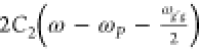 , teff(ω) =
  , respectively. This delay introduces a
relative phase between the Ak and Bk processes,
equal to ΔϕB–A = ωg′g′(teffBk(ω) – teff(ω)) = 2C2ωg′g2, which can be experimentally tuned by
means of the probe chirp.
, respectively. This delay introduces a
relative phase between the Ak and Bk processes,
equal to ΔϕB–A = ωg′g′(teffBk(ω) – teff(ω)) = 2C2ωg′g2, which can be experimentally tuned by
means of the probe chirp.
The above analysis clarifies that
a wealth of information on the
excited-state potentials is encoded in the dependence of the ISRS
signal on the probe wavelength. This dependence is complicated by
multiple interfering optical processes but can be controlled by varying
the chirp of the PP. Following this strategy, we determined the nuclear
displacements between the ground and excited PESs from the measured,
mode-dependent Raman excitation profiles (REPs), i.e., the change
in the intensities of the individual ISRS bands as a function of the
probe wavelength. In Figure 4, we report the cresyl violet REP  for different Raman modes
(
for different Raman modes
( 345, 470, 493, 525, 592, 675, 751, and
832 cm–1).
345, 470, 493, 525, 592, 675, 751, and
832 cm–1).  have been extracted by
fitting the ISRS
maps in the frequency domain (Figure 1d,e) as the sum of Lorentzian profiles, which have
been used to preliminary identify the normal-mode frequencies. The
REPs have been evaluated for three different values of the probe chirp
(−80, 40, and 360 fs2) to tune the relative phase
between A0–B1 processes: the experimental results are in good agreement
with signals modeled using eqs 1–4, which have been numerically
integrated using small steps of dz = 12.5 μm,
considering the sample absorption and the nonlinear contributions
separately.10 Because the
have been extracted by
fitting the ISRS
maps in the frequency domain (Figure 1d,e) as the sum of Lorentzian profiles, which have
been used to preliminary identify the normal-mode frequencies. The
REPs have been evaluated for three different values of the probe chirp
(−80, 40, and 360 fs2) to tune the relative phase
between A0–B1 processes: the experimental results are in good agreement
with signals modeled using eqs 1–4, which have been numerically
integrated using small steps of dz = 12.5 μm,
considering the sample absorption and the nonlinear contributions
separately.10 Because the  signal is evaluated over
a broad spectral
range, the PP chirp has been included in the simulation considering
also the third-order dispersion coefficient C3 (details on the calibration are reported in Methods and in the Supporting Information). In order to retrieve the displacements of the excited PES along
the normal modes, eqs 1–4 have been used to fit the experimental
signal is evaluated over
a broad spectral
range, the PP chirp has been included in the simulation considering
also the third-order dispersion coefficient C3 (details on the calibration are reported in Methods and in the Supporting Information). In order to retrieve the displacements of the excited PES along
the normal modes, eqs 1–4 have been used to fit the experimental  traces globally over
the data sets measured
at different PP chirps, considering fixed normal-mode frequencies
and dephasing times. The obtained normal-mode frequencies ν̃g′g and displacements d =
traces globally over
the data sets measured
at different PP chirps, considering fixed normal-mode frequencies
and dephasing times. The obtained normal-mode frequencies ν̃g′g and displacements d =  are summarized
in Table 1, while the projection of ground- and excited-state
potentials
along two normal modes (592 and 675 cm–1) are reported
in Figure 5. Importantly,
our results indicate a high displacement (d = 0.64
± 0.04) of the excited PES along the 592 cm–1 coordinate, pointing to an elongation of the Oxazine ring in the
excited state (Figure 5), due to a reorganization of the electronic density in the excited
state (as confirmed by the electronic density difference map reported
in the Supporting Information as Figure
S5), and small d (≤0.25) along the other investigated
vibrational degrees of freedom. We note that the values reported in Table 1 are lower than the
displacements obtained by spontaneous Raman spectroscopy for CV dissolved
in water,31 as expected in view of the
asymmetrically blue-shifted absorption spectrum of the aqueous CV
solutions (reported in the Supporting Information). In Figure 1c, we
show a comparison between the measured sample absorption spectrum
and the one calculated using the ISRS retrieved displacement, where
two additional contributions (at 300 and 1400 cm–1), which take into account for the Raman mode not accessed by the
present ISRS experiment, have been included in the simulation to fit
the experimental data. Interestingly, such a comparison indicates
that CV has high-frequency displaced normal modes outside the frequency
range investigated in the present work, in agreement with the results
reported for CV dissolved in aqueous solution.31
are summarized
in Table 1, while the projection of ground- and excited-state
potentials
along two normal modes (592 and 675 cm–1) are reported
in Figure 5. Importantly,
our results indicate a high displacement (d = 0.64
± 0.04) of the excited PES along the 592 cm–1 coordinate, pointing to an elongation of the Oxazine ring in the
excited state (Figure 5), due to a reorganization of the electronic density in the excited
state (as confirmed by the electronic density difference map reported
in the Supporting Information as Figure
S5), and small d (≤0.25) along the other investigated
vibrational degrees of freedom. We note that the values reported in Table 1 are lower than the
displacements obtained by spontaneous Raman spectroscopy for CV dissolved
in water,31 as expected in view of the
asymmetrically blue-shifted absorption spectrum of the aqueous CV
solutions (reported in the Supporting Information). In Figure 1c, we
show a comparison between the measured sample absorption spectrum
and the one calculated using the ISRS retrieved displacement, where
two additional contributions (at 300 and 1400 cm–1), which take into account for the Raman mode not accessed by the
present ISRS experiment, have been included in the simulation to fit
the experimental data. Interestingly, such a comparison indicates
that CV has high-frequency displaced normal modes outside the frequency
range investigated in the present work, in agreement with the results
reported for CV dissolved in aqueous solution.31
Figure 4.

Normalized cresyl violet ISRS Raman excitation profiles measured for different vibrational modes. The REP intensities have been obtained by fitting the frequency domain traces (Figure 1d,e) with a sum of Lorentzian profiles, evaluating the areas of the different Raman bands. The data (circles) and the model (solid lines) are compared as a function of the λP probe pulse wavelength for three different values of the probe chirp (reported on top of each column). All the y-axes range from 0 to 1.2, and the absolute intensities of each mode have been scaled by the factors reported on the axis.
Table 1. Peak Positions and Displacements between Ground- and Excited-State PESs Extracted by Fitting the ISRS Raman Excitation Profiles Reported with the Corresponding 90% Confidence Intervalsa.
| ν̃g′g (cm–1) |
 (DFT) (DFT) |
 (experimental) (experimental) |
Q (au) |
|---|---|---|---|
| 345 (2) | 0.27 | 0.18 (0.03) | 0.063 |
| 470 (2) | 0.24 | 0.21 (0.04) | 0.059 |
| 493 (2) | 0.13 | 0.15 (0.03) | 0.040 |
| 526 (2) | 0.18 | 0.26 (0.03) | 0.071 |
| 592 (2) | 0.60 | 0.63 (0.04) | 0.13 |
| 675 (2) | 0.19 | 0.19 (0.03) | 0.044 |
| 752 (2) | 0.11 | 0.17 (0.03) | 0.039 |
| 833 (2) | 0.17 | 0.17 (0.03) | 0.041 |
The results are compared with the displacements obtained by TD-DFT calculations performed with CAM-B3LYP functional and the 6-311++G(2d,2p) basis set. In the last column the extracted detected displacements are reported in atomic units.
Figure 5.

Cresyl violet potential energy surfaces along two normal modes (592 and 675 cm–1) measured by mapping the ISRS Raman excitation profiles. The experimental results are compared with time-dependent density functional theory calculations performed with CAM-B3LYP functional and the 6-311++G(2d,2p) basis set. The vertical dashed lines indicate the measured minima of ground- and excited-state parabolas. The excited-state potentials are vertically offset by a constant factor.
The experimental results are compared with ab initio time-dependent density functional theory (TD-DFT) calculations performed with CAM-B3LYP functional44 and the 6-311++G(2d,2p) basis set. Interestingly, the TD-DFT calculations reported in Table 1 and the ISRS extracted displacements are in good agreement, confirming the capability of the presented approach to access the details of the excited-state PES and of the normal modes with small displacements. A detailed comparison between the ISRS experimental results and the TD-DFT calculations is reported in the Supporting Information.
In conclusion, we have investigated the ISRS response in the presence of an off-resonant RP and a broadband resonant PP. A diagrammatic treatment of the pathways concurring with the signal generation has been employed to analyze the data. Taking advantage of the heterodyne background and fluorescence-free detection generated on the broadband probe pulse, Raman excitation profiles can be recorded in a single measurement, uncovering the vibrational response of systems that cannot be accessed by spontaneous or frequency domain approaches. The interaction with the resonant PP enables the projection of the molecular density matrix to the entire vibrational manifold in the excited electronic state, resulting in the interference between quantum pathways that concur with the generation of the experimental signal. Utilizing this molecular description, we have shown how to retrieve detailed information on the dipole moments over the different recorded Raman modes. As a benchmark of the proposed theoretical model and experimental scheme, we applied the two-color ISRS setup to map the PESs of cresyl violet dissolved in methanol, determining the nuclear displacements of excited-state PESs along the different monitored normal modes. Furthermore, our results establish a convenient experimental protocol, based on the use of a resonant chirped PP, to enhance the ISRS cross section of low scattering Raman modes. This protocol can be exploited for the identification of mode displacements for PESs involved in relaxation dynamics, being relevant for accessing the reaction coordinates that rule the initial stages of photoreactions. Upon adding an actinic pump beam, our approach can indeed be extended in a straightforward fashion for mapping the relative orientation between different excited transient PESs.
Methods
Experimental Setup. The experimental setup exploited for the measurements is based on a 1 kHz repetition rate Ti:sapphire laser source that generates 3.5 mJ, 35 fs pulses at 800 nm. The vertically polarized RP is synthesized by a noncollinear optical parametric amplifier (NOPA) that produces 15 fs broadband pulses centered at 680 nm, and its compression is controlled by a pair of chirped mirrors.45 The 10 nJ vertically polarized probe pulse is a white light continuum10 generated by focusing part of the source pulse on a sapphire plate and filtering the 800 nm component by means of a short-pass filter. The PP chirp can be reduced by a second pair of chirped mirrors or increased by introducing glass windows of different widths along the beam path. Both the pulses are focused on a 500 μm glass cuvette containing the cresyl violet solution, and then the transmitted PP is frequency dispersed by a spectrometer onto a CCD device. A synchronized chopper at 500 Hz blocks alternating RP pulses in order to measure the transmitted PP in both the presence and absence of the RP excitation. In such a way, the ISRS signal S(ω, ΔT) can be extracted as the normalized difference between the transmitted PP spectrum in the presence (IRP-on(ω, ΔT)) and in the absence (IRP-off(ω)) of the RP:
The measurement of the PP chirp and the calibration of the C2 and C3 terms in eq 3 can be obtained monitoring both the coherent artifact generated inside the sample as well as the slope of the coherent oscillations recorded in the time domain due to the 1040 cm–1 Raman mode of the solvent (details are reported in the Supporting Information). Taking advantage of the small period of such a mode (∼30 fs) this approach represents a convenient way to measure the probe pulse chirp simultaneously with the ISRS measurement. In addition, the chirp is directly measured in the very same spatial region where the ISRS signal is generated, hence directly taking into account the dispersion of the probe pulse during the propagation in the sample cuvette. Further details on the chirp calibration are reported in refs (15 and 40). The ISRS spectra of CV have been acquired for a 6.5 ps temporal window (ΔT spans from −0.5 to 6 ps), much higher than the dephasing time of the vibrational coherences (<3 ps for all the modes under consideration), with a 13 fs sampling interval. Further details on the experimental scheme are reported in ref (46).
Density Functional Theory Calculations. DFT and TD-DFT calculations reported in Figure 5 have been performed with the CAM-B3LYP44 functional and 6-311++G(2d,2p) basis set by using the Gaussian 09 software package. Upon optimization of the ground-state geometry by DFT, the normal mode eigenvectors calculated in the ground state have been exploited to generate displaced geometries along the normal modes under consideration, which in turn have been exploited to calculate the excited-state electronic potential by TD-DFT, with the same level of theory. Further details are provided in the Supporting Information.
Acknowledgments
G.B. and T.S. acknowledge the “Progetti di Ricerca Medi 2019” grant by Sapienza Universitá di Roma and the “MESPES: Mapping Excited State Potential Energy Surcafes” project by Italian Super Computing Resource Allocation. G.B. acknowledges Massimiliano Aschi for helpful support with computational resources and Emanuele Mai for constructive discussions. S.M gratefully acknowledges the support of NSF through Grant CHE-1953045. This project has received funding from the PRIN 2017 Project, Grant No. 201795SBA3-HARVEST, and from the European Union’s Horizon 2020 research and innovation program Graphene Flagship under Grant Agreement No. 881603.
Supporting Information Available
The Supporting Information is available free of charge at https://pubs.acs.org/doi/10.1021/acs.jpclett.1c02209.
Cresyl violet ISRS spectra obtained with a negatively chirped probe pulse; detailed derivation of the ISRS signals; details of the DFT and TD-DFT calculations; details of the calibration of the PP chirp; absorption spectrum of CV (PDF)
The authors declare no competing financial interest.
Supplementary Material
References
- Zewail A. H. Femtochemistry: Atomic-Scale Dynamics of the Chemical Bond. J. Phys. Chem. A 2000, 104, 5660–5694. 10.1021/jp001460h. [DOI] [PubMed] [Google Scholar]
- Long D. A.The Raman Effect; John Wiley & Sons, Ltd., 2002. [Google Scholar]
- Polli D.; Brida D.; Mukamel S.; Lanzani G.; Cerullo G. Effective temporal resolution in pump-probe spectroscopy with strongly chirped pulses. Phys. Rev. A: At., Mol., Opt. Phys. 2010, 82, 053809. 10.1103/PhysRevA.82.053809. [DOI] [Google Scholar]
- Kuramochi H.; Takeuchi S.; Tahara T. Femtosecond time-resolved impulsive stimulated Raman spectroscopy using sub-7-fs pulses: Apparatus and applications. Rev. Sci. Instrum. 2016, 87, 043107. 10.1063/1.4945259. [DOI] [PubMed] [Google Scholar]
- Liebel M.; Schnedermann C.; Wende T.; Kukura P. Principles and Applications of Broadband Impulsive Vibrational Spectroscopy. J. Phys. Chem. A 2015, 119, 9506–9517. 10.1021/acs.jpca.5b05948. [DOI] [PubMed] [Google Scholar]
- Mukamel S.Principles of Nonlinear Spectroscopy; Oxford University Press: New York, 1995. [Google Scholar]
- Potma E. O.; Mukamel S. In Coherent Raman Scattering Microscopy; Cheng J.-X., Xie X. S., Eds.; CRC Press: Boca Raton, FL, 2012. [Google Scholar]
- Rosca F.; Kumar A. T. N.; Ye X.; Sjodin T.; Demidov A. A.; Champion P. M. Investigations of Coherent Vibrational Oscillations in Myoglobin. J. Phys. Chem. A 2000, 104, 4280. 10.1021/jp993617f. [DOI] [Google Scholar]
- Kahan A.; Nahmias O.; Friedman N.; Sheves M.; Ruhman S. Following Photoinduced Dynamics in Bacteriorhodopsin with 7-fs Impulsive Vibrational Spectroscopy. J. Am. Chem. Soc. 2007, 129, 537–546. 10.1021/ja064910d. [DOI] [PubMed] [Google Scholar]
- Agrawal G.Nonlinear Fiber Optics; Academic Press, 2013. [Google Scholar]
- Kukura P.; McCamant D. W.; Mathies R. A. Femtosecond Stimulated Raman Spectroscopy. Annu. Rev. Phys. Chem. 2007, 58, 461. 10.1146/annurev.physchem.58.032806.104456. [DOI] [PubMed] [Google Scholar]
- Batignani G.; Bossini D.; Di Palo N.; Ferrante C.; Pontecorvo E.; Cerullo G.; Kimel A.; Scopigno T. Probing ultrafast photo-induced dynamics of the exchange energy in a Heisenberg antiferromagnet. Nat. Photonics 2015, 9, 506–510. 10.1038/nphoton.2015.121. [DOI] [Google Scholar]
- Virga A.; Ferrante C.; Batignani G.; De Fazio D.; Nunn A. D. G.; Ferrari A. C.; Cerullo G.; Scopigno T. Coherent anti-Stokes Raman spectroscopy of single and multi-layer graphene. Nat. Commun. 2019, 10, 3658. 10.1038/s41467-019-11165-1. [DOI] [PMC free article] [PubMed] [Google Scholar]
- Batignani G.; Pontecorvo E.; Bossini D.; Ferrante C.; Fumero G.; Cerullo G.; Mukamel S.; Scopigno T. Modeling the Ultrafast Response of Two-Magnon Raman Excitations in Antiferromagnets on the Femtosecond Timescale. Ann. Phys. 2019, 531, 1900439. 10.1002/andp.201900439. [DOI] [Google Scholar]
- Monacelli L.; Batignani G.; Fumero G.; Ferrante C.; Mukamel S.; Scopigno T. Manipulating Impulsive Stimulated Raman Spectroscopy with a Chirped Probe Pulse. J. Phys. Chem. Lett. 2017, 8, 966–974. 10.1021/acs.jpclett.6b03027. [DOI] [PubMed] [Google Scholar]
- Gdor I.; Ghosh T.; Lioubashevski O.; Ruhman S. Nonresonant Raman Effects on Femtosecond Pump–Probe with Chirped White Light: Challenges and Opportunities. J. Phys. Chem. Lett. 2017, 8, 1920–1924. 10.1021/acs.jpclett.7b00559. [DOI] [PubMed] [Google Scholar]
- Musser A. J.; Liebel M.; Schnedermann C.; Wende T.; Kehoe T. B.; Rao A.; Kukura P. Evidence for conical intersection dynamics mediating ultrafast singlet exciton fission. Nat. Phys. 2015, 11, 352–357. 10.1038/nphys3241. [DOI] [Google Scholar]
- Fujisawa T.; Kuramochi H.; Hosoi H.; Takeuchi S.; Tahara T. Role of Coherent Low-Frequency Motion in Excited-State Proton Transfer of Green Fluorescent Protein Studied by Time-Resolved Impulsive Stimulated Raman Spectroscopy. J. Am. Chem. Soc. 2016, 138, 3942–3945. 10.1021/jacs.5b11038. [DOI] [PubMed] [Google Scholar]
- Kim W.; Kim T.; Kang S.; Hong Y.; Würthner F.; Kim D. Tracking Structural Evolution during Symmetry Breaking Charge Separation in Quadrupolar Perylene Bisimide with Time-Resolved Impulsive Stimulated Raman Spectroscopy. Angew. Chem., Int. Ed. 2020, 59, 8571. 10.1002/anie.202002733. [DOI] [PubMed] [Google Scholar]
- Molesky B. P.; Guo Z.; Cheshire T. P.; Moran A. M. Perspective: Two-dimensional resonance Raman spectroscopy. J. Chem. Phys. 2016, 145, 180901. 10.1063/1.4966194. [DOI] [PubMed] [Google Scholar]
- Kuramochi H.; Takeuchi S.; Kamikubo H.; Kataoka M.; Tahara T. Fifth-order time-domain Raman spectroscopy of photoactive yellow protein for visualizing vibrational coupling in its excited state. Sci. Adv. 2019, 5, eaau4490. 10.1126/sciadv.aau4490. [DOI] [PMC free article] [PubMed] [Google Scholar]
- Fumero G.; Schnedermann C.; Batignani G.; Wende T.; Liebel M.; Bassolino G.; Ferrante C.; Mukamel S.; Kukura P.; Scopigno T. Two-Dimensional Impulsively Stimulated Resonant Raman Spectroscopy of Molecular Excited States. Phys. Rev. X 2020, 10, 011051. 10.1103/PhysRevX.10.011051. [DOI] [Google Scholar]
- Takeuchi S.; Ruhman S.; Tsuneda T.; Chiba M.; Taketsugu T.; Tahara T. Spectroscopic Tracking of Structural Evolution in Ultrafast Stilbene Photoisomerization. Science 2008, 322, 1073–1077. 10.1126/science.1160902. [DOI] [PubMed] [Google Scholar]
- Sun Y.; Benabbas A.; Zeng W.; Kleingardner J. G.; Bren K. L.; Champion P. M. Investigations of heme distortion, low-frequency vibrational excitations, and electron transfer in cytochrome c. Proc. Natl. Acad. Sci. U. S. A. 2014, 111, 6570–6575. 10.1073/pnas.1322274111. [DOI] [PMC free article] [PubMed] [Google Scholar]
- Johnson P. J. M.; Halpin A.; Morizumi T.; Prokhorenko V. I.; Ernst O. P.; Miller R. J. D. Local vibrational coherences drive the primary photochemistry of vision. Nat. Chem. 2015, 7, 980–986. 10.1038/nchem.2398. [DOI] [PubMed] [Google Scholar]
- Schnedermann C.; Muders V.; Ehrenberg D.; Schlesinger R.; Kukura P.; Heberle J. Vibronic Dynamics of the Ultrafast all- trans to 13- cis Photoisomerization of Retinal in Channelrhodopsin-1. J. Am. Chem. Soc. 2016, 138, 4757–4762. 10.1021/jacs.5b12251. [DOI] [PubMed] [Google Scholar]
- Batignani G.; Fumero G.; Kandada A. R. S.; Cerullo G.; Gandini M.; Ferrante C.; Petrozza A.; Scopigno T. Probing femtosecond lattice displacement upon photo-carrier generation in lead halide perovskite. Nat. Commun. 2018, 9, 1971. 10.1038/s41467-018-04367-6. [DOI] [PMC free article] [PubMed] [Google Scholar]
- Brennan M. C.; Herr J. E.; Nguyen-Beck T. S.; Zinna J.; Draguta S.; Rouvimov S.; Parkhill J.; Kuno M. Origin of the Size-Dependent Stokes Shift in CsPbBr3 Perovskite Nanocrystals. J. Am. Chem. Soc. 2017, 139, 12201–12208. 10.1021/jacs.7b05683. [DOI] [PubMed] [Google Scholar]
- Wang W.; Marshall M.; Collins E.; Marquez S.; Mu C.; Bowen K. H.; Zhang X. Intramolecular electron-induced proton transfer and its correlation with excited-state intramolecular proton transfer. Nat. Commun. 2019, 10, 1170. 10.1038/s41467-019-09154-5. [DOI] [PMC free article] [PubMed] [Google Scholar]
- Vogel E.; Gbureck A.; Kiefer W. Vibrational spectroscopic studies on the dyes cresyl violet and coumarin 152. J. Mol. Struct. 2000, 550–551, 177–190. 10.1016/S0022-2860(00)00385-9. [DOI] [Google Scholar]
- Leng W.; Kelley A. M. Resonance Raman Intensity Analysis of Cresyl Violet Bound to SiO2 Colloidal Nanoparticles. Langmuir 2003, 19, 7049–7055. 10.1021/la034638a. [DOI] [Google Scholar]
- Brazard J.; Bizimana L. A.; Gellen T.; Carbery W. P.; Turner D. B. Experimental Detection of Branching at a Conical Intersection in a Highly Fluorescent Molecule. J. Phys. Chem. Lett. 2016, 7, 14–19. 10.1021/acs.jpclett.5b02476. [DOI] [PubMed] [Google Scholar]
- Rafiq S.; Scholes G. D. Slow Intramolecular Vibrational Relaxation Leads to Long-Lived Excited-State Wavepackets. J. Phys. Chem. A 2016, 120, 6792–6799. 10.1021/acs.jpca.6b07796. [DOI] [PubMed] [Google Scholar]
- Batignani G.; Ferrante C.; Scopigno T. Accessing Excited State Molecular Vibrations by Femtosecond Stimulated Raman Spectroscopy. J. Phys. Chem. Lett. 2020, 11, 7805–7813. 10.1021/acs.jpclett.0c01971. [DOI] [PMC free article] [PubMed] [Google Scholar]
- Rahav S.; Mukamel S. Ultrafast Nonlinear Optical Signals Viewed from the Molecule’s Perspective: Kramers-Heisenberg Transition-Amplitudes versus Susceptibilities. Adv. At., Mol., Opt. Phys. 2010, 59, 223–263. 10.1016/S1049-250X(10)59006-2. [DOI] [Google Scholar]
- Dorfman K. E.; Fingerhut B. P.; Mukamel S. Time-resolved broadband Raman spectroscopies: A unified six-wave-mixing representation. J. Chem. Phys. 2013, 139, 124113. 10.1063/1.4821228. [DOI] [PMC free article] [PubMed] [Google Scholar]
- Batignani G.; Fumero G.; Mukamel S.; Scopigno T. Energy flow between spectral components in 2D broadband stimulated Raman spectroscopy. Phys. Chem. Chem. Phys. 2015, 17, 10454–10461. 10.1039/C4CP05361C. [DOI] [PMC free article] [PubMed] [Google Scholar]
- Fumero G.; Batignani G.; Dorfman K. E.; Mukamel S.; Scopigno T. On the Resolution Limit of Femtosecond Stimulated Raman Spectroscopy: Modelling Fifth-Order Signals with Overlapping Pulses. ChemPhysChem 2015, 16, 3438–3443. 10.1002/cphc.201500548. [DOI] [PubMed] [Google Scholar]
- Condon E. U. The Franck-Condon Principle and Related Topics. Am. J. Phys. 1947, 15, 365–374. 10.1119/1.1990977. [DOI] [Google Scholar]
- Batignani G.; Ferrante C.; Fumero G.; Scopigno T. Broadband impulsive stimulated Raman scattering based on a chirped detection. J. Phys. Chem. Lett. 2019, 10, 7789–7796. 10.1021/acs.jpclett.9b03061. [DOI] [PubMed] [Google Scholar]
- Batignani G.; Pontecorvo E.; Giovannetti G.; Ferrante C.; Fumero G.; Scopigno T. Electronic resonances in broadband stimulated Raman spectroscopy. Sci. Rep. 2016, 6, 18445. 10.1038/srep18445. [DOI] [PMC free article] [PubMed] [Google Scholar]
- Batignani G.; Fumero G.; Pontecorvo E.; Ferrante C.; Mukamel S.; Scopigno T. Genuine dynamics vs cross phase modulation artefacts in Femtosecond Stimulated Raman Spectroscopy. ACS Photonics 2019, 6, 492–500. 10.1021/acsphotonics.8b01467. [DOI] [Google Scholar]
- Zhou Y.; Constantine S.; Harrel S.; Ziegler L. D. The probe frequency dependence of nonresonant femtosecond pump–probe nuclear responses: Undercutting vibrational inhomogeneities. J. Chem. Phys. 1999, 110, 5893–5905. 10.1063/1.478489. [DOI] [Google Scholar]
- Yanai T.; Tew D. P.; Handy N. C. A new hybrid exchange–correlation functional using the Coulomb-attenuating method (CAM-B3LYP). Chem. Phys. Lett. 2004, 393, 51–57. 10.1016/j.cplett.2004.06.011. [DOI] [Google Scholar]
- Zavelani-Rossi M.; Cerullo G.; De Silvestri S.; Gallmann L.; Matuschek N.; Steinmeyer G.; Keller U.; Angelow G.; Scheuer V.; Tschudi T. Pulse Compression over a 170-THz Bandwidth in the Visible by Use of Only Chirped Mirrors. Opt. Lett. 2001, 26, 1155–1157. 10.1364/OL.26.001155. [DOI] [PubMed] [Google Scholar]
- Ferrante C.; Batignani G.; Pontecorvo E.; Montemiglio L. C.; Vos M. H.; Scopigno T. Ultrafast dynamics and vibrational relaxation in six-coordinate heme proteins revealed by Femtosecond Stimulated Raman Spectroscopy. J. Am. Chem. Soc. 2020, 142, 2285–2292. 10.1021/jacs.9b10560. [DOI] [PMC free article] [PubMed] [Google Scholar]
Associated Data
This section collects any data citations, data availability statements, or supplementary materials included in this article.