Summary
Multiple myeloma accounts for 10-15% of all hematologic malignancies, and 20% of deaths related to cancers of the blood and bone marrow. Diagnosis is defined by the presence of a serum monoclonal spike (M-spike) of more than 3 g/dL or more than 10% clonal plasma cells in the bone marrow and at least one myeloma-defining event, such as hypercalcemia, anemia, bone lesions, or renal impairment. The kidney is a major target organ, and renal impairment is frequently the first manifestation of the disease. Renal damage occurs in up to 40% of patients and 10-20% will require dialysis. Monoclonal immunoglobulin light chains are the major causes of renal complications in multiple myeloma. Glomerular disease, with the deposition of monoclonal immunoglobulins or their components, includes monoclonal immunoglobulin deposition disease, AL or AH amyloidosis, type I cryoglobulinemia, proliferative glomerulonephritis with monoclonal IgG deposits, immunotactoid glomerulopathy, and fibrillary glomerulonephritis. In addition, tubulointerstitial diseases with the deposition of monoclonal immunoglobulins or their components, are constituted by light chain cast nephropathy, light chain proximal tubulopathy, and crystal-storing histiocytosis.
We report the case of a 66-year-old woman who presented with albumin-predominant moderate proteinuria and renal failure. Serum and urine immunofixation electrophoresis showed monoclonal κ light chain in both. Renal biopsy confirmed κ-restricted crystal-storing renal disease involving proximal tubular epithelial cells and crystal storing histiocytosis. Multiple myeloma with crystal storing histiocytosis was discovered in bone marrow biopsy. Thus, we present an unusual case of a myeloma patient presenting light chain proximal tubulopathy and crystal-storing histiocytosis both in the kidney and in the bone marrow.
Key words: multiple myeloma, crystal-storing histiocytosis, bone marrow biopsy, light chains glomerulopathy, renal biopsy
Introduction
Multiple myeloma (MM) results from the clonal proliferation of plasma cells in the bone marrow and subsequent overproduction of immunoglobulins (Igs) or immunoglobulin fragments into the serum, and in urine, including free light chains (FLCs) 1. FLCs are low molecular-weight proteins normally produced by lymphoid tissues and 1-10 mg/day are excreted in the urine 2,3. Patients with paraprotein- related kidney disease present with various manifestations, including light chain cast nephropathy, monoclonal Ig deposition disease, light chain amyloidosis, light chain proximal tubulopathy (LCPT), and tubule-interstitial nephritis 3-5. A majority (approximately 70%) of pathogenic FLCs is tubulopathic, and about one-third interacts with glomerular structures (glomerulopathic FLCs) 6,7. Myeloma cast nephropathy (MCN), the most common Ig-related crystalline nephropathy, is characterized by crystalline precipitates of monoclonal light chain (either κ or λ) within distal tubules. On rare occasions, Igs crystallization occurs intracellularly within proximal tubular cells and in histiocytes, “crystal-storing histiocytosis” (CSH) 6-8. LCPT is classified into crystalline or non- crystalline, based on the presence or absence of crystal structures in the cytoplasm of epithelial tubular cells 3,8. Crystal-storing histiocytosis (CSH) is characterized by crystalline refractile eosinophilic inclusions in the cytoplasm of histiocytes. CSH involving lung, bone marrow 9, kidney 10, skin 11, ocular and periorbital areas 12 has been reported, usually with underlying lymphoplasmacytic disorders, most likely lymphoplasmacytic lymphoma or multiple myeloma.
Herein, we describe a myeloma patient presenting light chain proximal tubulopathy and crystal- storing histiocytosis both in the kidney and in the bone marrow.
Materials and methods
LIGHT MICROSCOPY
Kidney biopsy was fixed in buffered 10% formalin and embedded in paraffin. Serial 3-mm thick sections were cut and treated with hematoxylin and eosin, Jones methenamine silver, Masson trichrome, and periodic acid-Schiff reagent. In addition, Congo red staining was performed on 5-mm thick sections. Bone marrow biopsy was fixed in buffered 10% formalin, decalcified by EDTA, dehydrated in graded alcohols, and embedded in paraffin using standard techniques. Serial 3-mm thick sections were cut. Routine hematoxylin and eosin staining, Giemsa, Gomory and Prussian blue for Fedetection were used. In addition, Congo red staining was performed on 5-mm thick sections.
IMMUNOHISTOCHEMISTRY MICROSCOPY
The detection system for immunostaining is BOND Polymer Refine Detection on staining platform LEICA BOND III with 30 min at 100°C with BOND Epitope Retrievial sol. 1 for antibodies Lambda (polyclonal, Dako, 1:105) and CD138 (clone MI15, Dako, 1:300), 20 min for antibody Kappa (polyclonal, Dako, 1:50.000), antibody CD68R/PG-M1 (clone PG-M1, Dako, 1:500) use 30 min at 100°C with BOND Epitope Retrieval 2. The Roche’s CD14 antibody (clone EPR3653) are detected on platform BenchMark ULTRA using UltraView Universal DAB Detection Kit, 64 min of antigen retrieval with Cell Conditioning 1 and incubation for 32 min.
IMMUNOFLUORESCENCE MICROSCOPY
Samples were transported in Michel’s media, washed in buffer, and frozen in a cryostat. Sections, cut at 5 mm, were rinsed in buffer and reacted with fluorescein-tagged polyclonal rabbit anti-human antibodies to IgG, IgA, IgM, C3, C4, C1q, fibrinogen, and k-, and l-light chains (all from Dako, Carpenteria, CA, USA) for 1 h, rinsed, and a coverslip applied using aqueous mounting media.
ELECTRON MICROSCOPY
For electron microscopic examination, renal biopsy was fixed with glutaraldehyde, followed by post- fixation at 4°C in osmium tetroxide for 1 h. The sections were dehydrated by passing through a graded alcohol series, treated with aceton, and then embedded in epoxy resin. Semi-thin sections measuring 1-2 μm in thickness prepared using an ultramicrotome were stained with toluidine blue and then analyzed under a light microscope. Ultrathin sections measuring 70-80 nm in thickness placed on the grids were subjected to ultrastructural examination under a transmission electron microscope FEI TECNAI G2 SPRIT BIOTWIN c/o Virology Unit of Istituto Zooprofilattico Sperimentale della Lombardia e dell’Emilia-Romagna (IZSLER) of Brescia.
Case presentation
A 66-year-old normotensive woman was referred to our Renal Division to evaluate kidney dysfunction. At the presentation serum creatinine was 138 mmol/L (measured clearance 34 ml/min), and BUN 29 mg/dl. The patient had severe ipokalaemia (2,7 mEq/L) with a renal loss of potassium (excrete fraction 22%, urinary potassium 34 mEq/24h). Urine analysis showed absent glycosuria, proteinuria 1.95 g/24 h, red cells and granular cast at sediment examination. Serum protein electrophoresis showed a hypogammaglobulinemia (8.4%). Immune electrophoresis detected a Bence-Jones protein κ, and at serum κ and λ free light chain (κ 904 mg/dl, l 11,7 mg/dl; κ/λ ratio 77). ANA, ENA, hepatitis B and C markers, HIV antibodies, Quantiferon-TB were negative; C3 and C4 were normal, as were blood cell count and other electrolytes (calcium, phosphate, magnesium, sodium), except for potassium. Serum sodium bicarbonate was 23 mEq/L.
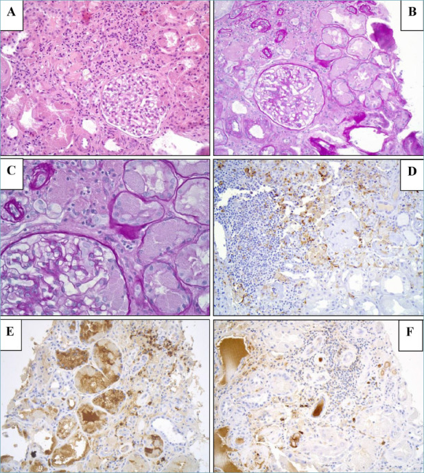
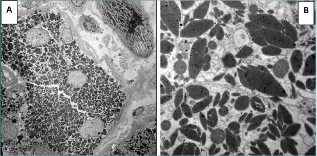
Percutaneous ultrasound-guided renal biopsy (Fig. 1) revealed a total of 9 glomeruli: 5 patents, without significant pathologic alterations; 4 globally sclerosed. Only one glomerulus showed macrophages with abnormal intracytoplasmic accumulation of crystals. Crystal-storing histiocytes also infiltrated the interstitium. Proximal tubular epithelial cells showed prominent intracytoplasmic inclusions, some of which were sharply demarcated and rhomboid in appearance. These inclusions were eosinophilic on the hematoxylin and eosin stain, pale on periodic acid Schiff stain. There was mild interstitial inflammation composed of mainly lymphocytes and rarer plasma cells. There were no associated with cast nephropathy or amyloid deposits. Immunohistochemical studies for κ and λ light chain on paraffin sections revealed κ monoclonal plasma cells, with variable staining of the intracytoplasmic material in tubular epithelial cells, while no staining was observed within histiocytes. Sampling for immunofluorescence consisted of 10 glomeruli and exhibited no significant glomerular positivity for IgG, IgM, IgA, C3, fibrinogen, k or λ light chains. Sampling for electron microscopy consisted of no glomeruli. Proximal tubular epithelial cells showed intracytoplasmic electron dense, polygonal-, rhomboid- (most frequent), or needle-shaped crystalline structures (Fig. 2). No electron dense deposits were observed in the tubular basal membrane.
Figure 1.

Renal biopsy. (A) (hematoxylin and eosin; 20x), (B) (PAS; 20x) and (C) (PAS; 40x) - Proximal tubule epithelial cells with prominent brightly eosinophilic intracytoplasmic inclusions and crystal - storing histiocytes present in the glomerulus and interstitium. (D) (immunohistochemical analyses; 20x) - Crystal-storing histiocytes were positive for CD14. (E) (immunohistochemical analyses; 20x) - κ light chain expressed in the majority of plasma cells and in the cytoplasm of proximal tubular epithelium. (F) (immunohistochemical analysis; 10x) - λ light chain expressed in rare plasma cells.
Figure 2.

(A, B). Tubular epithelial cells showed intracytoplasmic electron dense, polygonal-, rhomboid- (most frequent), or needle-shaped crystalline structures.
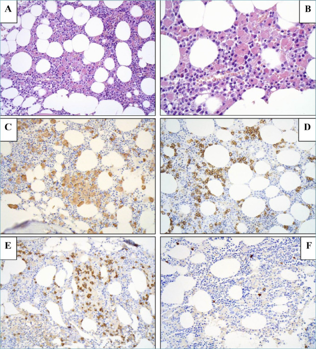
Bone marrow biopsy (Fig. 3) showed about 10-15% of mature plasma cells infiltrate with interstitial distribution. Plasma cells showed expression of CD138, CD20, and restriction of κ light chains by immunohistochemistry. No amyloid deposits were found in the bone marrow specimen by Congo red staining, also with examination in polarized light. Moreover, bone marrow specimen showed accumulation of histiocytes, positive by CD68R/PG-M1 immunostaining. The cytoplasm of the histiocytes was filled with eosinophilic crystalline material that was not immunohistochemically positive for kappa light chains. There were no intracytoplasmic crystals inside plasma cells. The hematopoietic matrix was normal for cellularity and maturation. Bone marrow aspiration showed normal cellularity and maturation of hematopoietic cells, with 12% of mature plasma cells. Hemophagocytic phenomenon was not observed. Cytogenetic evaluation showed a normal female karyotype. FISH analysis excluded the presence of IgH rearrangement (14q32.33) or the presence of del(17p)(p13.1).
Figure 3.

First bone marrow biopsy. (A) (hematoxylin and eosin; 10x) - Normocellular hematopoietic matrix with crystal-storing histiocytes. (B) (hematoxylin and eosin; 20x) - Hematopoietic matrix with crystal-storing histiocytes. (C) (immunohistochemical analysis; 10x) - Diffuse expression of CD68R by macrophages. (D) (immunohistochemical analysis; 10x) - Diffuse expression of CD138 by plasma cells. (E) (immunohistochemical analysis; 10x) - κ light chain expressed in the majority of plasma cells. (F) (immunohistochemical analysis; 10x) - λ light chain expressed in rare plasma cells.
The laboratory examination revealed normal blood counts; total circulating proteins were 6.8 g/dL. No monoclonal component was found, but hypo-gammaglobulinemia was detected. Free circulating light chain were 1030 mg/L (kappa) and 11 mg/L (lambda) with a ratio of 84. Urinary light chain kappa was 5600 mg/24h. Plasmatic calcium was normal. A low dose total body CT scan excluded skeletal lithic lesions.
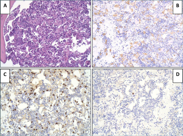
The diagnosis was consistent with micro-molecular kappa type multiple myeloma. The International Staging System 13 score was II. The presence of renal impairment was a sufficient criterion to start treatment. The patient underwent induction therapy with VTD (bortezomib - thalidomide - dexamethasone) triplet. Creatinine levels immediately before treatment were 1.9 mg/dL. After 4 cycles, the kappa/lambda free light chain ratio was 29, and urinary light chain kappa was 1536/24h. A new bone marrow biopsy was therefore performed (Fig. 4). It showed a light hypercellular matrix with persistence of about 8% of mature plasma cells infiltrate with interstitial distribution (Fig. 4). Plasma cells showed expression of CD138 and restriction of κ light chains by immunohistochemistry, mixed with a lot of crystal-storing histiocytes, positive by CD68R immunostaining.
Figure 4.

Second bone marrow biopsy. (A) (hematoxylin and eosin; 10x) - Light hypercellular matrix with scattered crystal-storing histiocytes. (B) (immunohistochemical analysis; 10x) - Diffuse expression of CD68R by crystal storing histiocytes. (C) (immunohistochemical analysis; 10x) - κ light chain expressed in the majority of plasma cells. (D) (immunohistochemical analysis; 10x) - λ light chain expressed in rare plasma cells.
According to these results, we confirmed that this patient with myeloma with crystal- storing histiocytosis exhibited simultaneous light chain proximal tubulopathy and crystal-storing histiocytosis, which were attributed to monoclonal κ light chains.
In a condition of partial remission according to IMWG 2006 recommendation 14, VTD therapy was continued until the sixth cycle. After the sixth cycle the urinary light chain kappa further lowered to 1010 mg/24h, Free light chain kappa/lamba ratio was reduced to 26. Creatinine levels were 1.36 mg/dL. At present, still in a condition of partial remission, the patient is waiting to proceed with autologous hematopoietic stem cells mobilization and harvest, followed by high dose chemotherapy with autologous transplantation.
Discussion
Multiple myeloma often involves the kidney and results in various light microscopic and ultrastructural entities falling under the umbrella of monoclonal gammopathy-associated renal pathology 10. Renal damage, most commonly detected as an asymptomatic elevation in serum creatinine, is the second leading cause of death in myeloma and 10-20% will require dialysis 4. Precipitated monoclonal immunoglobulins in the kidney exhibits various forms, including fibrils (amyloidosis, fibrillary glomerulopathy), microtubules (cryoglobulinemic glomerulonephritis, immunotactoid glomerulopathy), and crystals 10,15. Monoclonal crystals can take either of the following forms: (a) extracellular crystals within glomerular capillary loops or within interstitial vessel lumens, (b) intracellular crystals within one or more cell types of the kidney which may be glomerular (mesangial cell and visceral epithelial cell) and/or extra-glomerular (usually proximal tubular epithelial cell), and (c) infiltration of histiocytes containing monoclonal crystals in their cytoplasm (crystal-storing histiocytosis)10,16-18.
Usually, the overload of the proximal tubular epithelial cells lysosomal system is followed by acute tubular necrosis. On rare occasions, the reabsorbed pathogenic LCs form intracellular crystals that impair tubular function, which apart from the rise in serum creatinine, is typically associated with the occurrence of glycosuria, aminoaciduria, hyperuricosuria, hyperphosphaturia, hypercalciuria, and type 2 renal tubular acidosis, typical constituents of an acquired Fanconi syndrome 19-23. Light chain crystals can also accumulate in other cell types such as podocytes and macrophages, disease processes termed light chain podocytopathy and crystal storing histiocytosis, respectively 3,24-25.
The diagnosis of LCPT can be challenging, and is made by the identification of light chain-restricted intracytoplasmic inclusions within the proximal tubule epithelial cells, which may be quite focal. In the largest series of such cases, Stokes et al. 7 reported 46 cases of LCPT (40 with crystals and 6 without). All 40 cases with crystals were kappa light chain restricted. Stokes reported that most patients had proteinuria (98%) and renal insufficiency (83%). Urinary protein excretion was often heavy (> 1 g/day in 87% and > 3 g/day in 37%), but hypoalbuminemia was not seen. This means the proteinuria consisted predominantly of LC rather than albumin 7. As in our patient, in up to 85% of patients kidney biopsy showing LCPT is the initial clue to an underlying plasma cell dyscrasia before the disease has come to clinical attention 7. The underlying hematological disease is MM in 14-33% of cases with the rest being MGUS, SMM, WM, NHL, and CLL 10. Therefore, the diagnosis of LCPT is crucial for appropriate patient management. The renal biopsy of our patient showed light chain deposition disease, with proximal tubulopathy (κ light chain restricted) and CSH. The bone marrow biopsy showed κ monoclonal plasmacytosis and CSH. Renal immunofluorescence (IF) exhibited no significant glomerular positivity for IgG, IgM, IgA, C3, fibrinogen, k or λ light chains. However, in crystalline LCPT renal injury, IF staining of the light chain on frozen tissue is often negative, which may be due to the light chain epitope hiding in the crystal pattern. However, immunostaining for light chains on paraffin tissue after antigenic retrieval and immuno-electron microscopy study can show monoclonal light chain deposition in the crystals.
Crystal-storing histiocytosis presents with sheets of polygonal and occasionally spindle shaped histiocytes with eccentrically placed, hyperchromatic nuclei with fine chromatin. On close examination or occasionally with polarization, the cytoplasm of the histiocytes will contain needle- like to rhomboid shaped crystals. The intracytoplasmic crystals stain positively for kappa or lambda in most instances, or for heavy chains, although they may occasionally be polyclonal 26. In the majority of CSH (76-90%) there is an associated underlying lymphoplasmacytic neoplasm expressing κ light chain, although there are case reports with λ and polyclonal crystal formation 27. In the remaining 10-34% of cases, CSH is associated with other conditions, autoimmune disorders, reactive inflammatory conditions, metabolic disorders, and drugs (e.g. clofazimine, which is used to treat leprosy) 15,27-30. Crystal-storing histiocytosis is classified into two types, localized and generalized, based on the number of organs involved 10. The localized form of CSH (58-82% of cases) is confined to a single organ. Generalized CSH (18-42% of cases) involves multiple organ sites (bone marrow, lungs, kidney, lymph nodes, liver, spleen, and gastrointestinal tract) and tends to have a worse prognosis 27-30. About 20 cases have been reported of renal involvement in CSH up to the date 16,31,32. In the kidney CSH is rarely a stand-alone pathology, and is in fact, often found co- existing with other morphologic forms of monoclonal renal disease (both crystal and non-crystal forms), including LCPT, usually crystalline subtype, crystalloid glomerulopathy or nephropathy, and cast nephropathy 21. Conditions mimicking CSH include hemophagocytic lymphohistiocytic syndrome, storage diseases such as Gaucher’s, mycobacterial and fungal infections, xanthogranuloma, Langerhans histiocytosis 33. Similarly, pathologists should be aware that an underlying plasma cell neoplasm or B-cell lymphoma can be masked by the histiocytic infiltrates. The International Kidney and Monoclonal Gammopathy Research Group recommends chemotherapy or stem cell transplant for LCPT even before worsening hematologic disease occurs 34. Kidney function showed a general improvement trend in patients who were treated with chemotherapy alone or chemotherapy and stem cell transplantation 7.
In summary, we present a case of LCPT associated with CSH, which is a rare finding on renal biopsy and requires a high index of suspicion, often including immunohistochemical studies on the paraffin- embedded tissue for the diagnosis. Once diagnosis is established, it is necessary to test for underlying lymphoplasmacytic disorders. Correct diagnosis is crucial in driving proper patient management including chemotherapy and/or stem cell transplant for both control of the hematologic process and treatment of kidney disease.
Figures and tables
References
- 1.Swerdlow SH, Campo E, Lee Harris N, et al., eds. WHO classification of tumours of haematopoietic and lymphoid tissues (revised 4th edition). Lyon: IARC, 2017. pp. 465-482. [Google Scholar]
- 2.Nasr SH, Valeri AM, Sethi S, et al. Clinico-pathologic correlations in multiple myeloma: a case series of 190 patients with kidney biopsies. Am J Kidney Dis 2012;59:786-794. https://doi.org/10.1053/j.ajkd.2011.12.028 10.1053/j.ajkd.2011.12.028 [DOI] [PubMed] [Google Scholar]
- 3.Herlitz LC, D’Agati VD, Markowitz GS. Crystalline nephropathies. Arch Pathol Lab Med 2012;136:713-720. https://doi.org/10.5858/arpa.2011-0565-RA 10.5858/arpa.2011-0565-RA [DOI] [PubMed] [Google Scholar]
- 4.Korbet SM, Schwartz MM. Multiple myeloma. J Am Soc Nephrol 2006;17:2533-2545. https://doi.org/10.1681/ASN.2006020139 10.1681/ASN.2006020139 [DOI] [PubMed] [Google Scholar]
- 5.Doshi M, Lahoti A, Danesh FR, et al.; American Society of Nephrology Onco- Nephrology Forum. Paraproteinrelated kidney disease: kidney injury from paraproteins-what determines the site of injury? Clin J Am Soc Nephrol 20167;11:2288-2294. https://doi.org/10.2215/CJN.02560316 10.2215/CJN.02560316 [DOI] [PMC free article] [PubMed] [Google Scholar]
- 6.Stompór T, Perkowska-Ptasińska A, Wojciechowska M, et al. Unusual manifestation of crystalline light chain tubulopathy in patient with multiple myeloma: case report and review of the literature. Ren Fail 2014;36:795-799. https://doi.org/10.3109/0886022X.2014.884446 10.3109/0886022X.2014.884446 [DOI] [PubMed] [Google Scholar]
- 7.Stokes MB, Valeri AM, Herlitz L, et al. Light chain proximal tubulopathy: clinical and pathologic characteristics in the modern treatment era. J Am Soc Nephrol 2016;27:1555-1565. https://doi.org/10.1681/ASN.2015020185 10.1681/ASN.2015020185 [DOI] [PMC free article] [PubMed] [Google Scholar]
- 8.Basnayake K, Stringer SJ, Hutchison CA, et al. The biology of immunoglobulin free light chains and kidney injury. Kidney Int 2011;79:1289-1301. https://doi.org/10.1038/ki.2011.94 10.1038/ki.2011.94 [DOI] [PubMed] [Google Scholar]
- 9.Uthamalingam P, Mehta S. Crystal-storing histiocytosis: report of a rare case presenting with pathological fracture of femur. is there more to the entity? Int J Surg Pathol 2017;25:458-461. https://doi.org/10.1177/1066896917696746 10.1177/1066896917696746 [DOI] [PubMed] [Google Scholar]
- 10.Kousios A, Roufosse C. An update on paraprotein related renal pathology. Diagnostic Pathology 2019;25:10;408-421. [Google Scholar]
- 11.Li JJ, Henderson C.Cutaneous crystal storing histiocytosis: a report of two cases J Cutan Pathol. 2015February;42(2):136-43. https://doi.org/10.1111/cup.12413. 10.1111/cup.12413 [DOI] [PubMed] [Google Scholar]
- 12.Duquesne A, Werbrouck A, Fabiani B, et al., Complete remission of monoclonal gammopathy with ocular and periorbital crystal storing histiocytosis and Fanconi syndrome. Hum Pathol 2013;44:927-933. https://doi.org/10.1016/j.humpath.2012.10.012 10.1016/j.humpath.2012.10.012 [DOI] [PubMed] [Google Scholar]
- 13.Palumbo A, Avet-Loiseau H, Oliva S, et al. Revised International Staging System for Multiple Myeloma: A Report From International Myeloma Working Group. J Clin Oncol 2015;33:2863-2869. https://doi.org/10.1200/JCO.2015.61.2267 10.1200/JCO.2015.61.2267 [DOI] [PMC free article] [PubMed] [Google Scholar]
- 14.Durie BGM, Harousseau JL, Miguel JS, et al. International uniform response criteria for multiple myeloma. Leukemia 2006;20;1467-1473. https://doi.org/10.1038/sj.leu.2404284 10.1038/sj.leu.2404284 [DOI] [PubMed] [Google Scholar]
- 15.Gupta RK, Rosenberg AZ, Bagnasco SM, et al. Renal crystal-storing histiocytosis involving glomeruli - A comprehensive clinicopathologic analysis. Ann Diagn Pathol 2019;43:1-9. https://doi.org/10.1016/j.anndiagpath.2019.151403 10.1016/j.anndiagpath.2019.151403 [DOI] [PubMed] [Google Scholar]
- 16.Vankalakunti M, Bonu R, Shetty S, et al. Crystalloid glomerulopathy in monoclonal gammopathy of renal significance (MGRS). Clin Kidney J 2014;7:296-298. https://doi.org/10.1093/ckj/sfu025 10.1093/ckj/sfu025 [DOI] [PMC free article] [PubMed] [Google Scholar]
- 17.Bridoux F, Leung N, Hutchison CA, et al. Diagnosis of monoclonal gammopathy of renal significance. Kidney Int 2015; 87:698-711. https://doi.org/10.1038/ki.2014.408 10.1038/ki.2014.408 [DOI] [PubMed] [Google Scholar]
- 18.Wu CK, Yang AH, Lai HC, et al. Combined proximal tubulopathy, crystal storing histiocytosis, and cast nephropathy in a patient with light chain multiple myeloma. BMC Nephrology 2017;18:170. https://doi.org/10.1186/s12882-017-0584-8 10.1186/s12882-017-0584-8 [DOI] [PMC free article] [PubMed] [Google Scholar]
- 19.Raman SBK, Van Slyck EJ. Nature of intracytoplasmic crystalline inclusions in myeloma cells (morphologic, cytochemical, ultrastructural and immunofluorescent studies). Am J Clin Pathol 1983;80:224-228. https://doi.org/10.1093/ajcp/80.2.224 10.1093/ajcp/80.2.224 [DOI] [PubMed] [Google Scholar]
- 20.Messiaen T, Deret S, Mougenot B, et al. Adult Fanconi syndrome secondary to light chain gammopathy. Clinicopathologic heterogeneity and unusual features in 11 patients. Medicine (Baltimore). 2000;79:135-154. https://doi.org/10.1097/00005792-200005000-00002 10.1097/00005792-200005000-00002 [DOI] [PubMed] [Google Scholar]
- 21.El Hamel C, Thierry A, Trouillas P, et al., Crystal-storing histiocytosis with renal Fanconi syndrome: pathological and molecular characteristics compared with classical myeloma-associated Fanconi syndrome. Nephrol Dial Transplant 2010;25:2982-2990. https://doi.org/10.1093/ndt/gfq129. 10.1093/ndt/gfq129 [DOI] [PubMed] [Google Scholar]
- 22.Leboulleux M, Lelongt B, Mougenot B, et al. Protease resistance and binding of Ig light chains in myeloma-associated tubulopathies. Kidney Int 1995;48:72-79. https://doi.org/10.1038/ki.1995.269 10.1038/ki.1995.269 [DOI] [PubMed] [Google Scholar]
- 23.Herrera GA. Proximal tubulopathies associated with monoclonal light chains: the spectrum of clinicopathologic manifestations and molecular pathogenesis. Arch Pathol Lab Med 2014;138:1365-1380. https://doi.org/10.5858/arpa.2013-0493-OA 10.5858/arpa.2013-0493-OA [DOI] [PubMed] [Google Scholar]
- 24.Kowalewska J, Tomford RC, Alpers CE. Crystals in podocytes: an unusual manifestation of systemic disease. Am J Kidney Dis 2003;42:605-611. https://doi.org/10.1016/s0272-6386(03)00794-7 10.1016/s0272-6386(03)00794-7 [DOI] [PubMed] [Google Scholar]
- 25.Ma CX, Lacy MQ, Rompala JF, et al. Acquired Fanconi syndrome is an indolent disorder in the absence of overt multiple myeloma. Blood 2004;104:40-42. https://doi.org/10.1182/blood-2003-10-3400 10.1182/blood-2003-10-3400 [DOI] [PubMed] [Google Scholar]
- 26.Dogan S, Barnes L, Cruz-Vetrano WP. Crystal-storing histiocytosis: report of a case, review of the literature (80 cases) and a proposed classification. Head Neck Pathol 2012;6:11-20. https://doi.org/10.1007/s12105-011-0326-3 10.1007/s12105-011-0326-3 [DOI] [PMC free article] [PubMed] [Google Scholar]
- 27.Lebeau A, Zeindl-Eberhart E, Muller EC, et al. Generalized crystal-storing histiocytosis associated with monoclonal gammopathy: molecular analysis of a disorder with rapid clinical course and review of the literature. Blood 2002;100:1817-27. [PubMed] [Google Scholar]
- 28.Gupta V, El Ters M, Kashani K, et al. Crystalglobulin-induced nephropathy. J Am Soc Nephrol 2015; 26:525-529. https://doi.org/10.1681/ASN.2014050509 10.1681/ASN.2014050509 [DOI] [PMC free article] [PubMed] [Google Scholar]
- 29.Fang H, Chiu A, Reichard KK. Crystal-storing histiocytosis in bone marrow: a clinicopathologic study of eight cases and review of the literature. Am J Clin Pathol 2018;149:148-163. https://doi.org/10.1093/ajcp/aqx150 10.1093/ajcp/aqx150 [DOI] [PubMed] [Google Scholar]
- 30.Kanagal-Shamanna R, Xu-Monette ZY, Miranda RN, et al. Crystal-storing histiocytosis: a clinicopathological study of 13 cases. Histopathology 201668:482-491. https://doi.org/10.1111/his.12768 10.1111/his.12768 [DOI] [PMC free article] [PubMed] [Google Scholar]
- 31.Pitman SD, Wang J, Serros ER, Zuppan C. A 70-year-old woman with acute renal failure. Crystal-storing histiocytosis. Arch Pathol Lab Med 2006;130:1077-1078. https://doi.org/10.5858/2006-130-1077-AYWWAR 10.5858/2006-130-1077-AYWWAR [DOI] [PubMed] [Google Scholar]
- 32.Sethi S, Cuiffo BP, Pinkus GS, Rennke HG. Crystal-storing histiocytosis involving the kidney in a low-grade B-cell lymphoproliferative disorder. Am J Kidney Dis 2002;39:183-8. https://doi.org/10.1053/ajkd.2002.29914 10.1053/ajkd.2002.29914 [DOI] [PubMed] [Google Scholar]
- 33.Muppa P, Hussain F, Yasir S. Gastric crystal storing histiocytosis: clinicopathologic findings and morphologic mimics. Human Pathology: Case Reports 2018;14:78-80. https://doi.org/10.1016/j.ehpc.2018.09.002 10.1016/j.ehpc.2018.09.002 [DOI] [Google Scholar]
- 34.Fermand JP, Bridoux F, Kyle RA. How I treat monoclonal gammopathy of renal significance (MGRS). Blood 2013;122:3583-3590. https://doi.org/10.1182/blood-2013-05-495929 10.1182/blood-2013-05-495929 [DOI] [PubMed] [Google Scholar]


