
Figure 2: Deletion of BST2 leads to decreased MV a in primary neurons.
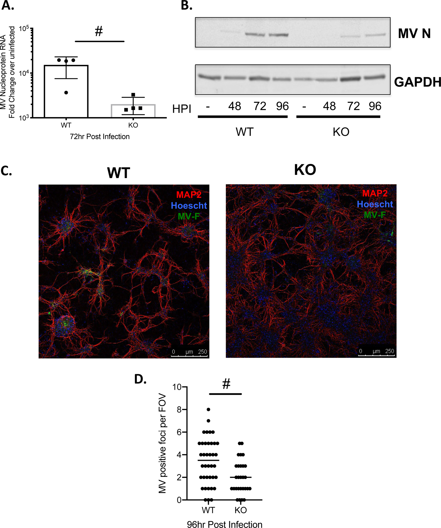
Primary neurons of the indicated genotype (WT: NSE-CD46+; KO: NSE-CD46+/BST2 KO) were infected with MV-Edmonston at an MOI=1. A) RNA was collected at the indicated time point (hours post-infection; hpi) and analyzed by RT-qPCR for MV nucleoprotein RNA. Results from at least 3 independent experiments are represented using the ΔΔCT method. B) Western blot analysis of protein collected at the indicated times post infection. Blots were probed with a polyclonal MV nucleoprotein antibody and an antibody to GAPDH as a loading control. C) Immunofluorescence staining of WT and KO primary neuronal cultures. Red- MAP2; neuronal marker. Blue-Hoescht; nuclei marker. Green- MV fusion protein. Representative images from each genotype are shown. D) MV positive foci were scored across multiple fields of view (FOV). # p <0.05 Unpaired T test.